
system useful for reporting protein-protein interactions in the bacterial periplasm

a protein-protein interaction and periplasm technology, applied in the field of system useful for reporting protein-protein interactions in the bacterial periplasm, can solve the problems of complicated interpretation of interaction data, high false positive rate of both y2h and b2h gal4-type assays, and inability to select genes

Active Publication Date: 2011-11-24
CORNELL UNIVERSITY
View PDF2 Cites 3 Cited by
  • Summary
  • Abstract
  • Description
  • Claims
  • Application Information

AI Technical Summary

Benefits of technology

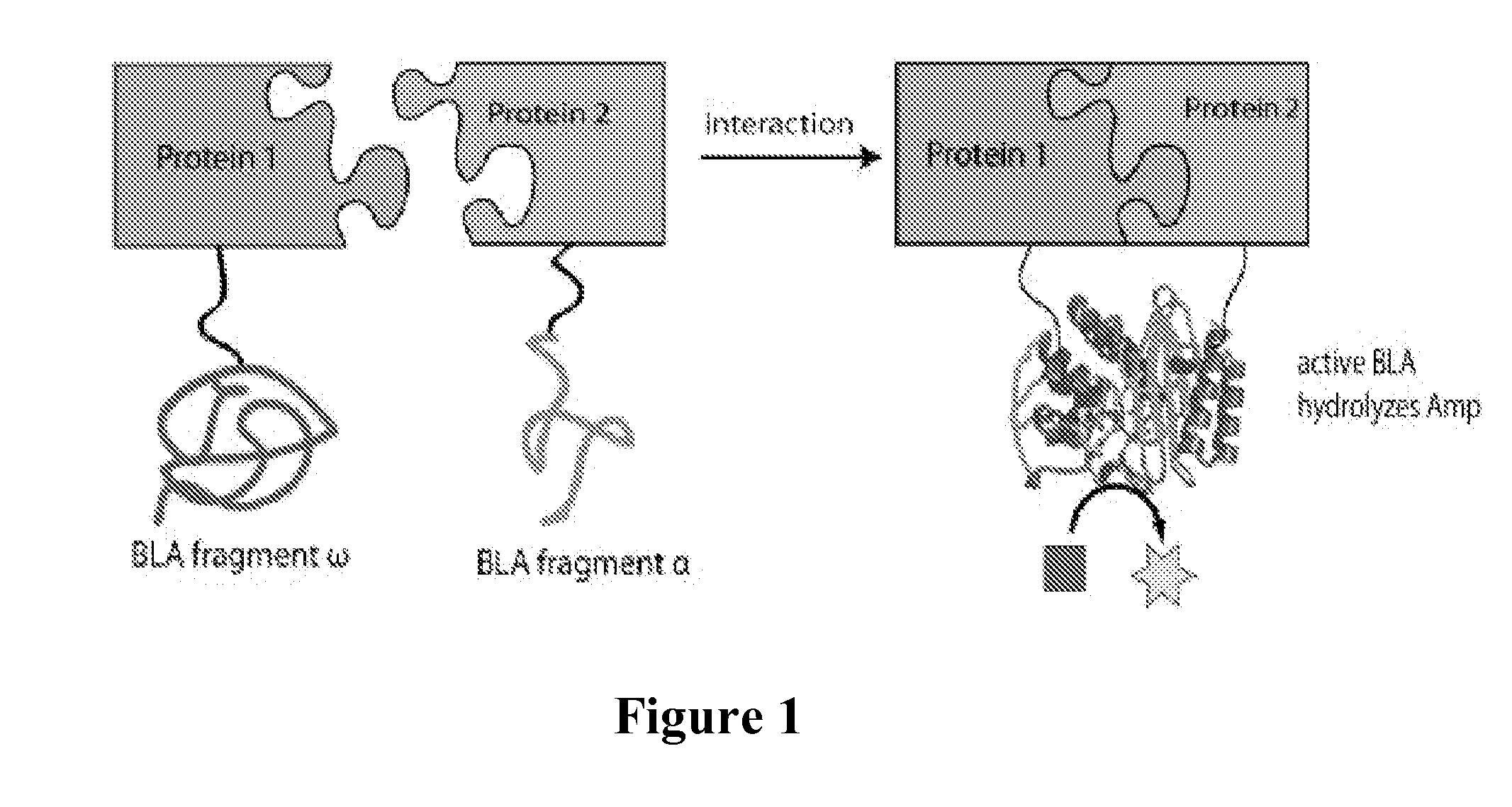
[0013]While numerous assays exist for detecting protein interactions in the cytoplasm of living cells, the major advantage of the present invention is that it can be used to assay for protein-protein interactions in the periplasmic compartment of bacteria. Such an assay would be significant because of the importance of the bacterial periplasm in a large number of biotechnological applications. This importance stems from several unique features of the periplasm relative to other sub-cellular compartments. For instance, proteins that require disulfide bonds for correct folding can only adopt a native conformation when localized in the oxidizing environment of the bacterial periplasm (disulfide bonds do not normally form in the cytoplasm of bacteria). Examples of disulfide-bond containing proteins of biotechnological relevance include proteins and protein fragments of the immunoglobulin family (e.g., IgGs). The ability to detect protein interactions in the periplasm can easily be combined with the ability to express full-length IgGs or fragments derived thereof (e.g., single-chain Fv (scFv)) in the periplasm and opens the door to a cell-based selection system for isolating and engineering virtually all types of antibodies or other immune / non-immune binding proteins. Since the technology described above only requires the antigen or domains of the antigen to be expressed in the periplasm, this technology should enable the selection of antibodies against both soluble and membrane proteins. Since many complex membrane proteins can be expressed in the inner membrane of E. coli (e.g., GPCRs, ion channels, efflux pumps, etc.), this technology should enable isolation of antibodies or other binding proteins that interact with exposed loops of membrane proteins. Such a feat is difficult or impossible using traditional antibody selection methods (e.g., cell surface display, phage display). More recently, the development of N-linked glycosylation in bacteria opens the bacterial periplasm as a compartment for attaching N-glycans to specific residues in recombinant proteins. Another advantage of the periplasm is that isolation of recombinant proteins from this compartment is greatly simplified compared to isolation from the more crowded cytoplasm. To accomplish this while also overcoming previous limitations of split β-lactamase systems in E. coli, a preferred embodiment of the present invention is co-expression of β-lactamase fragment fusions that are engineered with co-translational export signals (e.g., signal peptides from the E. coli DsbA protein). The advantage of such export signals is that at least one or both fragments of the reporter protein, and the interacting proteins (members of the putative binding pair) to which they are fused, are localized directly into the periplasm with little or no residence time in the cytoplasm. As a result, premature interaction between the proteins under investigation is effectively eliminated.
is that it can be used to assay for protein-protein interactions in the periplasmic compartment of bacteria. Such an assay would be significant because of the importance of the bacterial periplasm in a large number of biotechnological applications. This importance stems from several unique features of the periplasm relative to other sub-cellular compartments. For instance, proteins that require disulfide bonds for correct folding can only adopt a native conformation when localized in the oxidizing environment of the bacterial periplasm (disulfide bonds do not normally form in the cytoplasm of bacteria). Examples of disulfide-bond containing proteins of biotechnological relevance include proteins and protein fragments of the immunoglobulin family (e.g., IgGs). The ability to detect protein interactions in the periplasm can easily be combined with the ability to express full-length IgGs or fragments derived thereof (e.g., single-chain Fv (scFv)) in the periplasm and opens the door to a cell-based selection system for isolating and engineering virtually all types of antibodies or other immune / non-immune binding proteins. Since the technology described above only requires the antigen or domains of the antigen to be expressed in the periplasm, this technology should enable the selection of antibodies against both soluble and membrane proteins. Since many complex membrane proteins can be expressed in the inner membrane of E. coli (e.g., GPCRs, ion channels, efflux pumps, etc.), this technology should enable isolation of antibodies or other binding proteins that interact with exposed loops of membrane proteins. Such a feat is difficult or impossible using traditional antibody selection methods (e.g., cell surface display, phage display). More recently, the development of N-linked glycosylation in bacteria opens the bacterial periplasm as a compartment for attaching N-glycans to specific residues in recombinant proteins. Another advantage of the periplasm is that isolation of recombinant proteins from this compartment is greatly simplified compared to isolation from the more crowded cytoplasm. To accomplish this while also overcoming previous limitations of split β-lactamase systems in E. coli, a preferred embodiment of the present invention is co-expression of β-lactamase fragment fusions that are engineered with co-translational export signals (e.g., signal peptides from the E. coli DsbA protein). The advantage of such export signals is that at least one or both fragments of the reporter protein, and the interacting proteins (members of the putative binding pair) to which they are fused, are localized directly into the periplasm with little or no residence time in the cytoplasm. As a result, premature interaction between the proteins under investigation is effectively eliminated.

Problems solved by technology

Because such interactions mediate numerous disease states and biological mechanisms underlying the pathogenesis of bacterial and viral infections, identification of protein-protein interactions remains one of the most important challenges in the post-genomics era.
However, it has several drawbacks, including in the fact that it requires the nuclear environment of the eukaryotic yeast host, which may differ from the interaction environment of the proteins of interest.
Unfortunately, both Y2H and B2H GAL4-type assays are prone to a high frequency of false positives that arise from spurious transcriptional activation (Fields S., “High-throughput two-hybrid Analysis.
The Promise and the Peril,”FEBS J 272: 5391-5399 (2005)), and complicate the interpretation of interaction data.
One limitation of cytoplasmic expression is that cytoplasmic β-lactamase (Bla) is incapable of conferring resistance to ampicillin and thus genetic selection is not possible.
The limitation of this approach is that it uses post-translational Sec export signals.
A further limitation of this approach is the fact that only one pair of small peptides was tested for interaction; whether split Bla (β-Lactamase) could be used to report the interactions between globular proteins was not demonstrated.
Thus, to date there have been no reports detailing the use of split protein fragments for monitoring protein-protein interactions in the bacterial periplasm.

Method used


Image

  •  system useful for reporting protein-protein interactions in the bacterial periplasm
  •  system useful for reporting protein-protein interactions in the bacterial periplasm
  •  system useful for reporting protein-protein interactions in the bacterial periplasm

Examples


example 1

Construction of Vectors

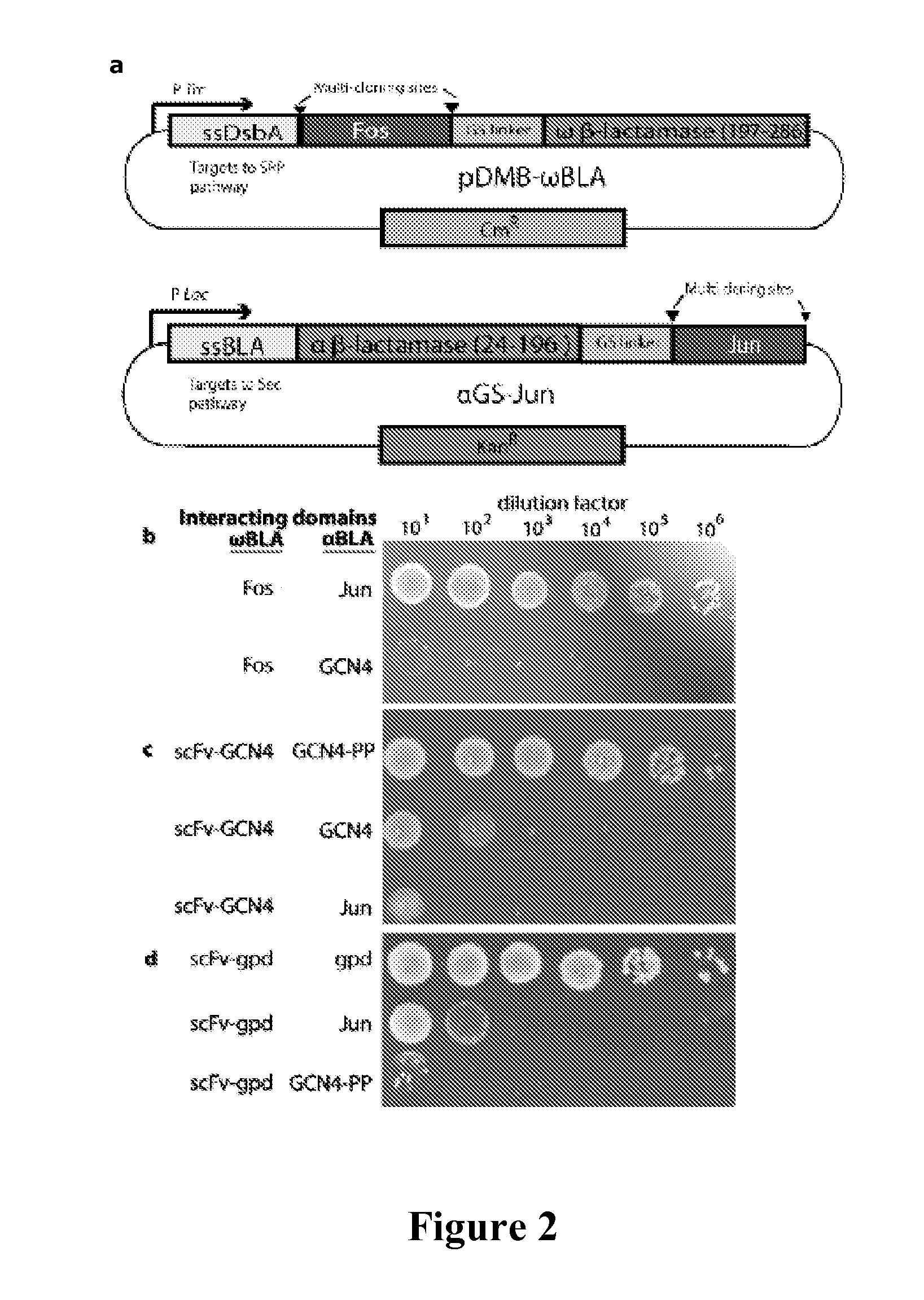
[0073]All cloning was performed using standard molecular biological techniques. A gene sequence comprising a (GGGGS)3-NGR linker sequence followed by residues 198-286 of TEM-1 BLA was cloned between the BamHI and HindIII sites of pDMB to create pDMB-ωBLA. Genes to be fused to the ωBLA fragment were then cloned into this vector between XbaI and SalI sites. Genes to be fused to the αBLA fragment were cloned between the KpnI and BamHI sites of vector αGS-Jun (Wehrman, T., et al., “Protein-protein Interactions Monitored in Mammalian Cells Via Complementation of Beta-lactamase Enzyme Fragments,”Proc Natl Acad Sci U S A 99:3469-3474 (2002), which is hereby incorporated by reference in its entirety). Fos and Jun leucine knockouts and scFv-GCN4 were as in (DeLisa, M. P., “Versatile Selection Technology for Intracellular Protein-protein Interactions Mediated by Bacterial Hitchhiker Transport Mechanism,”Proc Natl Acad Sci USA 106:3692-3697 (2009), which is hereby incorp...

example 2

Expression of Fusion Proteins and Cell Growth Assays

[0074]E. coli MC4100 cells were co-transformed with plasmids pDMB-P1-ωBLA (CmR) and aGS-P2 (KanR), where P1 and P2 represent proteins of interest. Cells were grown overnight at 37° C. in LB medium containing 20 μg / mL chloramphenicol (Cm) and 50 μg / mL kanamycin (Kan). Screening of cells on LB agar was performed by first normalizing overnight cultures by OD600 and then spotting 5 μL, of serially diluted (10-106-fold) cells on LB agar plates with 12, 25, 50, or 50 μg / mL of ampicillin (Amp). In all cases, the plates were incubated 16 h at 37° C. and then imaged using a ChemiDoc System (BioRad). For MBC / MIC determination, approximately 200 colony forming units (CFUs) of each clone were plated on LB agar plates containing 0, 3, 6, 12, 25, 50, 100, 200, 400, 800, or 1600 μg / mL Amp or 20 μg / mL Cm. The minimum bacteriocidal concentration was determined as the minimum Amp concentration at which no colonies appeared on the plates. Minimum inh...

example 3

Antibody-Antigen Interactions are Reported in Two Cases

[0076]To determine the ability of the assay of the present invention to detect and eventually engineer antibody-antigen interactions, interacting protein pairs as well as non-interacting proteins were cloned into two plasmids carrying the two inactive Bla fragments (FIG. 2A). In this study, two antibody-antigen pairs were examined. First, an scFv which is known to bind the GCN4 leucine zipper was tested. In addition to the scFv and wild-type GCN4 pair, a double proline mutant, GCN4-PP as an antigen for the scFv was also tested. GCN4 is normally a homodimer, but a double proline mutant of GCN4 (GCN4-pp) exhibits reduced dimerization, thereby reducing the amount of inactive α / α dimers in the assay. FIGS. 2B-D show the results of spot plating of the interacting pairs, with α-Jun as a negative control. The spot plates correspond to a 16-fold higher MBC (minimum bacteriocidal concentration, see Example 2 for full definition of MIC / MB...


PUM

PropertyMeasurementUnit
Protein activityaaaaaaaaaa
Transport propertiesaaaaaaaaaa
Affinityaaaaaaaaaa

Abstract

One aspect of the present invention relates to a reporter system for detection of protein-protein interactions in the periplasm of a prokaryotic host cell. The reporter system includes a first expression system which has a nucleic acid molecule encoding a first fragment of a reporter protein molecule, a nucleic acid molecule encoding a first signal sequence, and a nucleic acid molecule encoding a first member of a putative binding pair, where the nucleic acid molecule encoding the first fragment, the nucleic acid molecule encoding the first signal sequence, and the nucleic acid molecule encoding the first member of the putative binding pair are operatively coupled to permit their expression in a prokaryotic host cell as a first fusion protein. The reporter sys tem also includes a second expression system which has a nucleic acid molecule encoding a second fragment of the reporter protein molecule, a nucleic acid molecule encoding a second signal sequence, and a nucleic acid molecule encoding a second member of the putative binding pair, where the nucleic acid molecule encoding the second fragment, the nucleic acid molecule encoding the second signal sequence, and the nucleic acid molecule encoding the second member of the putative binding pair are operatively coupled to permit their expression in a prokaryotic host cell as a second fusion protein, where, when expressed in a prokaryotic host cell, at least one of the first and the second fusion proteins are co-translationally transported to the periplasm where, when present, the first and second members of the putative binding pair bind together and the first and second fragments of the reporter protein molecule are reconstituted, thereby producing an active reporter protein. The reporter system may be used to carry out methods of identifying candidate compounds which bind to a target protein, identifying a candidate gene which modulates binding between a first protein and second protein, and identifying a candidate compound which modulates binding between a first protein and second protein.

Description

[0001]This application claims the benefit of U.S. Provisional Patent Application Ser. No. 61 / 115,172, filed Nov. 17, 2008.[0002]This invention was made with government support under grant number 1R21CA132223 by the National Institutes of Health. The government has certain rights in this invention.FIELD OF THE INVENTION[0003]The present invention is directed to a system useful for reporting protein-protein interactions in the bacterial periplasm.BACKGROUND OF THE INVENTION[0004]Protein-protein interactions are key molecular events that integrate multiple gene products into functional complexes in virtually every cellular process. These cellular processes are mostly mediated by non-covalently interacting multi-protein complexes and can include, for example, transcription, translation, metabolic pathways or signal transduction pathways. Because such interactions mediate numerous disease states and biological mechanisms underlying the pathogenesis of bacterial and viral infections, iden...

Claims


Application Information

Patent Timeline
IPC IPC(8): C40B30/04C12Q1/68
CPCC07K2319/034C12N15/1055C07K2319/70C07K2319/61
Inventor DELISA, MATTHEW P.MANSELL, THOMAS
Owner CORNELL UNIVERSITY
Learn More