Assays using surface-enhanced raman spectroscopy (SERS)-active particles
a technology of raman spectroscopy and active particles, which is applied in auxillary shaping apparatus, group 4/14 element organic compounds, peptides, etc., can solve the problems of limitations of biological assays known in the art, and achieve the effects of improving the detection limit, increasing the signal, and increasing the signal
- Summary
- Abstract
- Description
- Claims
- Application Information
AI Technical Summary
Benefits of technology
Problems solved by technology
Method used
Image
Examples
example 1
Magnetic Capture SERS Assay with Reference Labels
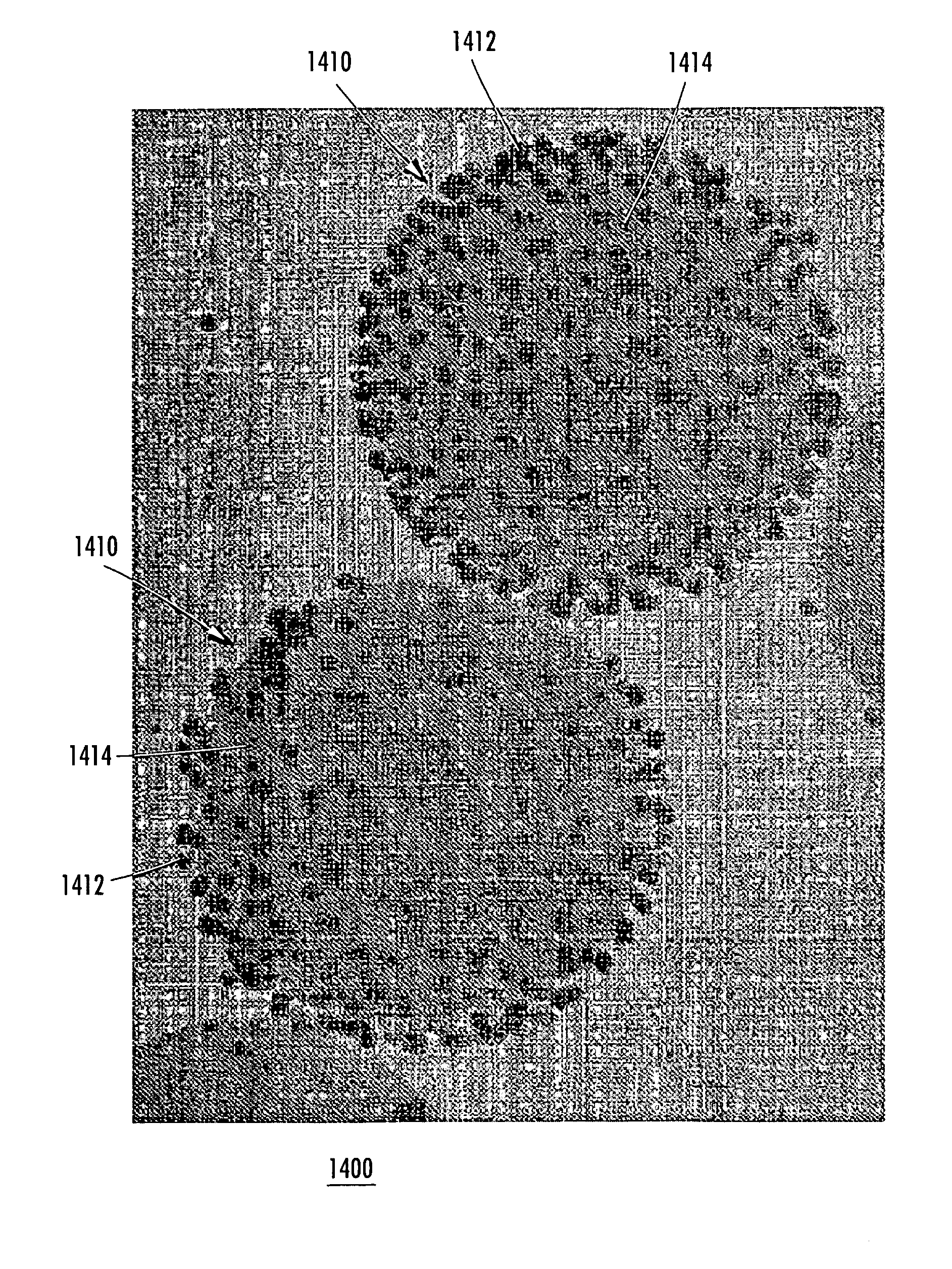
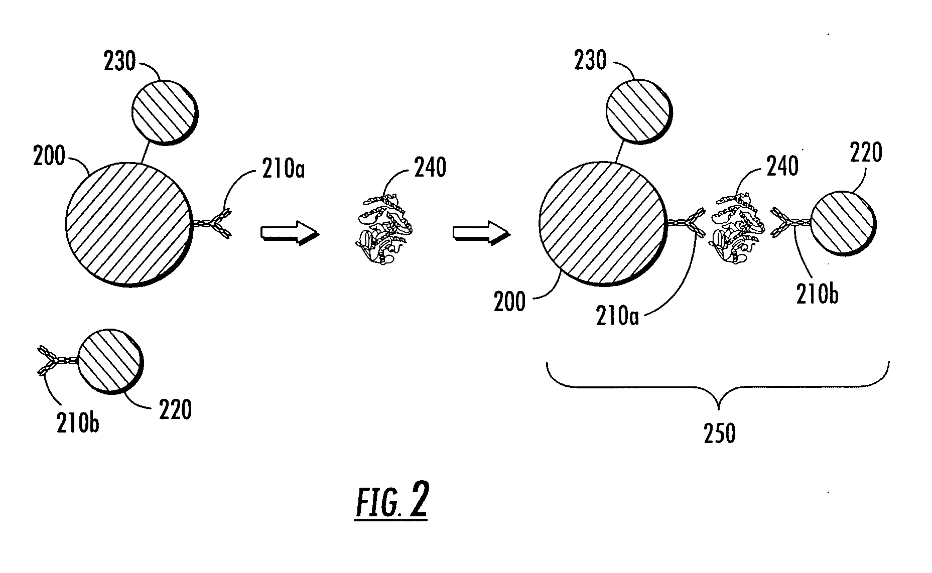
[0304]Nanoplex™ Biotags were purchased from Oxonica Inc. (Mountain View, Calif.). The particles were gold particles having a diameter of about 50 nm and labeled with Raman reporter selected from trans-1,2-bis(4-pyridyl)-ethylene (BPE) or 4,4′-dipyridyl (DPY) and encapsulated with glass as described herein. The glass encapsulant was biotinylated. Magnetic particles (approximately one micron in diameter) were purchased from Bangs Laboratories, Inc. (Fishers, Ind.) and labeled with streptavidin. Two solutions of biotinylated BPE- and DPY-labeled nanoparticles were mixed each with the streptavidin-coated magnetic particles. These two solutions were then mixed together in a ratio of 7:3 and a magnetic field was applied using a system of the type illustrated in FIG. 4, thereby forming a pellet at the bottom of the sample tube. The pellet was then repeatedly disrupted and reformed, and the Raman signal was measured for each pellet configurat...
example 2
Magnetic Capture SERS Assay with Lysis Reagent
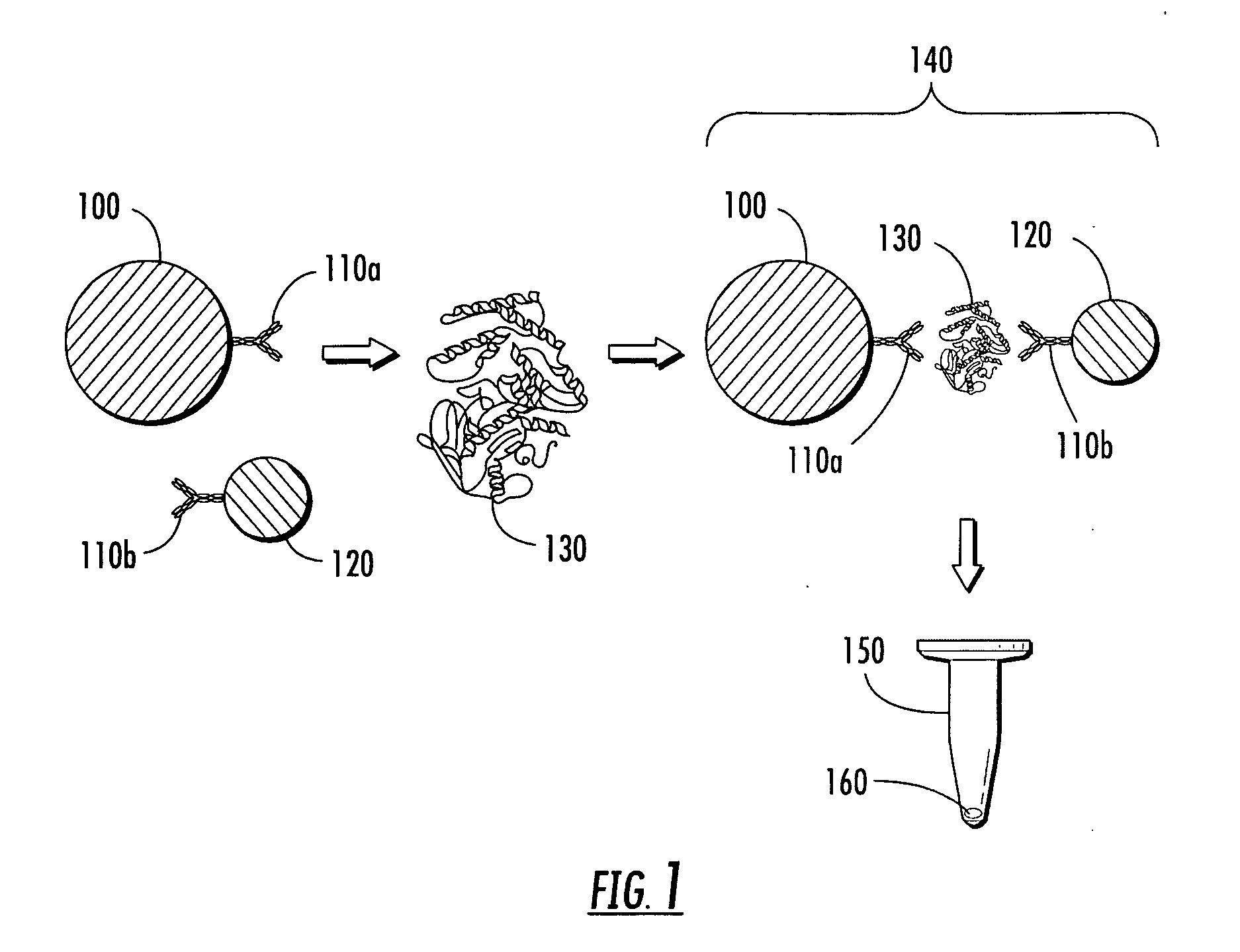
[0306]Oxonica Nanoplex™ nanoparticles with surface thiol groups were labeled with goat anti-human cTnI polyclonal antibodies (BiosPacific, Emeryville, Calif.) using Sulfo-SMCC (sulfosuccinimidyl-4-(N-maleimidomethyl)cyclohexane-1-carboxylate). Separately, magnetic particles (Bioclone 1-μm BcMag®Carboxyterminated magnetic beads) were labeled with anti-human cTnI monoclonal antibodies (BiosPacific, Emeryville, Calif.) using 1-ethyl-3-[3-dimethylaminopropyl]carbodiimide hydrochloride (EDC). A master mix of antibody-labeled nanoparticles and magnetic particles was created, which resulted in each assay of 162 μL containing 5.1×10⁸ nanoparticles and 15 μg magnetic particles. A lysing reagent solution also was prepared, containing 10 mM HEPES, 50 mM β-glycerophosphate, 70 mM NaCl, 2 mM EDTA, 1% Triton X100, and 1× Sigma Protease Inhibitor Cocktail P2714.
[0307]To test the impact of the lysing reagent on assay performance, samples were prepared c...
example 3
Pellet Formation by Rotation of Sample Tube
[0309]Magnetic particles from Bangs Laboratories (approximately one micron in diameter) dispersed in water were used to form a dense pellet as follows. A magnet (e.g., a rod) is mounted below a sample tube, where the center of the magnet is positioned off center in respect to the sample tube axis (see FIG. 5). After a few seconds, the magnet induced formation of a pellet at the bottom of the sample tube. The sample tube was rotated around its center axis, thereby modulating the magnetic field experienced by the pellet in such a way that the pellet becomes denser. The schematic in FIG. 5 shows the process of pellet formation by rotating the sample tube above an off-center mounted magnet.
[0310]After the magnet has been placed below the sample tube, particles are captured by the magnet in a manner of seconds. The formed pellet can be irregular in shape and can resemble the cross section of a torus. After turning the sample tube around its own ...
PUM
| Property | Measurement | Unit |
|---|---|---|
| diameter | aaaaa | aaaaa |
| thickness | aaaaa | aaaaa |
| thickness | aaaaa | aaaaa |
Abstract
Description
Claims
Application Information
 - R&D
- Intellectual Property
- Life Sciences
- Materials
- Tech Scout
- Unparalleled Data Quality
- Higher Quality Content
- 60% Fewer Hallucinations
Browse by: Latest US Patents, China's latest patents, Technical Efficacy Thesaurus, Application Domain, Technology Topic, Popular Technical Reports.
© 2025 PatSnap. All rights reserved.Legal|Privacy policy|Modern Slavery Act Transparency Statement|Sitemap|About US| Contact US: help@patsnap.com


