Increased resolution microscopy

a microscopy and resolution technology, applied in the field of increased resolution microscopy, can solve the problems of unnecessarily stressing the sample with radiation in the area, the current method cannot be used, and the disadvantage is considered, so as to achieve the effect of faster image production, faster acquisition speed, and faster acquisition speed

Active Publication Date: 2011-09-22
CARL ZEISS MICROSCOPY GMBH
View PDF14 Cites 21 Cited by
  • Summary
  • Abstract
  • Description
  • Claims
  • Application Information

AI Technical Summary

Benefits of technology

The invention is a method for improving the speed of image production in PAL microscopy by analyzing frames and adjusting the acquisition of frames accordingly. This method allows for faster image acquisition by evaluating frames and deriving a quality function based on the proportion of activated label molecules in the sample. The quality function is maximized to ensure optimal image acquisition speed. The method also adapts to changing boundary conditions and reduces the duration of the acquisition process overall. The invention ensures equipment protection and optimizes the speed of image acquisition.

Problems solved by technology

In this method also, it is to be considered disadvantageous that the sample is unnecessarily stressed with radiation in areas outside the detected focus, as the necessary structured illumination covers the whole sample volume.
Moreover, this method cannot currently be used in the case of thick samples, as extra-focally excited fluorescence also reaches the detector as a background signal and thus dramatically reduces the dynamic range of the detected radiation.
The result of this is that large data quantities are processed, and the measurement lasts a correspondingly long time.

Method used


Image

  • Increased resolution microscopy
  • Increased resolution microscopy
  • Increased resolution microscopy

Examples


Embodiment Construction

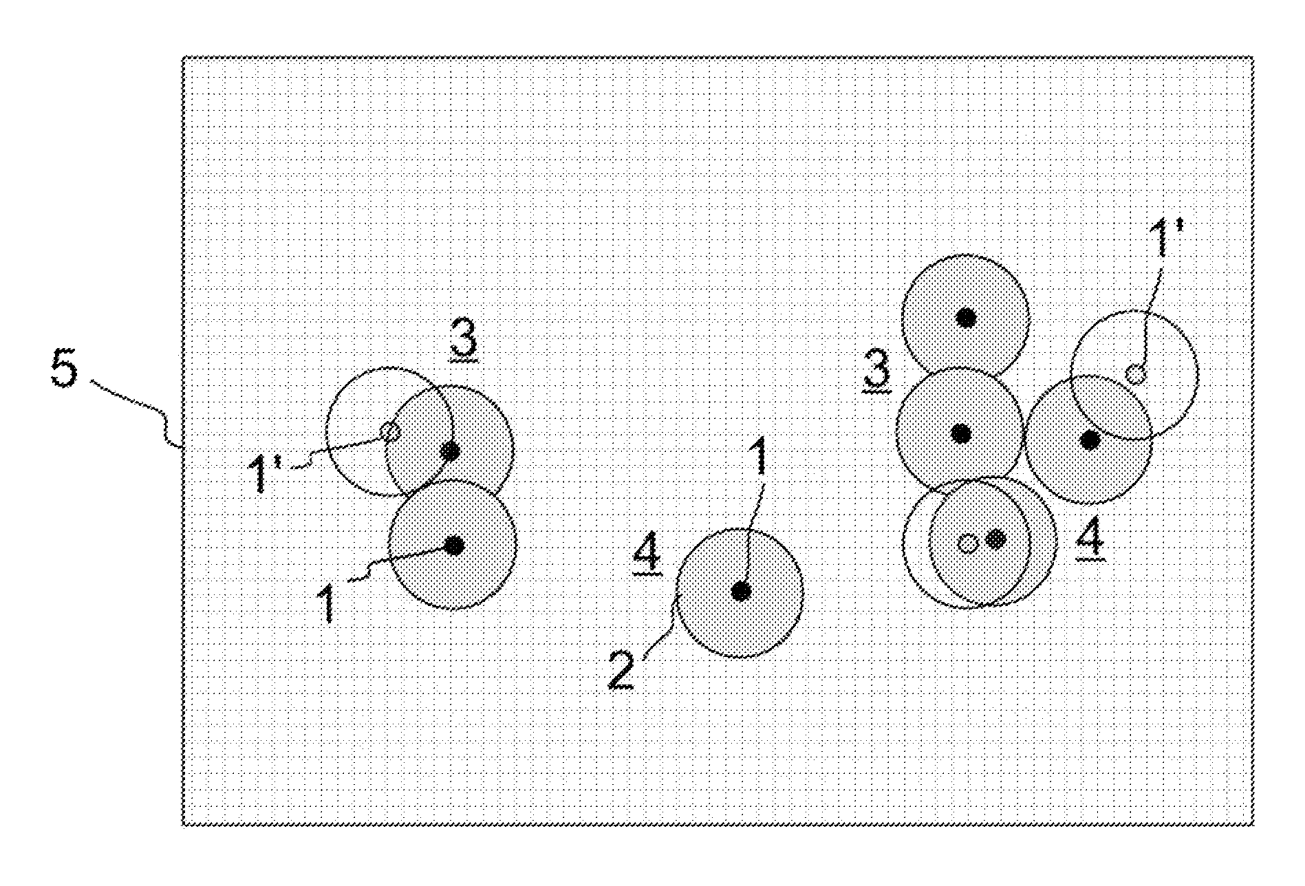
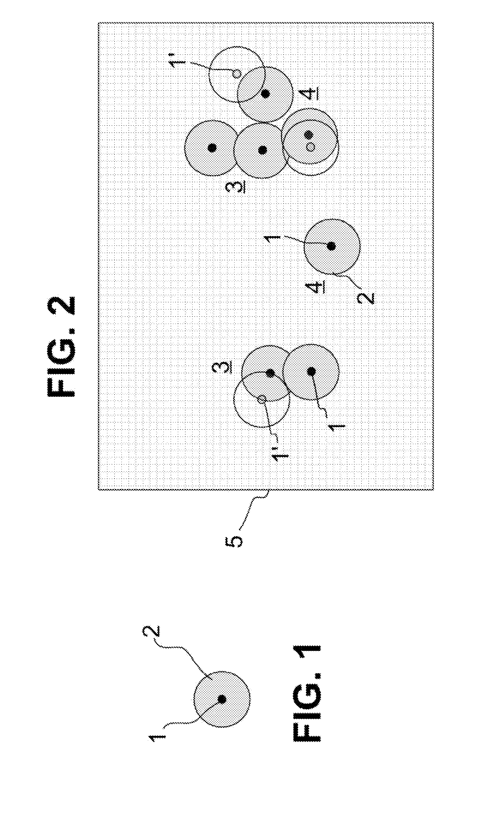
[0054]FIG. 1 schematically shows a label molecule 1 which has been excited to fluorescence. Of course, fluorescence detection requires a plurality of excitations, as each excitation delivers precisely one fluorescence photon and radiation detection requires an integration over many fluorescence photons. Due to the laws of physics, the fluorescence radiation emitted by the label molecule 1 can be detected in a microscope with only a limited optical resolution. Even if the microscope reaches the diffraction limit of the optical resolution, the photons of the fluorescent label molecule 1 are still always scattered due to diffraction and thus detected as an Airy disk 2. The microscope thus depicts in principle a larger object which is illustrated in FIG. 1 by the Airy disk 2, instead of the geometric extent of the label molecule 1 which is drawn schematically in FIG. 1 as a black circle. The size of the Airy disk 2 depends on the quality of the microscopy device used and is defined by t...


PUM

PropertyMeasurementUnit
wavelengthaaaaaaaaaa

Abstract

Method for spatially high-resolution luminescence microscopy in which label molecules in a sample are activated to emit luminescence radiation comprising activating only a subset of the label molecules in the sample, wherein activated label molecules have a distance to the closest activated molecules that is greater or equal to a length which results from a predetermined optical resolution, detecting the luminescence radiation, generating a frame from the luminescence radiation, identifying the geometric locations of the label molecules with a spatial resolution increased above the predetermined optical resolution, repeating the steps and forming a combined image, and controlling the acquisition of the several frames by evaluating at least one of the frames or a group of the frames and modifying at least one variable for subsequent repetitions of the steps of generating frames for combining into an image.

Description

PRIORITY CLAIM[0001]The present application is a National Phase entry of PCT Application No. PCT / EP2009 / 008117, filed Nov. 14, 2009, which claims priority from German Application Number 10 2008 059 328.1, filed Nov. 27, 2008, the disclosures of which are hereby incorporated by reference herein in their entirety.BACKGROUND OF THE INVENTION[0002]Method for spatially high-resolution luminescence microscopy of a sample which is labelled with label molecules which can be activated by a switching signal such that they can be excited to emit particular luminescence radiation only in the activated state, wherein the method has the following steps:[0003]a) introducing the switching signal onto the sample such that only a subset of the label molecules present in the sample are activated, wherein there are area parts in the sample in which activated label molecules have a distance to their closest neighbouring activated labelled molecules that is at least greater than or equal to a length whic...

Claims


Application Information

Patent Timeline
Patent Type & Authority Applications(United States)
IPC IPC(8): G01N21/64
CPCG01N21/6458G02B21/16G02B27/58G02B21/361G02B21/367
Inventor WOLLESCHENSKY, RALFKLEPPE, INGOKRAMPERT, GERHARDKEMPE, MICHAEL
Owner CARL ZEISS MICROSCOPY GMBH
Why Eureka
Try Eureka