Method for producing artificial skin

a skin and artificial technology, applied in the field of artificial skin production, can solve the problems of a potential risk of unknown infections, and a two-dimensional culture of only cells, and achieve the effects of no cytotoxic effect, no cytotoxic effect, and easy mixing

Inactive Publication Date: 2011-03-03
SHOWA UNIVERSITY
View PDF0 Cites 10 Cited by
  • Summary
  • Abstract
  • Description
  • Claims
  • Application Information

AI Technical Summary

Benefits of technology

[0024]According to the method for producing artificial skin of the invention, artificial skin that does not contain any animal-derived material or pathogen and has excellent biocompatibility can be produced. Moreover, in the method of the invention, a culture medium that is free of any animal-derived component, such as Fetal Bovine Serum (FBS), is used together with the above-mentioned scaffold, thereby enabling the production of a hybrid artificial skin material wherein neither the culture medium nor scaffold contain components derived from an animal or another person's tissue.
[0025]Further, in the method of the invention, the peptide hydrogel used as a scaffold can be readily mixed with cells and bioactive molecules (growth factors) during self-assembly, and is also unlikely to induce an immunological reaction because of its low molecular weight. Furthermore, the peptide hydrogel has a physiological affinity for tissues, and has no cytotoxic effects because it is degraded to amino acids, which are inherently present in large amounts within tissues.
[0026]Furthermore, in known methods for producing artificial skin using collagen gel, the collagen gel remains even after long-term culture; by contrast, in the method of the invention, the peptide hydrogel used as a scaffold for grafting is degraded after the passage of a necessary period of time, and therefore, does not remain in tissues. This provides the advantage of promoting the migration, proliferation, and differentiation of the cultured cells. In summary, the method of the invention is useful for growing skin in vivo, and the artificial skin produced by the method of the invention is particularly suitable for clinical graft applications.
[0027]The artificial skin produced by the method of the invention uses a synthetic material consisting of amino acids as a scaffold. This eliminates the costs that are incurred from removing potentially contained pathogens when using an animal-derived material as a scaffold; therefore, the artificial skin of the invention can be prepared at low cost.
[0028]Furthermore, excluding cells, the artificial skin produced by the method of the invention uses synthetic materials, so that it can be produced in large amounts with uniform quality. Furthermore, the artificial skin produced by the method of the invention contains no bioactive molecules (growth factors), which are endogenous substances that become problematic when artificial skin contains natural materials.

Problems solved by technology

Recently, however, the fact that it is difficult to completely reproduce what is happening in vivo by two-dimensional culture of only cells has been recognized.
However, hybrid artificial skin containing living fibroblasts and epidermal keratinocytes, which is currently being researched and developed, often uses an animal-derived substance as a scaffold, thus involving a potential risk of unknown infections.
The administration or transplantation of a substance derived from an animal or another person's tissue, even if it appears to be safe at the time, poses a potential risk of unknown infections, not to mention cases of HIV infections caused by the administration of blood products to leukemia patients, and the development of Creutzfeld-Jacob disease due to the use of imported dried dura mater.

Method used


Image

  • Method for producing artificial skin
  • Method for producing artificial skin
  • Method for producing artificial skin

Examples


example 1

Method for Producing Artificial Skin

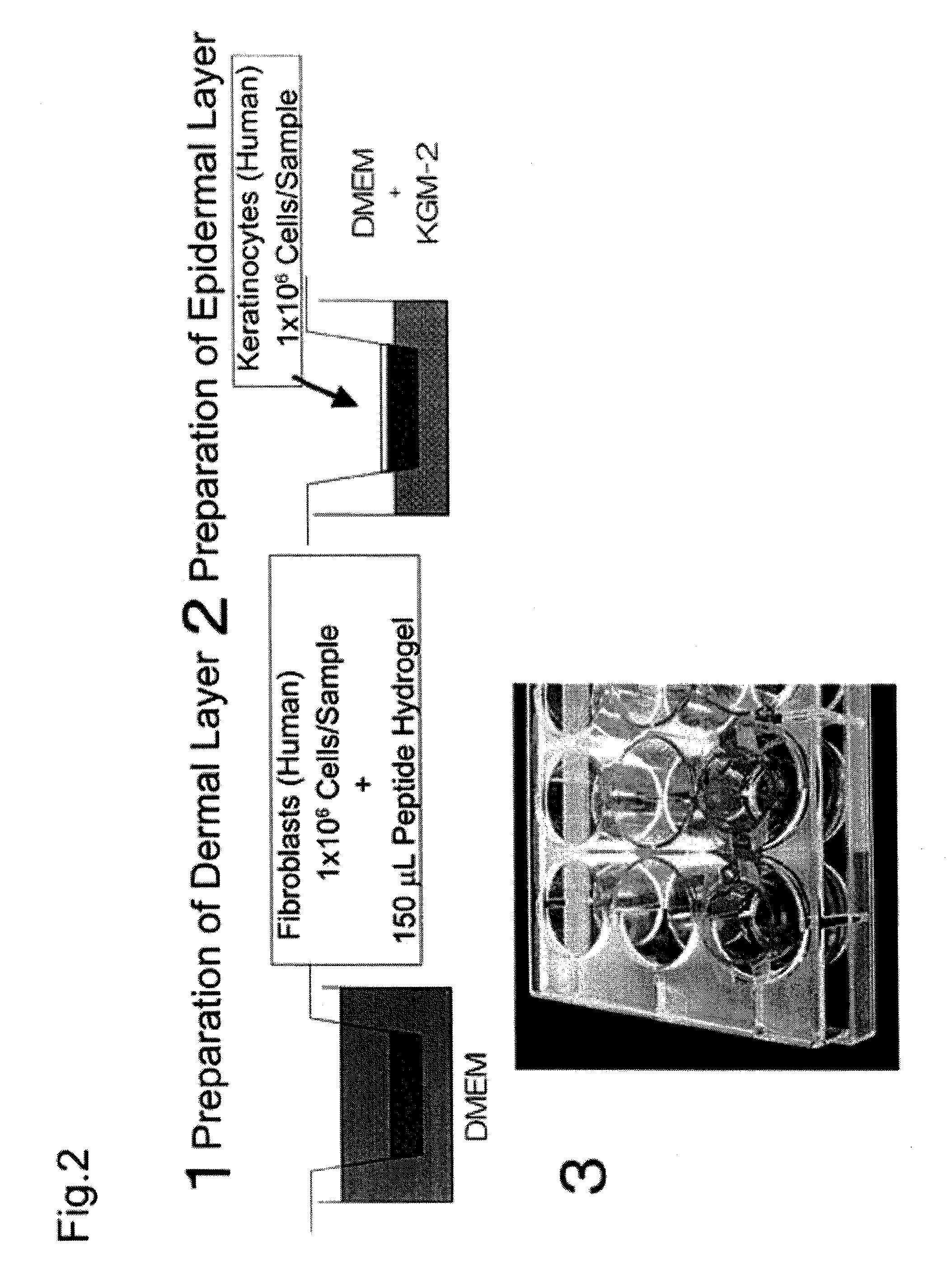
[0061](1) Cell expansion (cell culture)

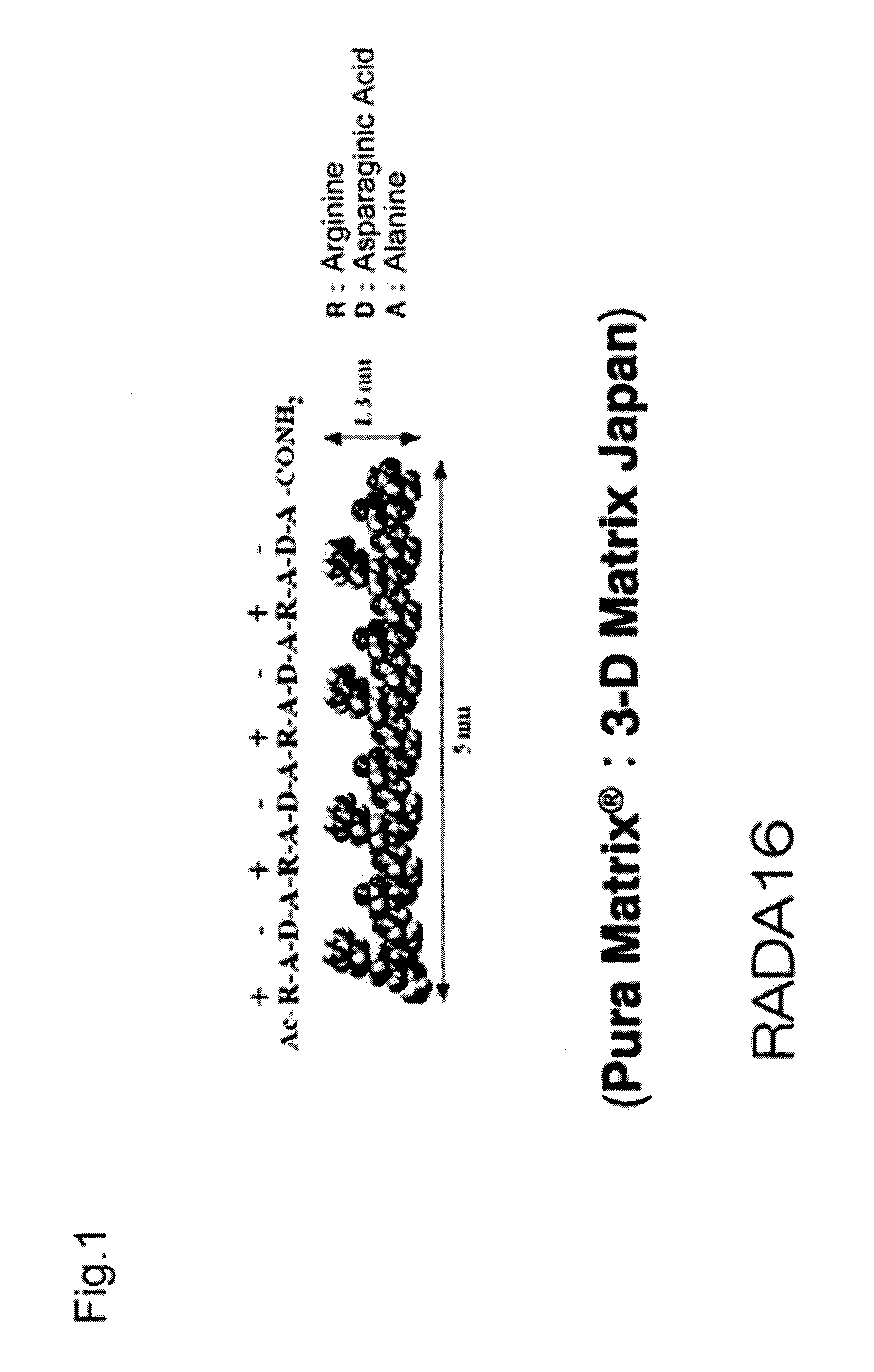
[0062]Neonatal human dermal fibroblasts (Lonza Walkersville, Walkersville, Md.) were subcultured 8 to 10 times in a culture flask using D-MEM medium (Lonza Walkersville, Walkersville, Md.) supplemented with 10% FBS (Invitrogen, Carlsbad, Calif.), and the cultured fibroblasts were used for the experiment. Neonatal human epidermal keratinocytes (Lonza Walkersville, Walkersville, Md.) were subcultured 4 or 5 times in a culture flask using KGM-2 medium (Lonza Walkersville, Walkersville, Md.), and the cultured keratinocytes were used for the experiment. Table 1 gives a summary of specific materials, reagents, and samples.

TABLE 1MaterialsPura Matrix (Peptide hydrogel): 3D matrix CompanyNormal human dermal fibroblasts (neonatal skin): Lonza CompanyHigh glucose Dulbecco's Modified Eagle's Medium (D-MEM): InvitrogenCorporationFetal Bovine Serum (FBS): Invitrogen CorporationPenicillin: Invitrogen CorporationStreptomyci...

example 2

Grafting of Artificial Dermis

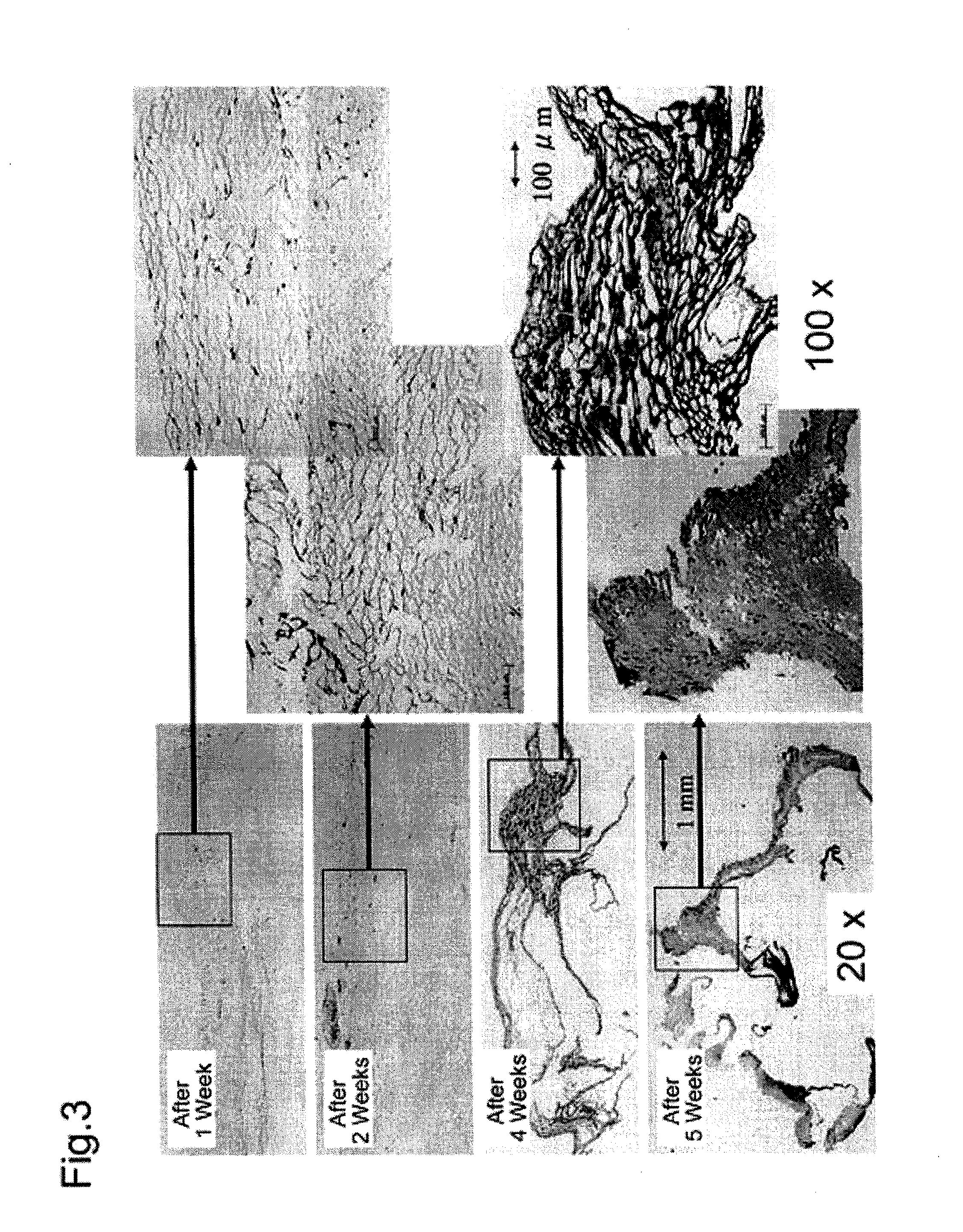
[0092]Following the same method as described in Example 1, skin was collected from the dorsal region of male Hairless rats weighing 250 to 300 g, and fibroblasts and epidermal keratinocytes were collected and grown, thereby preparing a hybrid artificial dermal material. A 5 mm long incision was placed in the dorsal region of the rats, and subcutaneous tissue was removed to prepare a pocket. The artificial dermis was inserted into the pocket, and then the incision was sutured. The rats were divided into five groups, each group containing three rats. After embedding the artificial dermis, the embedded portions and peripheral tissue were collected from these five groups after 1, 2, 3, 4 and 5 weeks, respectively, and a histopathological evaluation was made.


PUM

PropertyMeasurementUnit
pHaaaaaaaaaa
pore sizeaaaaaaaaaa
volumeaaaaaaaaaa

Abstract

An object of the invention is to provide artificial skin that does not contain any animal-derived material or pathogen and has excellent biocompatibility. The invention provides, as a solution means, a method for producing artificial skin comprising the steps of: (A) forming a dermal layer by solidifying a mixture of dermal fibroblasts and a peptide hydrogel having a fibrous structure; and (B) forming an epidermal layer by seeding skin keratinocytes onto the dermal layer obtained in Step (A), and culturing the epidermal keratinocytes.

Description

TECHNICAL FIELD[0001]The present invention relates to a method for readily producing safe hybrid artificial skin.BACKGROUND ART[0002]Ex-vivo culture of cells collected from living tissues is now commonly practiced using culture flasks in the laboratory. This has revealed the characteristics of various cells, providing a deeper understanding of our tissues and organs, thereby contributing to medical science. Recently, however, the fact that it is difficult to completely reproduce what is happening in vivo by two-dimensional culture of only cells has been recognized.[0003]Some cells spreading two-dimensionally in the culture flask adhere to the flask, some adhere to neighboring cells, and others are directly exposed to the culture medium. Therefore, nutrients, various growth factors, and cytokines contained in the culture medium directly act upon individual cells. In-vivo cells are arranged three-dimensionally, with an extracellular matrix filling between the cells; therefore, nutrien...

Claims


Application Information

Patent Timeline
Patent Type & Authority Applications(United States)
IPC IPC(8): A61K9/14C12N5/00A61K35/36A61P17/02A61K47/42A61L27/00A61L27/22A61L27/40A61L27/44A61L27/60C07K7/08C07K14/00
CPCA61L27/22A61L27/227A61L27/3813A61L27/52C12N2533/50A61L2430/34C12N5/0698C12N2502/094C12N2502/1323A61L27/60A61P17/02
Inventor KAO, BUNSHOHOSAKA, YOSHIAKIIIJIMA, AYUMI
Owner SHOWA UNIVERSITY
Why Eureka
Try Eureka