
Method of diagnosis and agents useful for same

a neoplastic cell and screening technology, applied in the field of screening for neoplastic cells, can solve the problems of unsustainable immunological approaches to cancer treatment, severe side effects, and cancer likely to become the most common fatal disease in these countries

Inactive Publication Date: 2010-12-09
AUSHEALTH CORP PTY LTD
View PDF0 Cites 1 Cited by
  • Summary
  • Abstract
  • Description
  • Claims
  • Application Information

AI Technical Summary

Problems solved by technology

As treatments for infectious diseases and the prevention of cardiovascular disease continues to improve, and the average life expectancy increases, cancer is likely to become the most common fatal disease in these countries.
However, immunological approaches to the treatment of cancer have been attempted for over a century with unsustainable results.
Further, these treatments are associated with severe side effects including disfigurement and scarring from surgery (e.g. mastectomy or limb amputation), severe nausea and vomiting from chemotherapy, and most significantly, the damage to normal tissues such as the hair follicles, gut and bone marrow which is induced as a result of the relatively non-specific targeting mechanism of the toxic drugs which form part of most cancer treatments.
However, when cytotoxic chemotherapeutic agents are used for the treatment of advanced cancer, the degree of cell kill and thus the response of the tumour to the first treatment is frequently difficult to assess soon after administration.
Usually, only a minority of patients with advanced cancer responds to cytotoxic drugs and so patients may experience the side effects of treatment without obtaining benefit.
Knowing whether the tumour is responding early would spare the majority of patients from ineffective and potentially toxic treatment.

Method used


Image

  • Method of diagnosis and agents useful for same
  • Method of diagnosis and agents useful for same
  • Method of diagnosis and agents useful for same

Examples


example 1

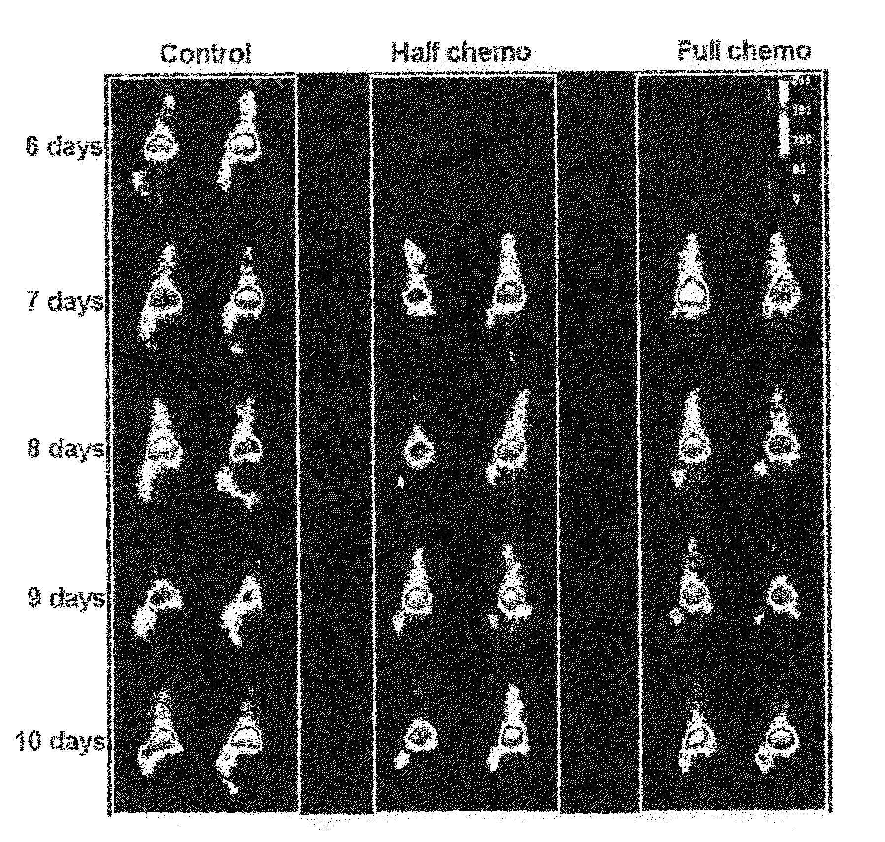
In Vivo Targetting of the Ribonucleoprotein La in a Mouse Tumour Model

Materials and Methods

Materials

[0197]Cell culture media, RPMI-1640, DMEM and Ham's F12, and fetal calf serum (FCS) were all purchased from JRH Biosciences Inc. (KS, USA). Trypsin-EDTA solution, trypan blue, propidium iodide (PI), bovine serum albumin (BSA), hydrocortisone and staurosporine (STS) were obtained from Sigma-Aldrich Co. (MO, USA). Hybond-P membrane (PVDF), ECF™ substrate, L-[U-14C]Leucine, D[U-14C]Glucose and Protein G purification columns were purchased from Amersham Biosciences, (NJ, USA). The miniPERM bioreactor was obtained from Vivascience (Hannover, Germany) and the BCA Protein Reagent Assay from Pierce Biotechnology Inc. (IL, USA). Solvable™ and UltimaGold™ were purchased from PerkinElmer Inc. (MA, USA). H2O2. The anti-poly(ADP-ribose) polymerase (PARP) monoclonal antibody (IgG1 mAb) clone C-2-10 was obtained from Oncogene™ Research Products (EMD Biosciences Inc., CA, USA). The anti-actin (N-20) ...

example 2

Analysis of Gene Expression Profiling Data to Identify Suitable Targets for Imaging and TCS

Introduction

[0215]Oncomine™ is a cancer-specific database containing microarray data from 962 studies of which 209 were analysed. The database contains 14,177 microarrays from 35 cancer types (information publicly available at the website www.oncomine.org). Several cancer signatures have been deduced from large scale analysis of data held in the database (Hampton, Jama 292(17): 2073, 2004; Rhodes et al., Proc Natl Acad Sci USA 101(25): 9309-14, 2004a; Rhodes et al., Neoplasia 6(1): 1-6, 2004b; Rhodes and Chinnaiyan, Nat Genet. 37 Suppl: S31-7, 2005; Rhodes et al., Nat Genet. 37(6): 579-83, 2005). 209 studies in the database as described below were analysed in order to investigate certain malignancy signatures, which may provide useful targets for the present invention

Method

[0216]In the catalogue of the database Oncomine™ on www.oncomine.org, the tissue of interest was selected and only analyse...

example 3

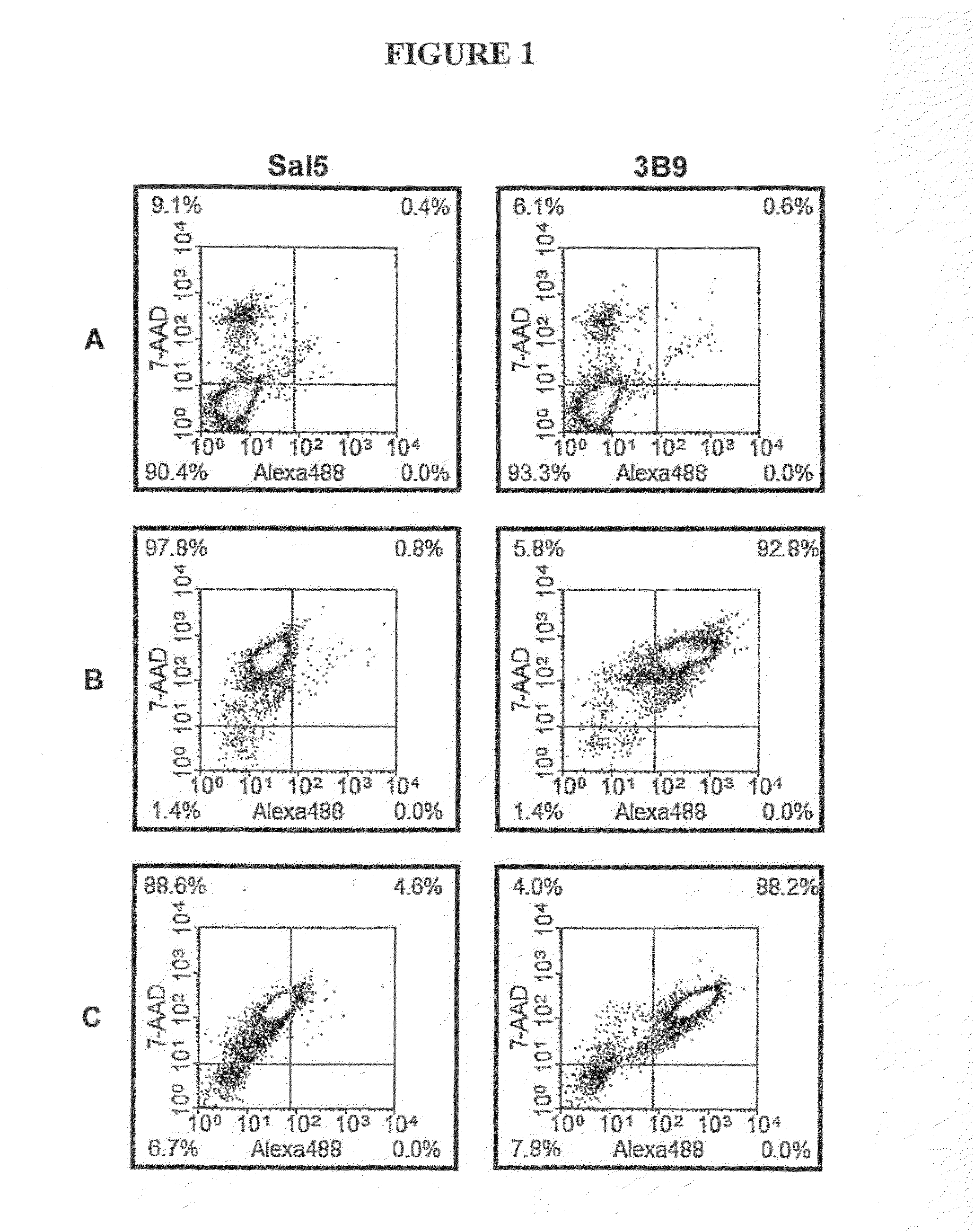
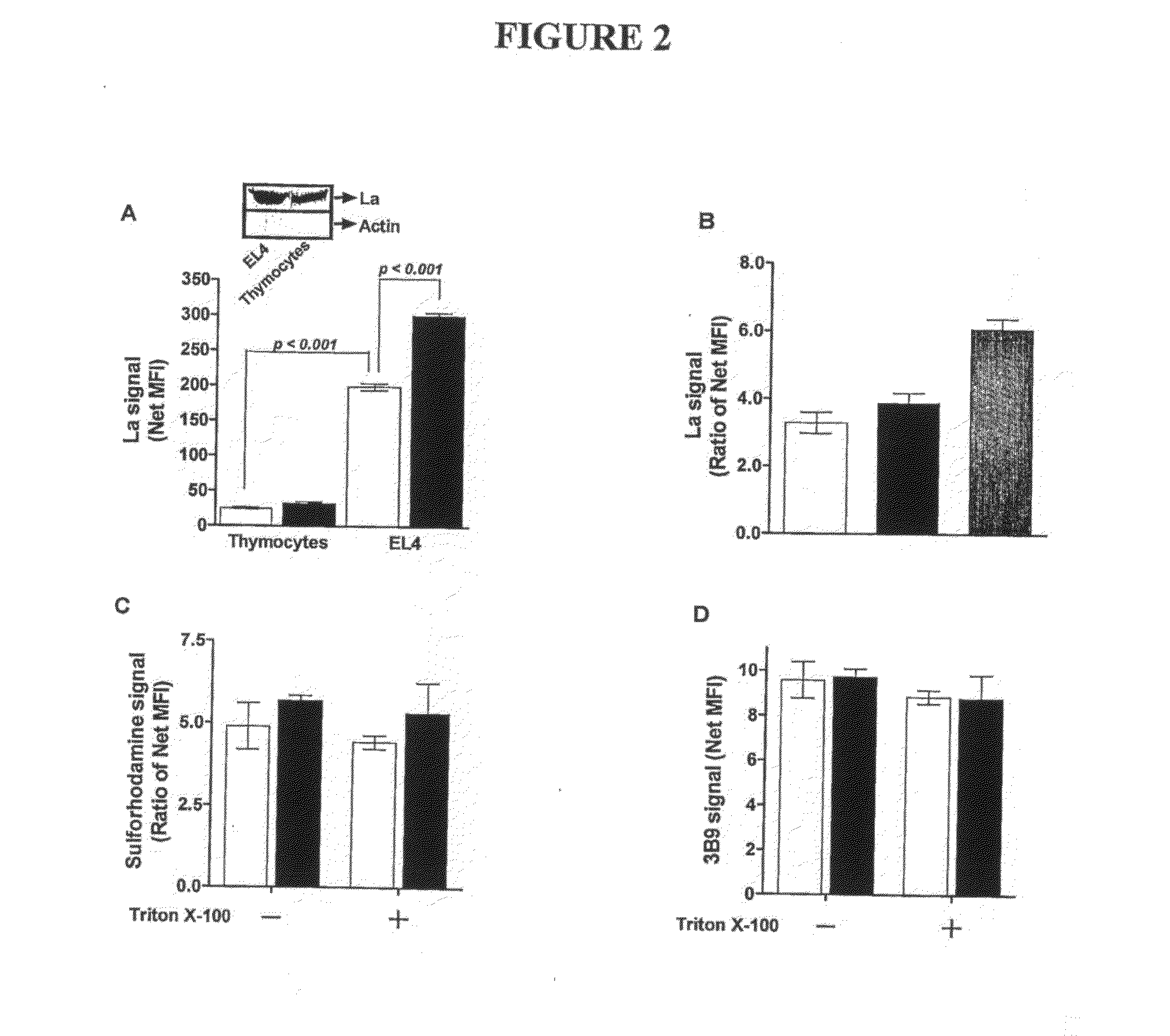
In Vitro Rationale for Targetting of the Ribonucleoprotein La in a Mouse Tumour Model

Materials and Methods

Materials

[0219]The suppliers of the materials are identified in brackets after each material. Cell culture media, RPMI-1640, DMEM and Ham's F12, and fetal calf serum (FCS) (JRH Biosciences Inc., Lenexa, Kans.); Trypsin-EDTA solution, trypan blue, propidium iodide (PI), bovine serum albumin (BSA), BCIP / NBT premixed substrate solution for alkaline phophatase (AP), hydrocortisone, monodansylcadaverine (MDC) and staurosporine (STS) and mouse anti-human β-tubulin mAb (TUB 2.1) (Sigma-Aldrich Co., St. Louis, Mo.). Hybond-P membrane (PVDF) and protein G purification columns (Amersham Biosciences, Piscataway, N.J.). BCA Protein Reagent Assay (Pierce Biotechnology Inc., Rockford, Ill.). Anti-poly(ADP-ribose) polymerase (PARP) monoclonal antibody (mAb) clone C-2-10 and anti-proliferating cell nuclear antigen (PCNA) mAb clone PC10 (Oncogene Research Products, Cambridge, Mass.). Trichostati...


PUM

PropertyMeasurementUnit
timeaaaaaaaaaa
time pointsaaaaaaaaaa
lengthaaaaaaaaaa

Abstract

The present invention relates generally to a method of screening for a neoplastic cell in a subject. More particularly, the present invention provides a method of screening for both viable neoplastic cells and, still further, cytotoxin induced neoplastic cell death by detecting the level of expression of La protein and / or gene by a cellular population in said subject or in a biological sample derived from said subject. The method of the present invention is useful in a range of applications including, but not limited to, diagnosing, prognosing or assessing a neoplastic condition, monitoring the progression of such a condition, assessing the effectiveness of a therapeutic agent or therapeutic regime and predicting the likelihood of a subject either progressing to a more advanced disease state or entering a remissive state. The present invention also provides diagnostic agents useful for detecting La protein and / or nucleic acid molecules.

Description

FIELD OF THE INVENTION[0001]The present invention relates generally to a method of screening for a neoplastic cell in a subject. More particularly, the present invention provides a method of screening for both viable neoplastic cells and, still further, cytotoxin induced neoplastic cell death by detecting the level of expression of La protein and / or gene by a cellular population in said subject or in a biological sample derived from said subject. The method of the present invention is useful in a range of applications including, but not limited to, diagnosing, prognosing or assessing a neoplastic condition, monitoring the progression of such a condition, assessing the effectiveness of a therapeutic agent or therapeutic regime and predicting the likelihood of a subject either progressing to a more advanced disease state or entering a remissive state. The present invention also provides diagnostic agents useful for detecting La protein and / or nucleic acid molecules.BACKGROUND OF THE I...

Claims


Application Information

Patent Timeline
Patent Type & Authority Applications(United States)
IPC IPC(8): A61K51/00A61K49/00A61K49/06C12Q1/68G01N33/53
CPCC12Q1/6886C12Q2600/106G01N33/57496G01N2800/52C12Q2563/101C12Q2545/114C12Q2563/125C12Q2563/131C12Q2600/158
Inventor BROWN, MICHAEL PAULAL-EJEH, FARESDARBY, JOCELYN MARGARET
Owner AUSHEALTH CORP PTY LTD
Learn More