
Polypeptides, Cyclic Polypeptides and Pharmaceutical Comprising Thereof for Non Invasive Specific Imaging of Fibrosis

a fibrosis and fibrosis technology, applied in the direction of peptides, cyclic peptide ingredients, drug compositions, etc., can solve the problems of destroying the affected structure, affecting the health of patients, and imposing an enormous health burden

Inactive Publication Date: 2010-12-02
INST NAT DE LA SANTE & DE LA RECHERCHE MEDICALE (INSERM) +1
View PDF0 Cites 2 Cited by
  • Summary
  • Abstract
  • Description
  • Claims
  • Application Information

AI Technical Summary

Benefits of technology

[0083]In another embodiment, the polypeptides of the invention are PEGylated. It has been widely demonstrated that the conjugation of polyethylene glycol PEG to peptides significantly increases their duration of biological activity (half-life). PEG provides a more stable conformation and increases the size and the weight of the molecule, thereby prolonging the circulation time of the peptide.
[0092]Oligomerization increases the size and the weight of biological activity, thereby prolonging the circulation time (half-life) of the peptide.
[0126]The “biocompatible carrier” is a fluid, especially a liquid, in which the imaging agent is suspended or dissolved, such that the composition is physiologically tolerable, i.e. can be administered to the mammalian body without toxicity or undue discomfort. The biocompatible carrier medium is suitably an injectable carrier liquid such as sterile water for injection; an aqueous solution such as saline; an aqueous solution of one or more tonicity-adjusting substances (e.g. salts of plasma cations with biocompatible counterions), sugars (e.g. glucose or sucrose), sugar alcohols (e.g. sorbitol or mannitol), glycols (e.g. glycerol), or other non-ionic polyol materials (e.g. polyethyleneglycols, propylene glycols and the like).

Problems solved by technology

Excessive fibrosis following physical, thermal, metabolic, ischemic, infectious, inflammatory, or immunological injury can occur in any part of the body, and can cause destruction of the affected structures such as lung, kidney, heart, liver, vascular system, skin, eye, bone marrow.
Fibrosis is increasingly recognized as an important feature of many chronic diseases, and as such, represents an enormous health burden.
As a result, fibrosis may accumulate through increased production of tissue and proteins like collagen and decreased degradation of these compounds so that the function of liver is impaired.
When the plaque is vulnerable, collagen becomes exposed to the blood flow and triggers arterial thrombosis and ischemia of downstream tissues.
Several biomarkers have been searched for diagnosing fibrosis, but very few methods have been developed for the in vivo visualization of fibrosis in an organ.
But these techniques are not specific for fibrosis.
Also they may suffer from a lack of sensitivity since large amounts of fibrosis are necessary to impair tissue elasticity.

Method used


Image

  • Polypeptides, Cyclic Polypeptides and Pharmaceutical Comprising Thereof for Non Invasive Specific Imaging of Fibrosis
  • Polypeptides, Cyclic Polypeptides and Pharmaceutical Comprising Thereof for Non Invasive Specific Imaging of Fibrosis
  • Polypeptides, Cyclic Polypeptides and Pharmaceutical Comprising Thereof for Non Invasive Specific Imaging of Fibrosis

Examples


example 1

Identification and Characterization of a Peptidomimetic of Human Platelets Glycoprotein VI with Collagen Binding Activity. Its Use for Molecular Imaging of Fibrosis

[0149]Material & Methods:

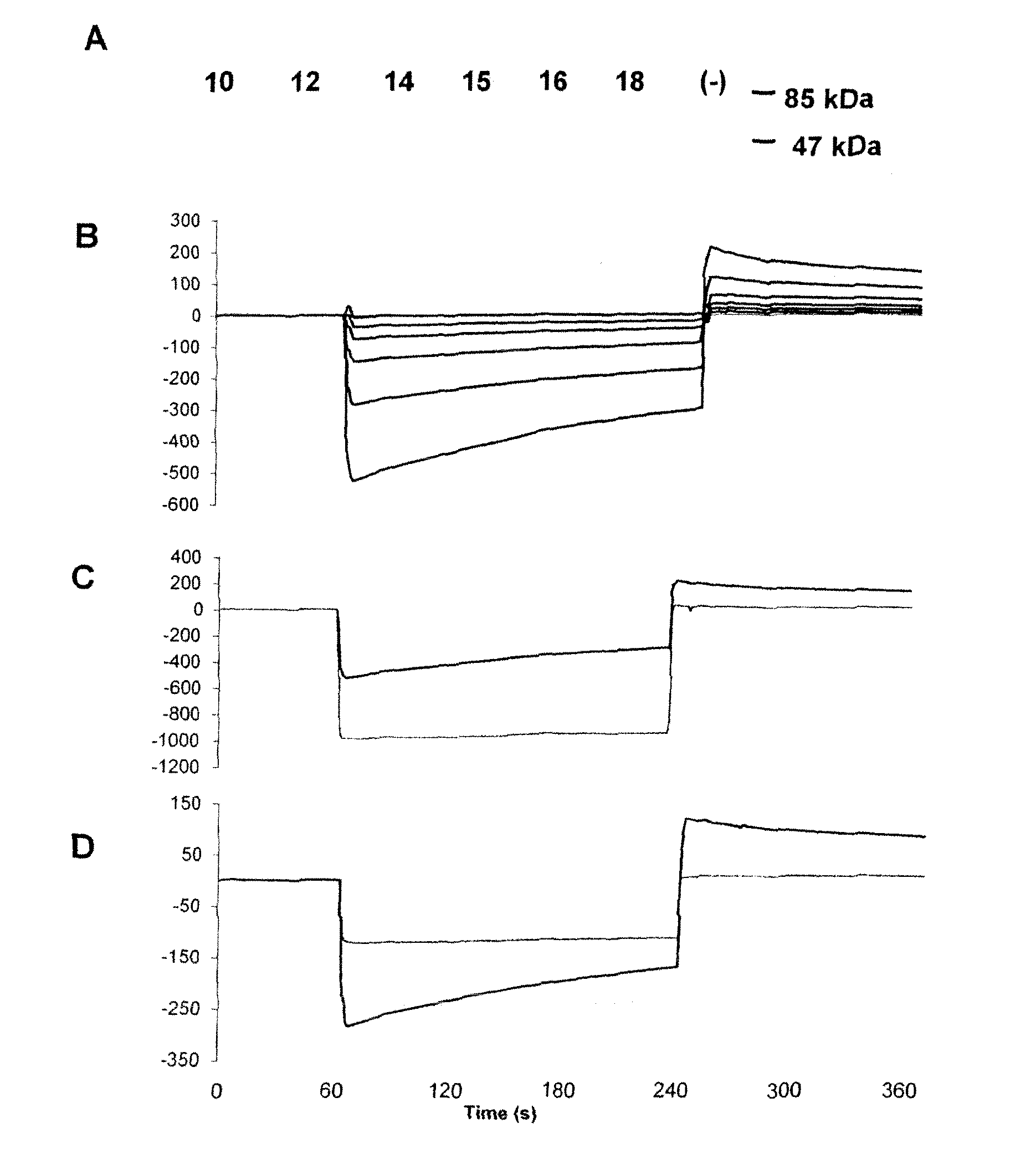
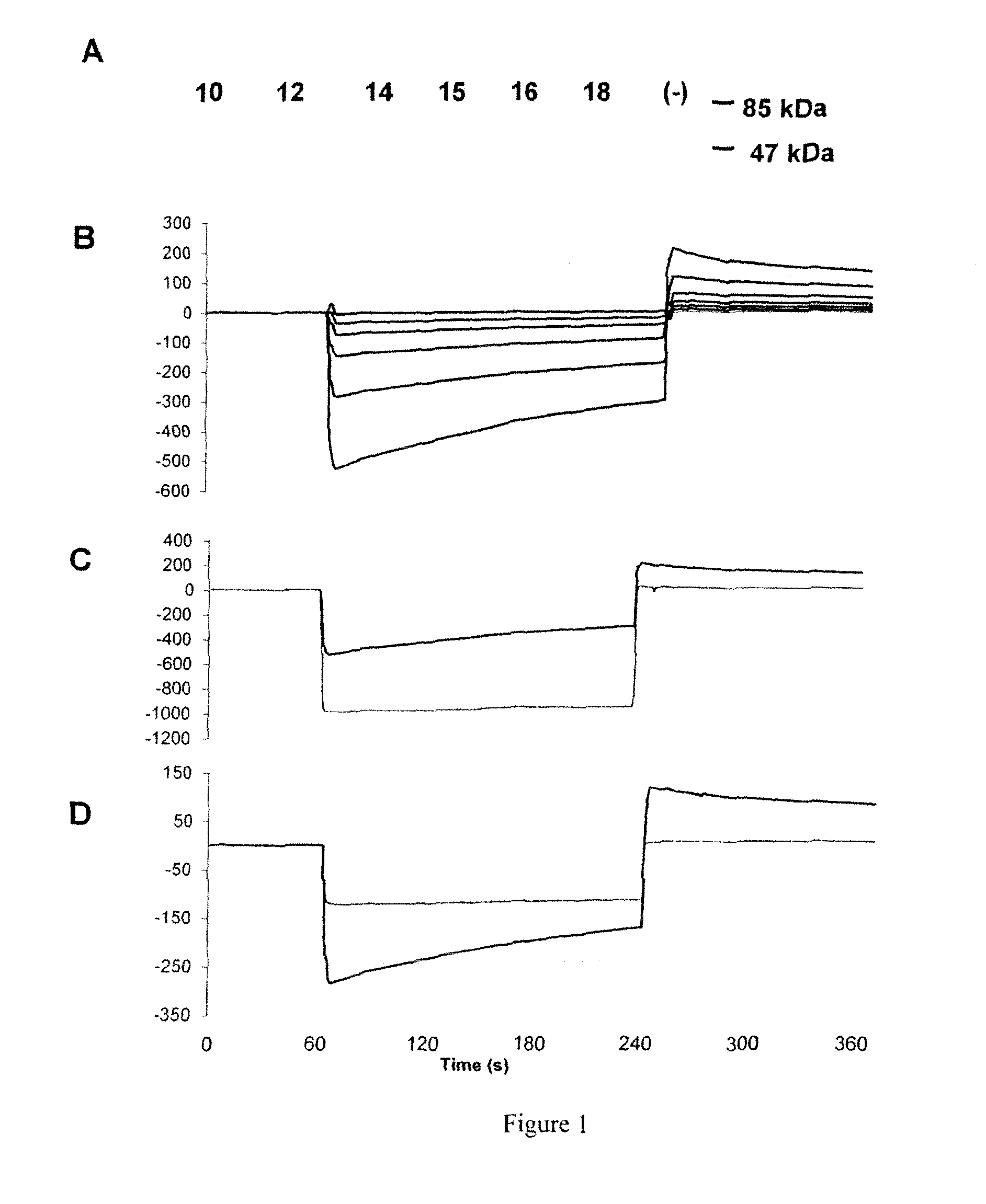
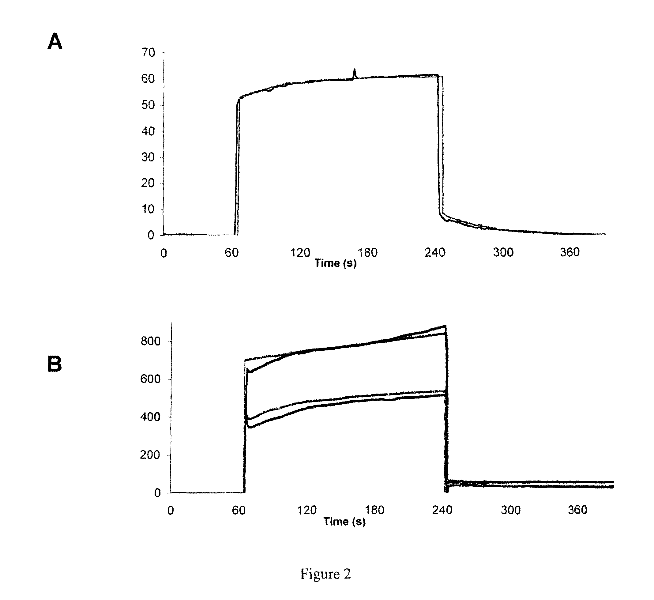
[0150]FliTrx™ peptide library and anti-GPVI antibody: The FliTrx™ Random Peptide Display Library is an E. coli-based system allowing the screening of peptide interactions. The FliTrx™ Library was constructed in the pFliTrx™ vector (Lu Z. et al. 1995). A diverse library of random dodecapeptides (108) is positioned in the active site loop of the thioredoxin protein (trxA), inside the dispensable region of the bacterial flagellin gene (fliC). The resultant recombinant fusion protein (FLITRX) is exported and assembled into partially functional flagella on the bacterial cell surface. The dodecapeptides are displayed on the cell surface in a conformationally constrained by a disulfide bridge. The FliTrx™ random peptide library, based on the system was obtained from Invitrogen (San Diego, Calif.).

[0151]T...

example 2

Imaging of Pulmonary Fibrosis by Scintigraphy Using 99MTC-Labeled Collagelin

[0192]Methods:

[0193]Male C57BL / 6J mice, aged 6-7 weeks were kept in accordance with INSERM rules. On day 0, mice were administered 80 μg of bleomycin hydrochloride (Bleomycine Bellon, Aventis, France) intratracheally. Mortality was assessed daily over a 14 day period. Naïve mice were used as controls.

[0194]At day 14 mice received one intravenous injection of 99 mTc-B-collagelin or of 99 mTc-B-Pc (3 MBq). Then planar whole-body scintigraphic imaging (60 min duration) was performed 1 h after tracer injection, using Biospace Lab dedicated small animal gamma camera.

[0195]At the end of the experiment, animals were sacrificed and lung were dissected for gamma counting, autoradiography, and histology (Sirius red coloration).

[0196]Results:

[0197]Scintigraphy: Significant 99 mTc-B-collagelin uptake was observed in pulmonary areas of the mice that received bleomycin (lung / muscle background activity ratio: 3.65±0.34), w...


PUM

PropertyMeasurementUnit
molecular weightaaaaaaaaaa
concentrationaaaaaaaaaa

Abstract

The present invention relates to diagnostic imaging and in particular to the diagnostic imaging of fibrosis. More particularly, the present invention provides a polypeptides, cyclic polypeptides and pharmaceutical compositions suitable for the non-invasive visualization of fibrosis. The polypeptide of the invention may comprise an amino acid sequence consisting of: X1-X2-M-H-G-L-X7-L-X9-X10-D-E wherein amino acid X1 is R, F or P; amino acid X2 is F or V; amino acid X7 is Q, H or L; amino acid X9 is W or G and amino acid X10 is A or D.

Description

FIELD OF THE INVENTION[0001]The present invention relates to diagnostic imaging and in particular to the diagnostic imaging of fibrosis. More particularly, the present invention provides polypeptides, cyclic polypeptides and pharmaceutical compositions which specifically target collagen and are suitable for the non-invasive specific imaging of fibrosis.BACKGROUND OF THE INVENTION[0002]Fibrosis is defined pathologically as inappropriate repair by connective tissue and is caused by increased synthesis and decreased degradation of matrix proteins, most notably collagen types I and III.[0003]Fibrosis can affect virtually every tissue and organ system. Excessive fibrosis following physical, thermal, metabolic, ischemic, infectious, inflammatory, or immunological injury can occur in any part of the body, and can cause destruction of the affected structures such as lung, kidney, heart, liver, vascular system, skin, eye, bone marrow. Fibrotic tissue is characterized by a loss of normal arch...

Claims


Application Information

Patent Timeline
Patent Type & Authority Applications(United States)
IPC IPC(8): A61K49/00C07K7/08C07H21/00C12N15/63C12N5/10A61P43/00
CPCC07K14/70503A61K51/08A61P43/00
Inventor JANDROT-PERRUS, MARTINEMUZARD, JULIENBILLIALD, PHILIPPELE GULUDEC, DOMINIQUESARDA, LAUREMEULEMANS, ALAIN
Owner INST NAT DE LA SANTE & DE LA RECHERCHE MEDICALE (INSERM)
Learn More