
Methods for providing personalized medicine test ex vivo for hematological neoplasms

a technology of hematological neoplasms and personalized medicine, which is applied in the direction of biocide, drug composition, instruments, etc., can solve the problems of limiting the use of personalized medicine tests, undesirable limitations of itrts of cell death, and inability to study the ability of drugs to slow or arrest neoplastic cell growth, etc., and achieves the effect of quick analysis

Inactive Publication Date: 2010-11-25
VIVIA BIOTECH SL
View PDF102 Cites 15 Cited by
  • Summary
  • Abstract
  • Description
  • Claims
  • Application Information

AI Technical Summary

Benefits of technology

[0014]The present invention relates to the development of a personalized medicine test for a patient. In a general embodiment, the present invention is directed to compositions, methods, and systems for analyzing cellular responses to drugs using an ex vivo assay. Described herein are methods of analyzing whole blood samples, manipulating a large number of variables, and quickly completing analyses.
[0029]In addition to individual drug effects, detailed analyses of drug interactions, including the Combination Index and Dose Reduction Index, can be used to identify effective polytherapy regimens. Estimates of accuracy of both indexes can be calculated with accurate algebraic estimation algorithms (i.e., Monte Carlo simulations) based on the Median Effect methods described by Chou and Talalay (Chou et al., Adv Enzyme Regul 1984, 22:27-55). The Combination Index (CI) is a quantitative measure of the degree of drug interaction in terms of additive effect, where synergism is indicated by a CI <1, additive effect is indicated by a CI˜1, and antagonism is indicated by a CI>1. A dose-reduction index (DRI) is a measure of how much the dose of each drug in synergistic combination may be reduced at a given effect level compared with the dose of each drug alone. More recently, the MixLow method (Boik et al., BMC Pharmacol 2008, 8:13; Boik, Stat Med 2008, 27(7):1040-61) has been proposed as an alternative to the Median-Effect method of Chou and Talalay (Chou et al., Adv Enzyme Regul 1984, 22:27-55) for estimating drug interaction indices. One advantage of the MixLow method is that the nonlinear mixed-effects model used to estimate parameters of concentration-response curves can provide more accurate parameter estimates than the log linearization and least-squares analysis used in the Median-Effect method. One of skill in the art will know that these calculations and related methods can be used to analyze drug interactions for mixed drug treatments as described herein. In some embodiments, the combination of more than one drug is assessed for potentiation, synergy, or dose reduction. In some embodiments, a combination identified as demonstrating a drug interaction is selected for treatment.
[0030]The limited amount of sample that can be extracted from patient limits the number of drug compositions that can be tested for the personalized medicine test. However, recent developments have provided mouse models that can propagate the primary cells of patients with hematological malignancies through multiple mice becoming a continuous source of patient cells (Pearson et al., Curr Top Microbiol Immunol. 2008, 324:25-51; Ito et al., Curr Top Microbiol Immunol. 2008, 324:53-76). These models may enable ex vivo sampling of many more drug compositions, and in particular drug combinations, than a recently extracted patient sample. It is contemplated that these models can be used in the methods described herein. For example, the samples may be drawn from an animal model, such as a mouse model. In particular, these models may enable exploring the efficacy of concomitant or adjuvant medicines, given to patients to palliate the effects of chemotherapy. These models may also enable exploration of the potential efficacy of approved non-cytotoxic safe drugs, which in the future could be added to treatments for an individual patient to increase the probability of therapeutic efficacy. Furthermore, the efficacy of any drug combination of a drug composition identified in ex vivo testing using human patient cells, directly from a patient sample or propagated by a mouse models, could be tested in mouse models in vivo.

Problems solved by technology

Initial ITRTs designed to study the ability of a drug to slow or arrest neoplastic cell growth (e.g., clonogenic assays) did not work well.
Even with good clinical correlations, currently available ITRTs of cell death suffer from undesirable limitations that restrict their use as personalized medicine tests.
Measuring total cell death limits the ability of an ITRT to distinguish between a drug's effect on tumor cells versus normal cells.
However, hematological cells start to lose important properties after only 24 to 48 hours outside the human body.

Method used


Image

  • Methods for providing personalized medicine test ex vivo for hematological neoplasms
  • Methods for providing personalized medicine test ex vivo for hematological neoplasms
  • Methods for providing personalized medicine test ex vivo for hematological neoplasms

Examples


example 1

Flow Cytometric Detection of Apoptotic Normal and Neoplastic Cells

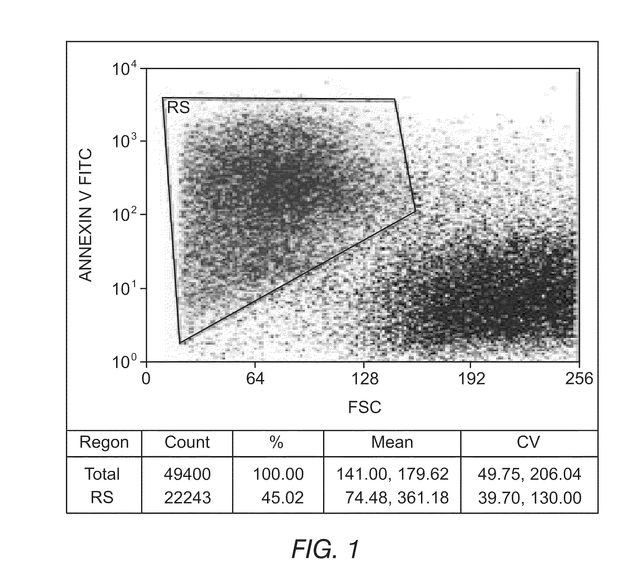
[0148]An ex vivo therapeutic index can be determined by measuring the ability of a drug composition to induce apoptosis. FIGS. 1 and 2 depict the ability to detect apoptotic cells and differentiate between normal and tumor phenotypes using flow cytometry. In FIG. 1, the reagent Annexin V coupled to Fluorescein Isothiocyanate (FITC) was used to detect phosphatidylserine expression on apoptotic cells. Fluorescein intensity is displayed on the y-axis, and cell size is displayed on the x-axis. FIG. 1 illustrates the ability to identify apoptotic cells (upper left box) and live cells (lower right cluster) and demonstrates that the simultaneous use of appropriate combinations of monoclonal antibodies and multiparametric analysis strategies can allow for the discrimination of leukemic cells from residual normal cells present in samples from patients with hematological disorders. FIG. 2 depicts a precursor B-ALL adult case di...

example 2

Protocol for the Ex Vivo Evaluation of Drug Compositions

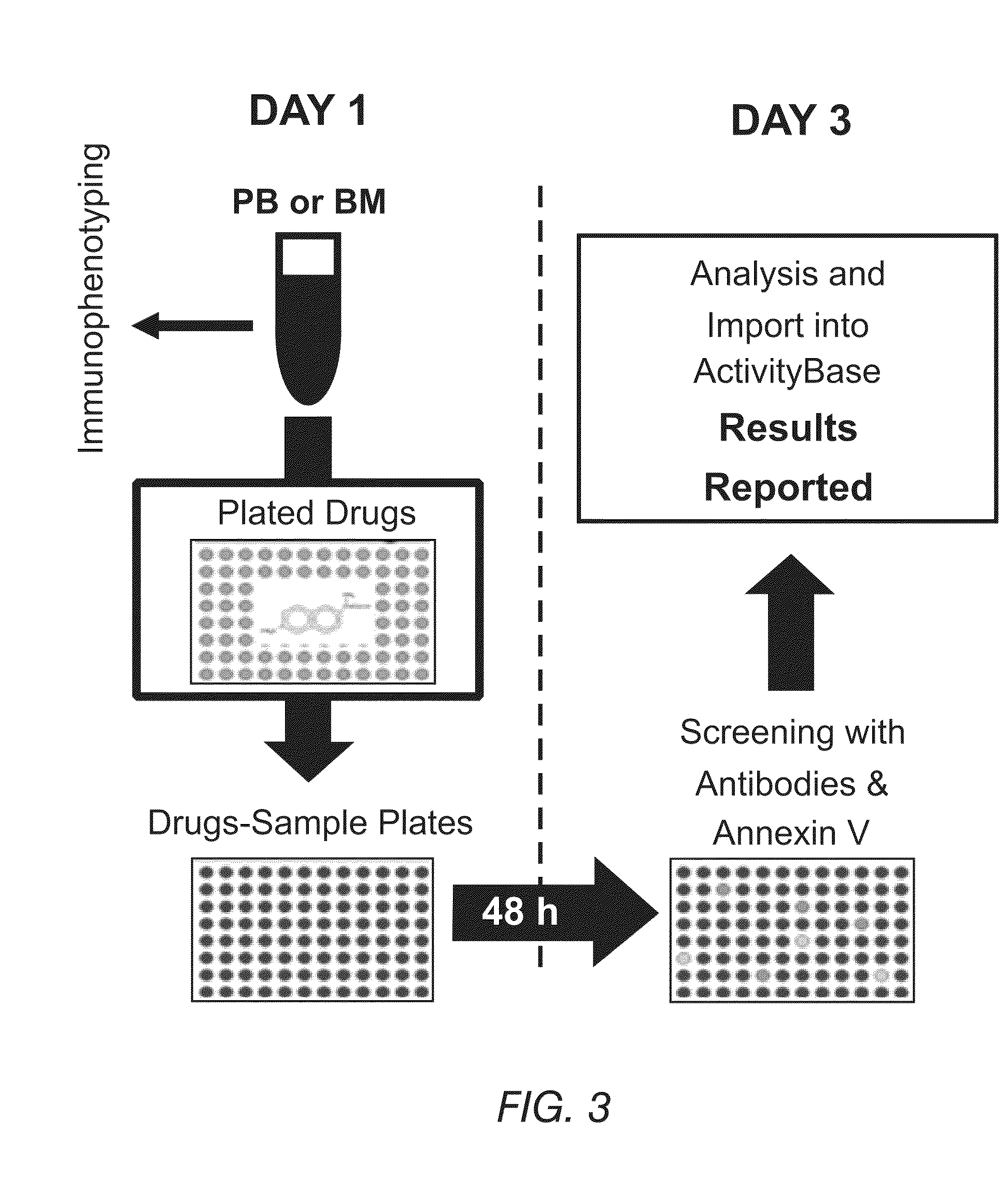
[0149]An ex vivo screening process for drug compositions is schematically shown in FIG. 3. A sample of blood can be split into small aliquots that are distributed into well plates of any suitable size. These well plates contain individual drugs or drug combinations, each at various concentrations. To facilitate optimal assay development, a sample is diluted in RPMI media and concentrated at about 20,000 leukemic cells per well. In parallel, another aliquot is tested for immunophenotypic identification using flow cytometry for the identification of normal and pathologic cells and the detection of basal apoptosis. Control wells without any drug can be included (not shown) to identify the spontaneous level of apoptosis not associated with drug treatment.

[0150]After approximately 48 hours, each well with the sample exposed to the drugs is treated with a buffer to lyse the erythrocyte population and concentrate the leukocyte populat...

example 3

Individual Patient Responses Demonstrate the Cytotoxic Effects of Different Drugs Currently Approved for Chronic Lymphocytic Leukemia Treatment

[0152]The present methods have been used to analyze 30 μM concentrations of chlorambucil, cyclophosphamide, vincristine, mitoxantrone, and doxorubicin—five drugs currently approved for chronic lymphocytic leukemia (CLL)—in various patients. The results of the efficacy of individually approved cytotoxic drugs for inducing apoptosis in malignant cells of 9 ex vivo patient samples are provided in FIG. 4. FIG. 4 demonstrates that there is a high person-to-person variability in the drug responses, highlighting an important use for the personalized medicine tests described herein.

[0153]Regarding patient response to individual drug treatments at 30 μM concentrations, several drugs generally had poor patient response, defined as inducing less than 60% apoptosis in patient samples as measured by Annexin V positive cells. Additionally, FIG. 4 indicates...


PUM

PropertyMeasurementUnit
Timeaaaaaaaaaa
Timeaaaaaaaaaa
Timeaaaaaaaaaa

Abstract

Described herein are methods, devices, and compositions for providing personalized medicine tests for hematological neoplasms. In some embodiments, the methods comprise measuring the efficacy of inducing apoptosis selectively in malignant cells using any number of potential alternative combination drug treatments. In some embodiments, the ex vivo testing is measured using a recently extracted patient hematological samples. In other embodiments, the efficacy is measured ex vivo using an automated flow cytometry platform. For example, by using an automated flow cytometry platform, the evaluation of hundreds, or even thousands of drugs and compositions, can be made ex vivo. Thus, alternative polytherapy treatments can be explored. Non-cytotoxic drugs surprisingly induce apoptosis selectively in malignant cells ex vivo. In some embodiments, the methods described herein comprise evaluating non-cytotoxic drugs.

Description

RELATED APPLICATIONS[0001]This application claims priority to U.S. Provisional Patent Application Ser. No. 61 / 179,685, filed on May 19, 2009, which is incorporated by reference herein in its entirety.BACKGROUND OF THE INVENTION[0002]This invention relates to the use of a screening platform to determine a cytotoxic drug sensitivity profile for multiple drugs and drug combinations using specimens from cancer patients. Described herein is a cell-based screening platform that incorporates both automated sample preparation and automated evaluation by flow cytometry that is useful as a personalized medicine test because of its rapid data acquisition, analysis, and reporting of results, even from very large numbers of drugs and drug combinations. Also disclosed are particular combinations of drugs useful in the treatment of proliferative lymphoid disease.DESCRIPTION OF THE RELATED ART[0003]There are many methods available to evaluate the cytotoxic drug sensitivity profiles of tumor cells i...

Claims


Application Information

Patent Timeline
IPC IPC(8): A61K31/7076A61P35/02C12Q1/02C12M1/34
CPCG01N33/57407G01N2800/52G01N33/57426A61P35/02A61P43/00G01N33/5011
Inventor BALLESTEROS, JUANBENNETT, TERESAPRIMO, DANIELORFAO, ALBERTOJACKSON, COYTLAGO, SANTIAGOMATOSES, MARIASUAREZ, LILIASAPIA, SANDRABOSANQUET, ANDREWGORROCHATEGUI, JULIANTUDELA, CONSUELOHERNANDEZ, PILARCAVEDA, LUIS IGNACIO
Owner VIVIA BIOTECH SL
Learn More