
Brain-derived stem cells for repair of musculoskeletal system in vertebrate subjects

a technology of musculoskeletal system and brain stem cells, which is applied in the field of brain stem cells for musculoskeletal system repair in vertebrate subjects, to achieve the effects of preventing massive structural and functional damage, restoring normal or near normal structure of injured people, and ensuring the integrity of the body

Inactive Publication Date: 2010-08-19
CELAVIE BIOSCI
View PDF73 Cites 2 Cited by
  • Summary
  • Abstract
  • Description
  • Claims
  • Application Information

AI Technical Summary

Benefits of technology

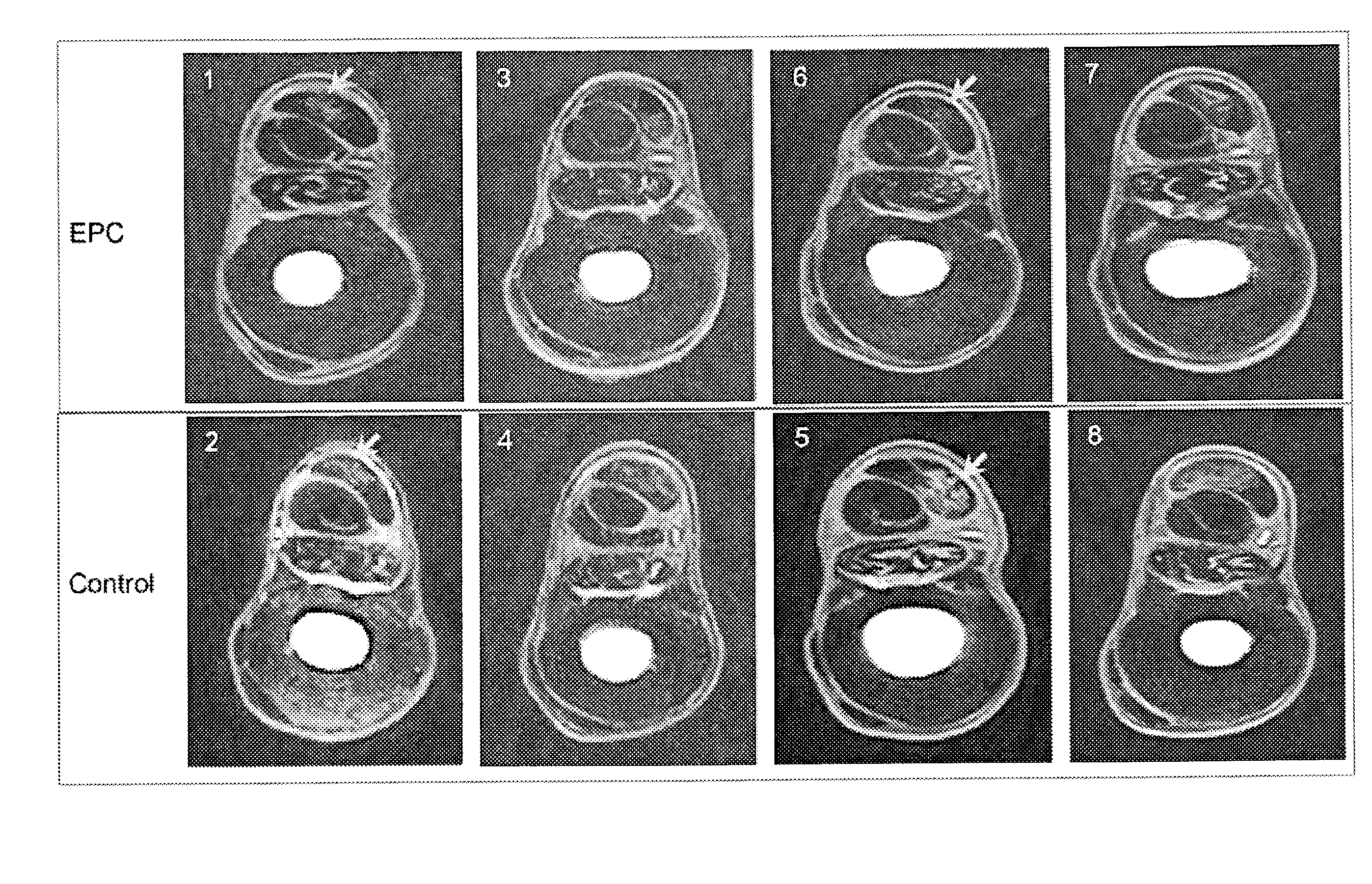
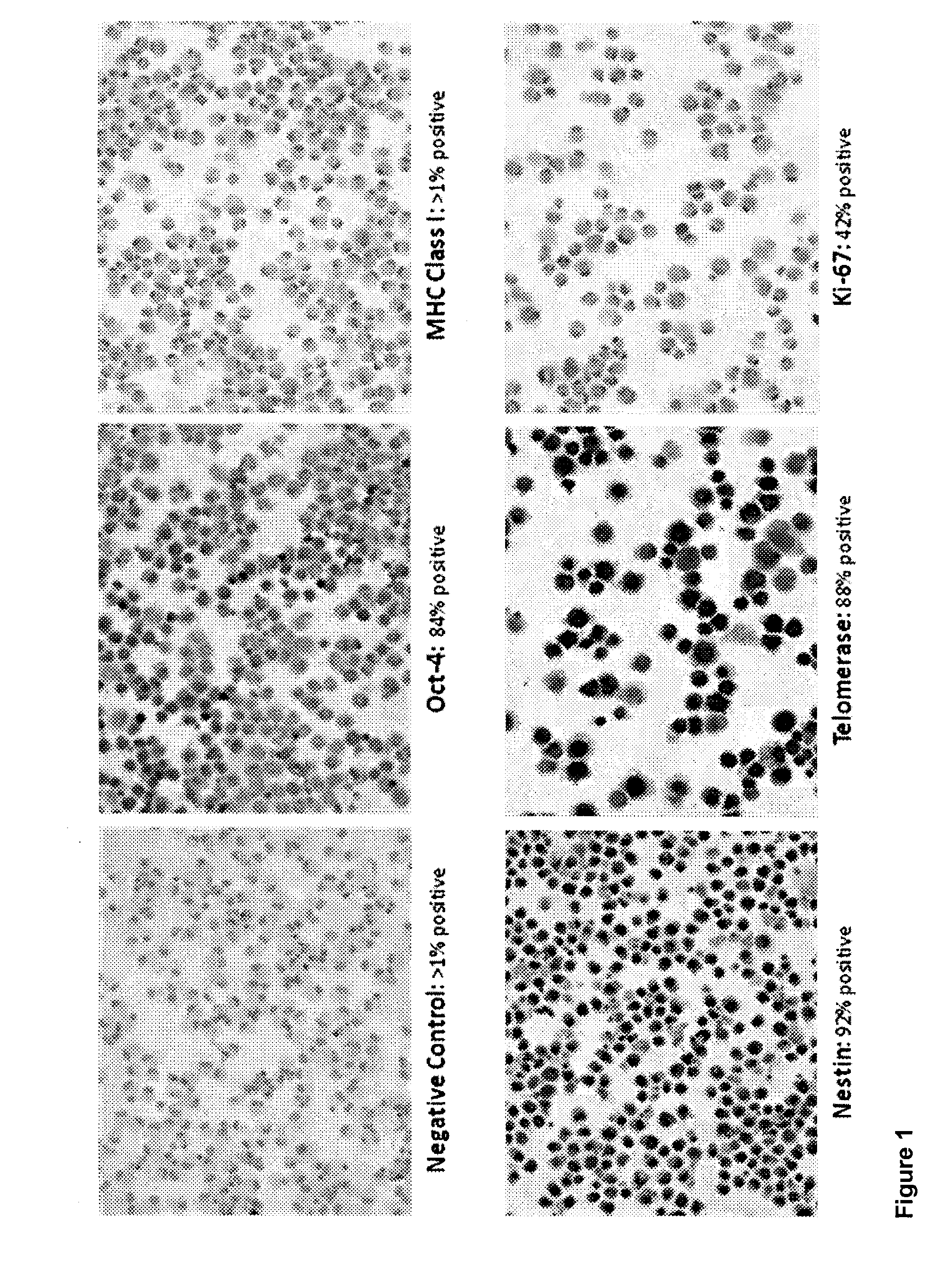
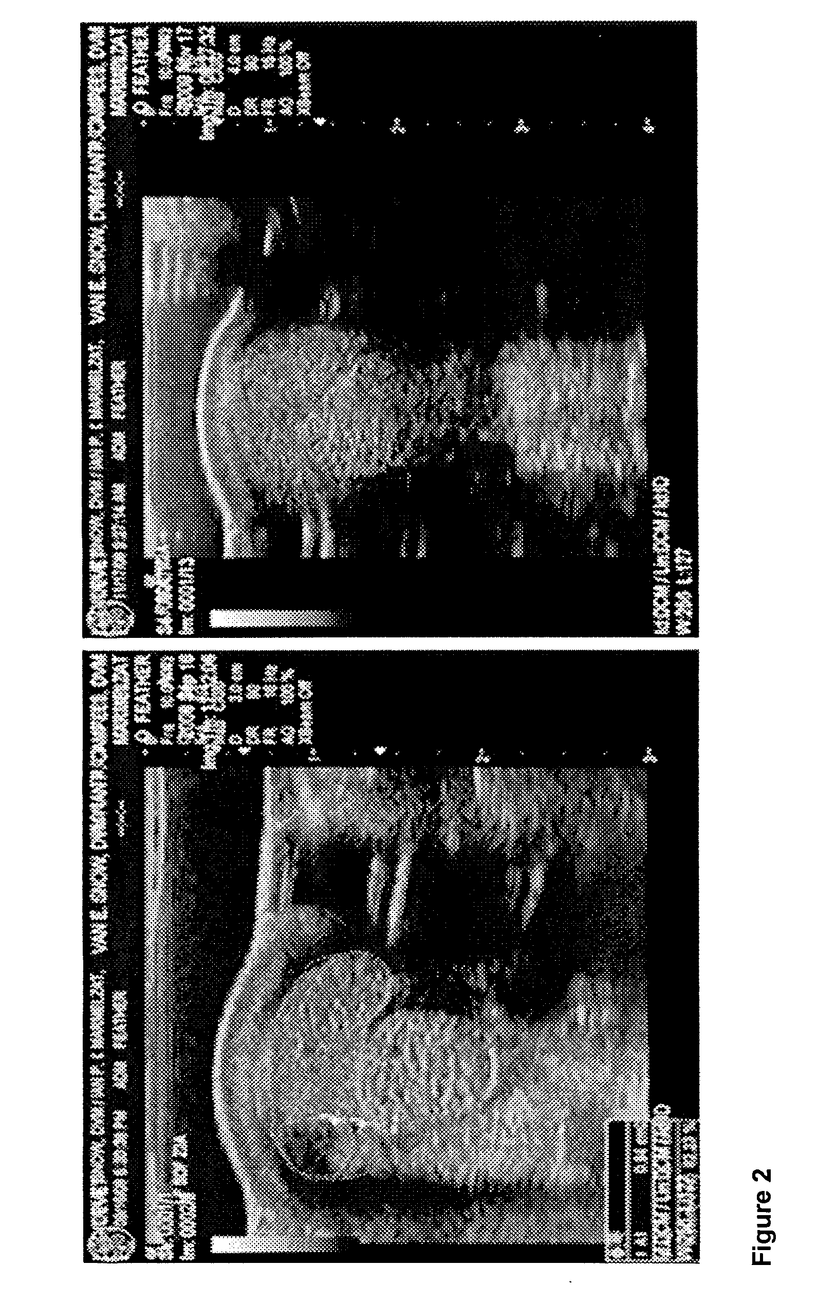
[0008]The invention provides stem cells that can be propagated and maintained for extended periods of time in culture in the absence of a feeder layer, and that can be used to repair tissue damage. Although these cells are derived from different fetal tissues (brain, heart, liver, etc.), they are able to repair injured or diseased tissues of the musculoskeletal system as well as the central nervous system of vertebrate subjects with significantly greater efficacy than stem cells derived from adult tissues. These cells are hypoimmunogenic, as they do not express MHC, and can be used for allogeneic transplantation to vertebrate hosts having disease and / or damage in musculoskeletal, central nervous system (CNS), and other tissues. The ability to repair damage has been documented for the musculoskeletal system of competitive and companion horses and dogs, and can be adapted to cats and other species. The ability to repair damage has also been documented for the CNS of rodent subjects transplanted with human stem cells, and has been extended to canine subjects. Once injected in the injured site, the stem cells of the invention differentiate according to a phenotype most appropriate to the nature of the injured part of the musculoskeletal system and restore normal or near normal structure of the injured or diseased tissue. Moreover, the stem cells of the invention have been found to have an ability to protect against massive structural and functional damage.

Problems solved by technology

The pluripotent nature of these cells renders it unnecessary to genetically modify the cells to be transplanted, and also obviates concerns about selecting the appropriate phenotype of cells, or predifferentiating cells prior to transplantation.

Method used


Image

  • Brain-derived stem cells for repair of musculoskeletal system in vertebrate subjects
  • Brain-derived stem cells for repair of musculoskeletal system in vertebrate subjects
  • Brain-derived stem cells for repair of musculoskeletal system in vertebrate subjects

Examples


example 1

Preparation of Progenitor Cells

[0108]The preparation of brain-derived pluripotent stem cells (BPC) is described in U.S. patent application Ser. No. 11 / 002,933, published Jun. 2, 2005 as publication number US2005-0118561. These same preparations, initially characterized as BPC, were later determined to have features and express markers associated with pluripotent cells. The BPC were derived from the telencephalon (T lines), mesencephalon (M lines) or whole fetal brain (B lines). Due to little or no differences observed between T, M and B lines, separate cultures for T and M lines proved unnecessary and cultures have henceforth been prepared using whole brain.

[0109]This example describes the preparation of cells from equine fetal brain. Subsequent studies have shown that the same preparation and culturing techniques are successful when used with fetal canine brain. Those skilled in the art will appreciate that the same techniques could likewise be adapted for use with other species, i...

example 2

Characterization of Source Tissue

[0111]Stem cells were obtained from a horse fetus. The fetal tissue was dissected under sterile conditions in Hanks Balanced Salt Solution (HBSS) supplemented with Gentamycin and Fungizone. After multiple washes (at least ten times in anti-microbial and anti-fungal HBSS), the tissue was minced with microscissors under a dissecting microscope in a laminar flow hood (Thermo Scientific, Fisher) and then triturated with sterile fire-polished Pasteur pipettes until a single cell suspension was obtained. Cell counts were performed using a hemocytometer. Cells were cultured in an incubator (Thermo Scientific, Fisher), approximately 10,000,000 cells per flask, for one week to confirm that there was no apparent contamination, as determined by examination under a light microscope. Samples of the cell culture were sent to an outside laboratory for karyotyping, and for genetic, microbial and viral screening. If results were negative, cell cultures remained in th...

example 3

Dosage & Transportation of the Cells

[0116]Stem cells are counted and repackaged in an optimal dose of 1.2 million cells / 2 mL of proprietary culture medium for transportation. The amount of cells has been validated as optimal in our pilot clinical study in soft tissue injuries. Vials with stem cells are packaged with ice packs in a STYROFOAM™ container and shipped to the veterinarian for delivery within 24 hours.

[0117]The optimal dose for most injuries is 1.2 million cells, and will be referred to by that number throughout this application. The dose of 1.2 million cells is designed to allow for retention of about 1 million cells after some cells are lost in transport. Viability tests have shown that about 85% of the cells remain viable at 48 hours after coast-to-coast transport via overnight delivery in a STYROFOAM™ cooler. However, the final dose is always to be determined by the nature of the injury and the veterinarian.

[0118]The equine brain-derived PC cultured in the medium of th...


PUM


Abstract

The stem cells that can be propagated and maintained for extended periods of time in culture in the absence of a feeder layer, and can be used to repair tissue damage. These cells are derived from fetal tissues and are able to repair different types of damage in musculoskeletal system, with significantly greater efficacy than stem cells derived from adult tissues. These cells are hypoimmunogenic and can be used for allogeneic transplantation to vertebrate hosts having disease and / or damage in musculoskeletal and other tissues. The cells can be administered by direct injection to the site in need of repair or by systemic (e.g., intravenous) administration. The stem cells of the invention are capable of migrating to the sites in need of repair, and of adopting a phenotype most appropriate to the nature of the damage, injury or disease.

Description

[0001]This application claims the benefit of provisional patent application No. 61 / 152,498, filed Feb. 13, 2009, the entire contents of which are incorporated herein by reference.[0002]This application is related to U.S. patent application Ser. No. 11 / 755,224, filed May 30, 2007, which application claims the benefit of provisional patent application No. 60 / 803,619, filed May 31, 2006; and is a continuation-in-part of U.S. patent application Ser. No. 11 / 002,933, filed Dec. 2, 2004, which claims priority to provisional application No. 60 / 526,242, filed Dec. 2, 2003, the entire contents of each of which are incorporated by reference herein. Throughout this application various publications are referenced. The disclosures of these publications in their entireties are hereby incorporated by reference into this application in order to describe more fully the state of the art to which this invention pertains.BACKGROUND OF THE INVENTION[0003]Standard treatments for musculoskeletal injury (MS...

Claims


Application Information

Patent Timeline
Patent Type & Authority Applications(United States)
IPC IPC(8): A61K35/12
CPCA61K35/12A61P19/00A61P19/10A61P21/00
Inventor KOPYOV, OLEG V.
Owner CELAVIE BIOSCI
Learn More