
Methods of Analysing Cell Behaviour

a cell behaviour and cell technology, applied in the field of cell behaviour and cell behaviour research, can solve the problems of bulge stem cells not supporting it is not known whether bulge stem cells actually support the other pools of stem cells found in the epidermis, and the prior art has not been able to address this question. , to achieve the effect of stable expression of the marker

Inactive Publication Date: 2009-12-10
MEDICAL RESEARCH COUNCIL
View PDF1 Cites 1 Cited by
  • Summary
  • Abstract
  • Description
  • Claims
  • Application Information

AI Technical Summary

Benefits of technology

[0024]Prior art techniques have not permitted the visualisation of clonal cells / clonal cell lines. Prior art techniques have been based on whole tissue X-gal staining which leaches and permeates the tissue rather than being associated with individual cells-expressing the marker gene. Prior art techniques have not enabled the tracing of individual cells derived from a common ancestor since the only wholemount techniques employed which could theoretically cover enough tissue have been crude low-resolution analyses which have served the purposes of the prior art investigations. There has been no need and no motivation in the art to go beyond low-resolution imaging such as. dissecting microscope imaging. Furthermore, this would be impossible in prior art settings such as the gut since the cells of interest are underlain by opaque tissue layers and thus in any case cannot be analysed as taught by the present inventors. A key advantage of the present invention is the capacity to analyse single clonal cells / clonal cell lines which has not been possible in the prior art.

Problems solved by technology

However, it was not possible to follow the behaviour of these stem cells such as their pattern of proliferation or differentiation over time.
Furthermore, although it is known that stem cells in the bulge can go on to produce hair, skin or sebaceous tissue, it is not known whether bulge stem cells actually support the other pools of stem cells found in the epidermis.
The techniques and materials available in the prior art have so far not been able to address this question.
However, it is not clear whether such migration occurs in uninjured adult epidermis.
Unfortunately, it is not possible to trace lineages to individual cells in this style of investigation.
Furthermore, it is frequently unclear if clusters, of marked cells are clonal or arise from multiple adjacent infected cells.
Characterisation of epidermal transit amplifying (TA) cells has been limited.
In retro viral marking and transgenic mouse studies in which the epidermis has been analysed using conventional histological sectioning; it is not possible to detect clusters of 2-32 cells such as would be expected to be produced by TA cells in vivo.
Thus, prior art techniques for studying TA cells are problematic.
Thus, despite advances in understanding stem cell / transit amplifying cell behaviour, problems remain.
Whilst the label retaining cell approach has been successful in delineating the location of slowly cycling cells, the location of proliferating stem and transit cells and the fate of their progeny cannot be defined by this approach.
It is a problem in the art that there is no satisfactory model of cancer beginning from a one cell (stem cell) stage.
Currently, toxicity testing for carcinogenesis is a very animal intensive process.
These animals clearly come at a large economic and moral cost.

Method used


Image

  • Methods of Analysing Cell Behaviour
  • Methods of Analysing Cell Behaviour
  • Methods of Analysing Cell Behaviour

Examples


example 1

Imaging a Clonal Cell Line in a Test Animal

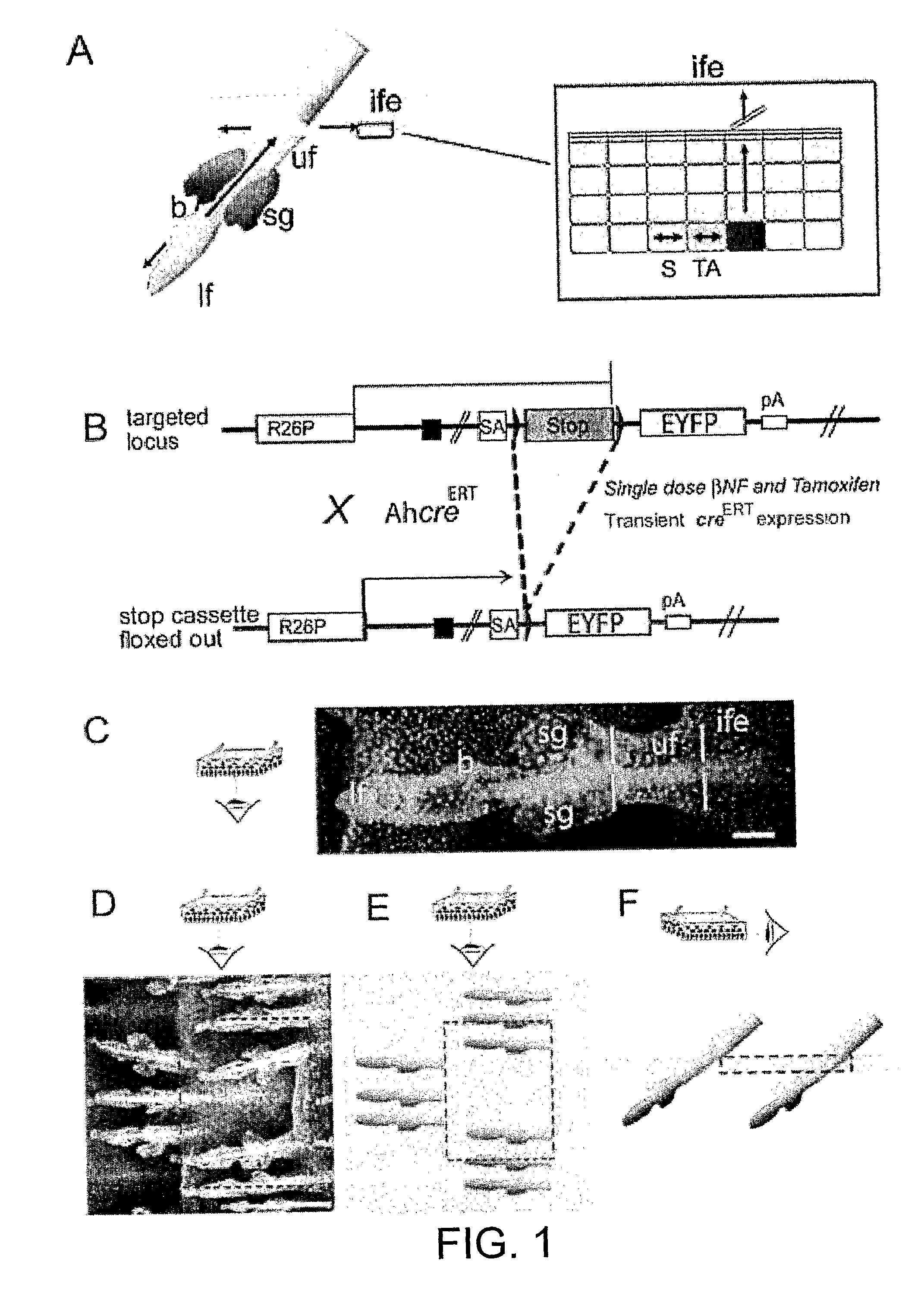
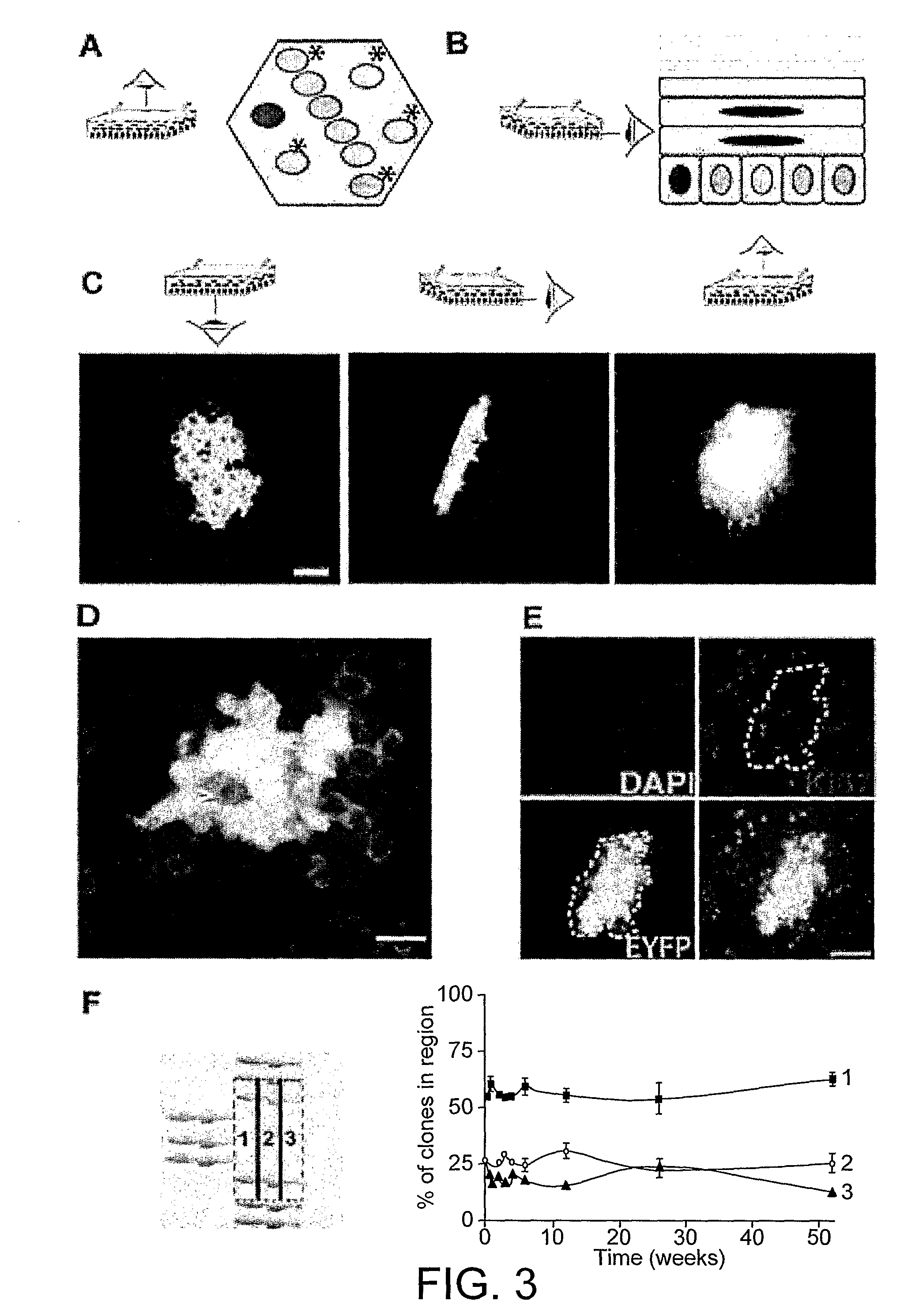
[0146]Mice transgenic for an inducible form of cre recombinase (AhcreERT) were crossed onto the R26EYFP / EYFP reporter strain in which a conditional allele of Enhanced Yellow Fluorescent Protein (EYFP) has been targeted to Rosa26 locus by homologous recombination (FIG. 1B, Kemp et al (2004) Nucleic Acids Res 32, e92; Srinivas et al (2001) BMC Dev Biol 1, 4). In the resultant AhcreERT R26EYFP / wt heterozygote animals, EYFP is expressed in the epidermal cells following a single injection of βNF and tamoxifen at 6-9 weeks of age. At intervals after induction, mice were sacrificed for analysis. Cells expressing EYFP (EYFP+) and their EYFP+ progeny were detected by confocal microscopy and reconstruction of wholemount epidermis, in which the entire epidermis is detached from the underlying dermis, allowing all cells in the tissue to be imaged at single cell resolution (Braun et al (2003) Development 130, 5241-5255). In this study we used tail skin...

example 2

Inducible Clonal Targeting in Adult Mouse Epidermis

[0165]We show a system that allows the controlled induction of specific mutations in individual epidermal cells in a wild type background, and enables the mutated clones and their progeny to be followed over an extended period. To do this we exploited a transgenic mouse line (AhcreERT) which expresses a tamoxifen regulated ere reeombinase-mutant oestrogen receptor fusion protein under the control of the CYPA1A promoter (Kemp et al. 2004). CYPA1A is normally tightly repressed, out is induced following administration of the non genetoxic xenobiatic B-napthoflavone (B-NF) in several tissues including the epidermis and the squamous oesophagus. Dual transcriptional and post-translational control of cre expression results in no detectable background recombination in adult epidermis in the absence of treatment with the inducing drugs. High doses of the inducing drugs result in widespread recombination in the upper hair follicle and interfo...

example 3

Clonal Modelling of Cancer

[0178]Cancer is hypothesised to evolve from oncogenic mutation in individual progenitor cells in a background of wild type cells. The mouse model system described above is ideally suited to test this hypothesis in the epidermis and analyse the changes to clonal behaviour that occur during tumour development.

Development of a Clonal Model of Basal Cell Carcinoma

[0179]Basal cell carcinoma (BSC) is the commonest career in Caucasians in the western world. It can cause significant morbidity through local invasion at the tumour site and requires treatment with surgery, cryoablation or radiotherapy. BCC development is linked strongly to over activation of the Hedgehog (HH) signalling pathway. HH ligands, including Sonic Hedgehog (SHH) bind to a transmembrane receptor Patched (PTCH, 1 on FIG. 9). Ligand binding relieves the inhibition of a second transmembrane protein, Smoothened (SMO), by Patched (2). Derepression of Smoothened leads to Gli transcription factors, h...


PUM

PropertyMeasurementUnit
timeaaaaaaaaaa
timeaaaaaaaaaa
timeaaaaaaaaaa

Abstract

The invention related to a method of imaging a clonal cell line comprising providing a test animal comprising a marker gene, inducing inheritable activation of said marker in at least one cell of said test animal, wherein inheritable activation is induced in fewer than 1 in 27 cells in the tissue of interest, incubating the test animal, and visualising those clonal cells which express the marker gene as a result of the inheritable activation. In particular the invention concerns-methods where the tissue is epidermis, and wherein the visualisation is by confocal microscopy such as wholemount confocal microscopy. The invention also relates to toxicity and carcinogenicity testing using such methods.

Description

FIELD OF THE INVENTION[0001]The invention is in the field of the study of cell behaviour and modelling same. In particular the invention is in the field of modelling cancer and determination of carcinogenicity / toxicity.BACKGROUND TO THE INVENTION[0002]Skin mounts a robust recovery on wounding. This recovery is based on the expansion and differentiation of stem cell populations in the skin, including stem cell populations in the inter-follicular epidermis (IFE) as well as stem cell populations associated with the follicles themselves. This regeneration is clinically important from the perspective of recovery from wounding, and also from the perspective of injury to the skin caused in the process of treatment for example following radiological treatments where skin burning and / or skin scorching can occur.[0003]As well as a clinical interest in the regeneration, process, understanding stem cell behaviour is important in modelling cancer.[0004]Stem cells have been identified in the bulg...

Claims


Application Information

Patent Timeline
Patent Type & Authority Applications(United States)
IPC IPC(8): G01N33/48
CPCA61K49/0006G01N33/5088G01N33/5017A61K49/0008
Inventor JONES, PHILIP H.
Owner MEDICAL RESEARCH COUNCIL
Learn More