Gating With Anatomically Varying Durations

Active Publication Date: 2009-08-13
SPECTRUM DYNAMICS LLC
View PDF106 Cites 66 Cited by
  • Summary
  • Abstract
  • Description
  • Claims
  • Application Information

AI Technical Summary

Benefits of technology

[0056]However, a known problem of such a method is the high computational load that is needed for the execution thereof. Even when using an ordered set method, such as the aforementioned Hudson et al. method, which is capable of reducing the computational load while giving si

Problems solved by technology

Yet, since the annihilation gamma is of 0.511 Mev, regardless of the radioisotope, PET imaging does not provide spectral information, and does not differentiate between radioisotopes.
1. The dedicated algorithms associated with the single pixel cannot reach the accuracy of a two-dimensional image of a plurality of single pixel detectors;
2. The single-pixel detector is a rigid unit, which does not have the flexibility of motion of a plurality of small detectors, each with independent motion; and
3. A single hot spot may cause the single pixel detector of the Anger camera to saturate, whereas when a plurality of single pixel detectors is employed, saturation is localized to a few pixels and does not affect the whole image.
The reconstruction process is generally complex, due to the large quantity of data that must be processed in order to obtain an accurate reconstruction.
In many medical imaging situations, the positioning of the detecting unit relative to the emitting object is constrained, so that complete measured data is not available.
Reconstruction based on filtered back-projection is therefore of limited use for medical imaging.
Any reconstruction from a limited range of projections potentially results in spatial distortions (artifacts) in the image.
While it is known that the severity of the artifacts increases with the increasing angular range of the missing projections, limited-angle tomography does not provide information on which projections should be used in order to most effectively reconstruct the image.
There is currently no analytic way to solve Eqn. 1 for the maximum of the likelihood function.
Limited computational resources (i.e., when the entire projection matrix Φ cannot be kept in memory) may require breaking the update computation according to a partition of Φ into a set of sub-matrices (Φi).
Since during medical testing, measurements are generally performed at many locations as the detecting unit is moved relative to the observed organ, the testing procedure generally requires a long time and is physically and emotionally difficult for the patient.
Additionally, reconstruction is based upon a large quantity of data, and is a lengthy and computationally complex process.
However, a known problem of such a method is the high computational load that is needed for the execution thereof.
Such a high computational load results in a longer reconstruction time that reduces the throughput of the processing unit and requires a more expensive processing hardware.

Method used


Image

  • Gating With Anatomically Varying Durations
  • Gating With Anatomically Varying Durations
  • Gating With Anatomically Varying Durations

Examples


Embodiment Construction

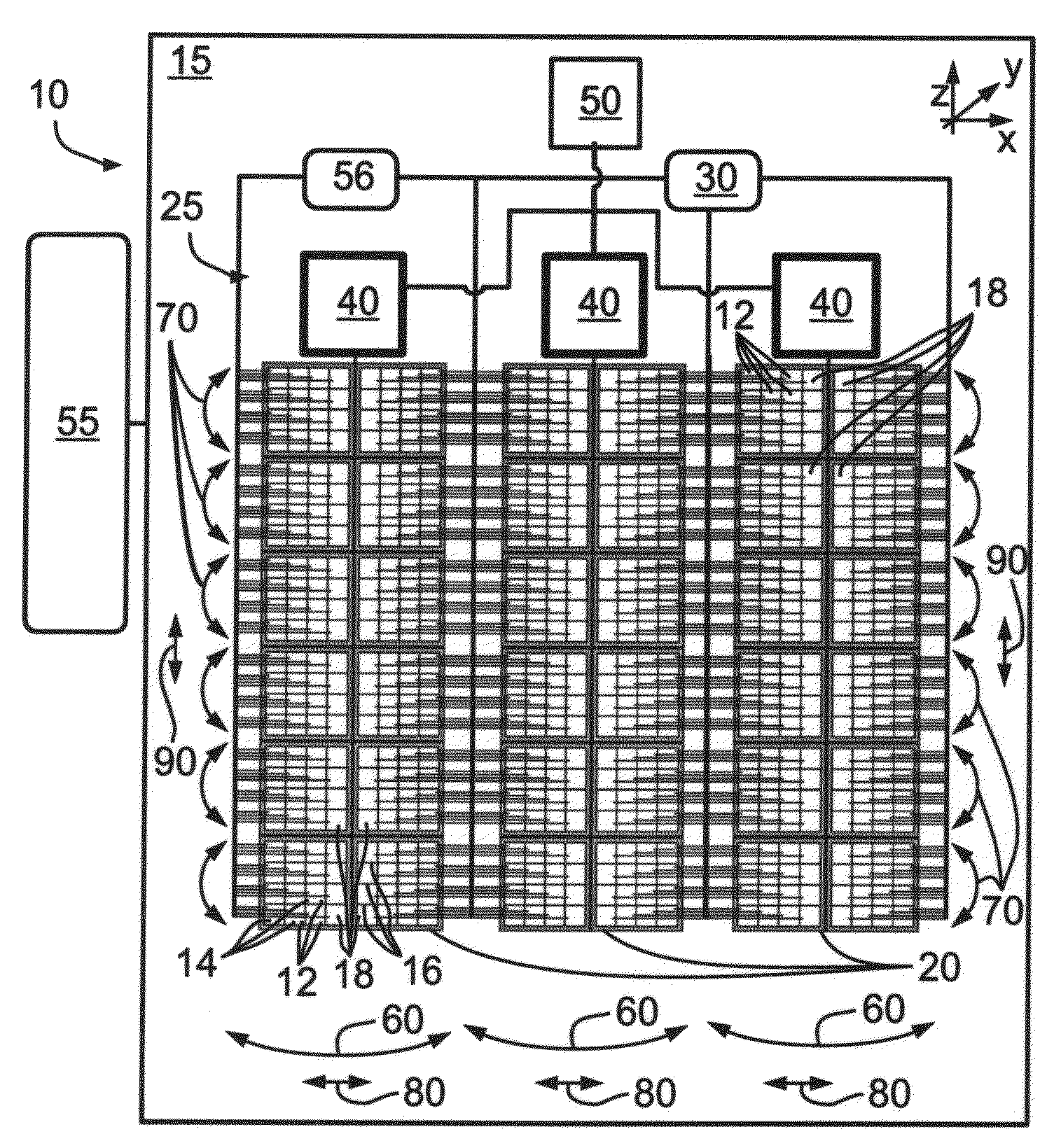
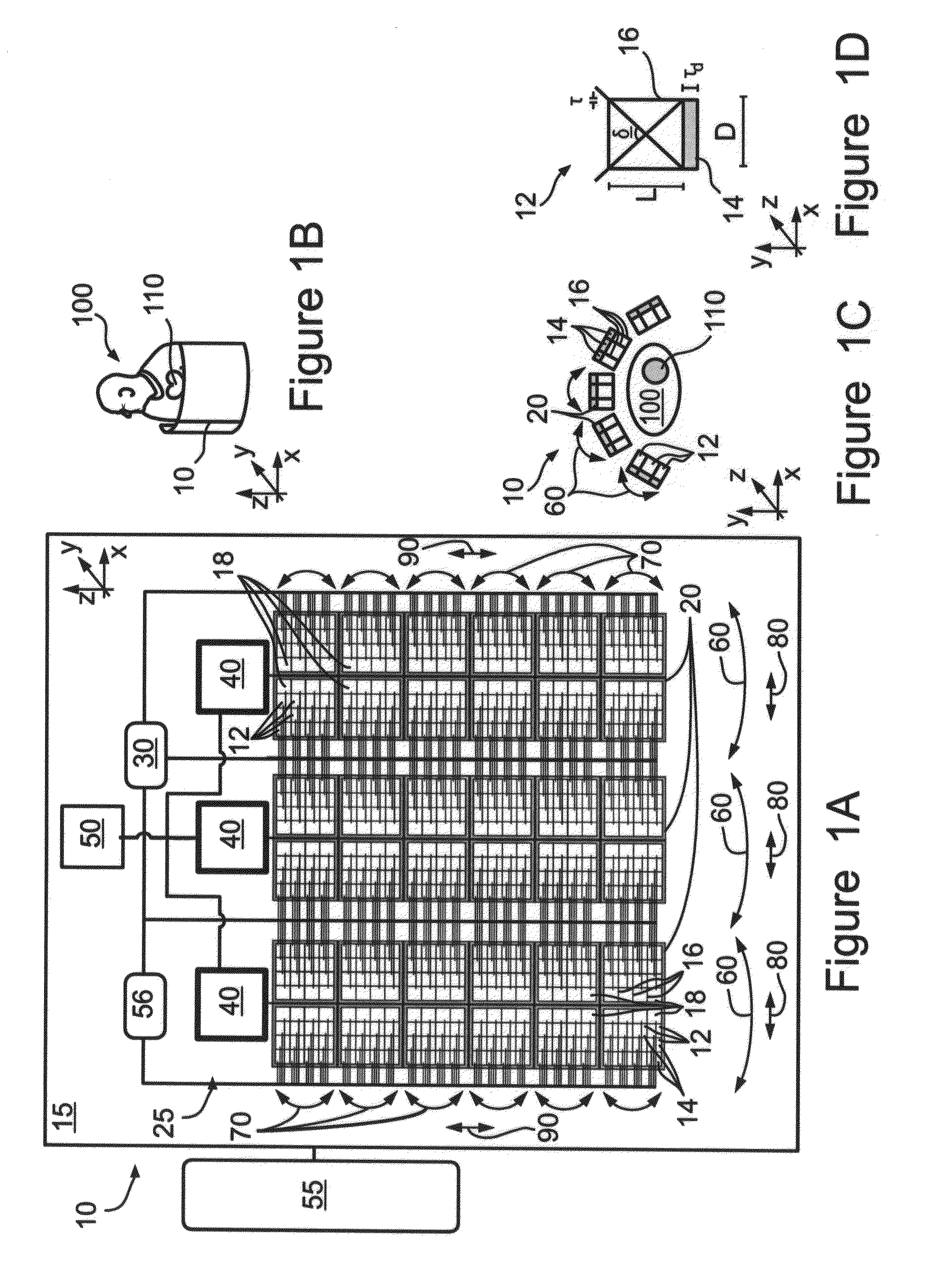
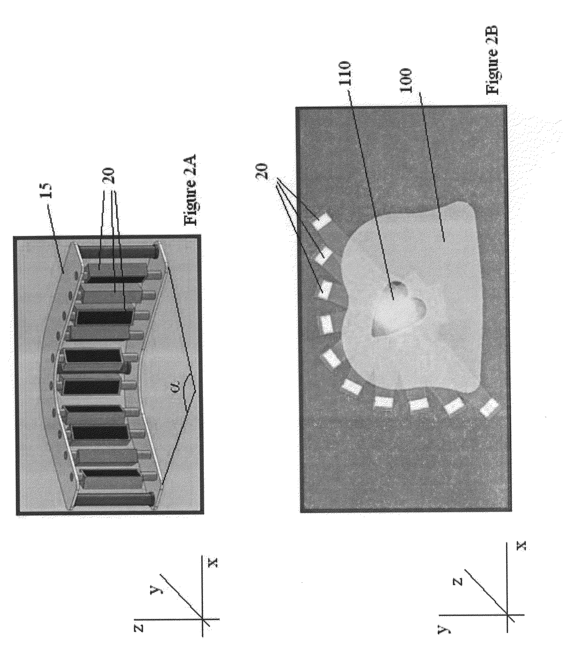
[0084]The present invention relates to an apparatus and a method for reconstructing a radioactive emission image of a overall volume having dynamic and static volumetric regions. The reconstructing is based on gated images with anatomically varying time-bin lengths. The apparatus and the method are designed to allow the segmentation of the radioactive emission image to gated and non-gated regions. In such a manner, the reconstructions radioactive emissions from the dynamic volumetric region and the static volumetric region are carried out separately. Thus, the high computational throughput that is needed in order to reconstruct a dynamic volumetric region, such as the heart, using time binning techniques has less or no effect on the reconstruction of the static volumetric region, as further described below. The disclosed apparatus comprises a number of detectors, such as PET or SPECT detectors, which are designed for obtaining radioactive emissions from the overall volume and an ima...


PUM


Abstract

A method for reconstructing a radioactive emission image of an overall volume having first and second volumetric regions, each volumetric region having respectively independent dynamic characteristics. The method comprises the following steps: a) obtaining radioactive emissions from the overall volume, including the volumetric regions, b) reconstructing an initial radioactive emission image of the volumetric region according to the radioactive emissions, c) segmenting the initial radioactive emission image to delineate the first and second volumetric regions, and d) separately reconstructing the first and the second volumetric regions according to the respectively independent dynamic characteristics.

Description

FIELD AND BACKGROUND OF THE INVENTION[0001]The present invention relates to a method and an apparatus for image reconstruction in nuclear medicine imaging and, more particularly, but not exclusively to image reconstruction in nuclear medicine imaging using gating techniques.[0002]Radionuclide imaging aims at obtaining an image of a radioactively labeled substance, that is, a radiopharmaceutical, within the body, following administration, generally, by injection. The substance is chosen so as to be picked up by active pathologies to a different extent from the amount picked up by the surrounding, healthy tissue in consequence; the pathologies are operative as radioactive-emission sources and may be detected by radioactive-emission imaging. Pathology may appear as a concentrated source of high radiation, that is, a hot region, as may be associated with a tumor, or as a region of low-level radiation, which is nonetheless above the background level, as may be associated with carcinoma.[...

Claims


Application Information

Patent Timeline
IPC IPC(8): G06T17/00G06T15/00G06K9/00
CPCA61B5/055A61B5/415A61B5/416A61B5/418A61B5/7285A61B6/037G06T2211/436A61B6/541A61B8/0883A61B8/543G01T1/1644G06T11/006A61B6/503G01T1/1663G06T11/005G06T11/008G06T2207/10104G06T2207/30048
Inventor ZIV, OMERRAVHON, RANDICHTERMAN, ELIGLUHOVSKY, LEONIDBEN-HAIM, SHIOMOROUSSO, BENNY
Owner SPECTRUM DYNAMICS LLC
Why Eureka
Try Eureka