
In vivo fluorescence sensors, systems, and related methods operating in conjunction with fluorescent analytes

Inactive Publication Date: 2009-07-16
VTQ IP HLDG
View PDF12 Cites 4 Cited by
  • Summary
  • Abstract
  • Description
  • Claims
  • Application Information

AI Technical Summary

Benefits of technology

[0010]Embodiments of the present invention can provide cost-effective methods, systems, devices and computer program products that can individualize and customize therapy to improve response and outcome, assess an internal dose received at a target site, and / or otherwise monitor therapeutic response or delivery of fluorescent analytes or compounds in the body. In certain embodiments, the methods, systems, devices and computer program products can provide data regarding the impact of therapies on tumors or normal (non-targeted) tissue.
[0013]Advantageously, in certain embodiments, the present invention can provide cost-effective minimally invasive methods, systems, devices and computer program products that can evaluate, in substantially real-time, one or more selected biokinetic parameters or predictor variables of a subject using fluor-data obtained in vivo. Certain embodiments of the devices and systems can be configured to identify the differences in response between normal and malignant tissue and / or the differences in the physiology and biology of individual tumors or tumor sites (or the same tumor site at different times) and to utilize the identified information regarding same to develop individualized treatment decisions, and / or to predict therapeutic outcome and / alter a therapy to improve tumor response.
[0015]Other embodiments may allow improved individualized treatment protocols based on an in vivo detected uptake, trapping and / or other desired response (over a selected time) of a non-therapeutic dose of a drug, typically evaluated before and proximate in time to the delivery of the therapeutic dose) to predict the response of the subject to a therapeutic dose of a drug in advance of administration thereof. Such pre-delivery assessment capability may allow an improved patient customized selection of chemical or treatment drug, reducing unnecessary ineffective administration of cytotoxic agents, which are unlikely to be clinically effective. Thus, embodiments of the present invention can identify, by measuring detected fluorescence associated with the uptake and / or retention of a fluorescent analyte, the sensitivity or receptiveness of a tumor for a particular treatment, proximate in time to the planned delivery or administration of same. In addition, the data can be used to determine when a subsequent therapy should be administered, based on the uptake or retention or non-retention or decrease in the therapeutic analyte at the target site. Such data may allow a customized treatment plan rather than the delivery of treatments based on established standards.

Problems solved by technology

Most of this work, however, deals with ex vivo samples and application to living tissue in vivo is difficult given the fluorescence microscopy tools presently available.
These compounds are restricted to treating refractory diseases and are not presently used as a first-line treatment.
A factor in this disposition of Ab therapies is the lack of clear knowledge of effect.
There is no currently viable or acceptable method by which such measurements of clinical effect can be made in vivo in target tissue.
Some compounds of interest, e.g., DOXORUBICIN, are naturally fluorescent, though the strength of the fluorescence may not be optimal for monitoring purposes.
Unfortunately, following detailed kinetics with endoscopic techniques of this sort would likely be commercially undesirable.
However, due to the strong attenuation and scattering of visible light by tissue, this technique is limited to tumors within a few millimeters of the skin surface.
Again, this technique is hampered by the attenuation of the light signal by tissue and is thus limited to mice with tumors near the surface.

Method used


Image

  • In vivo fluorescence sensors, systems, and related methods operating in conjunction with fluorescent analytes
  • In vivo fluorescence sensors, systems, and related methods operating in conjunction with fluorescent analytes
  • In vivo fluorescence sensors, systems, and related methods operating in conjunction with fluorescent analytes

Examples


examples

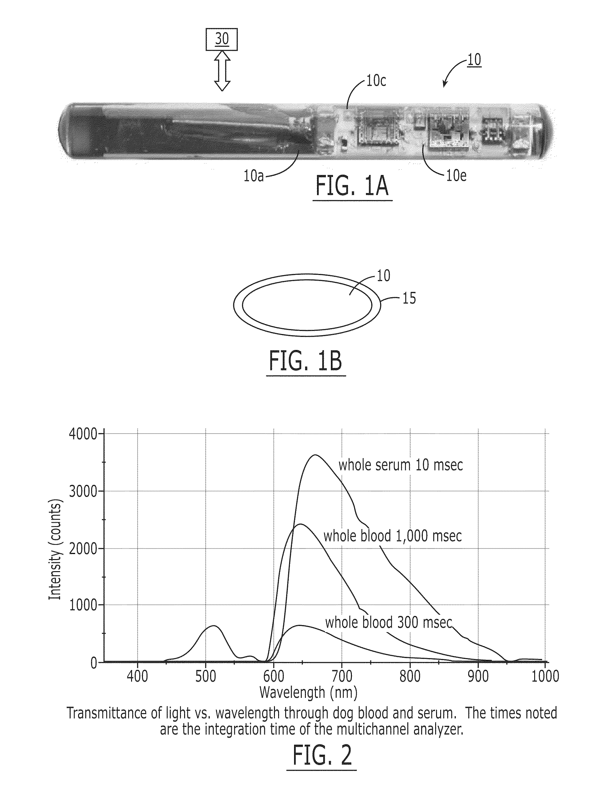
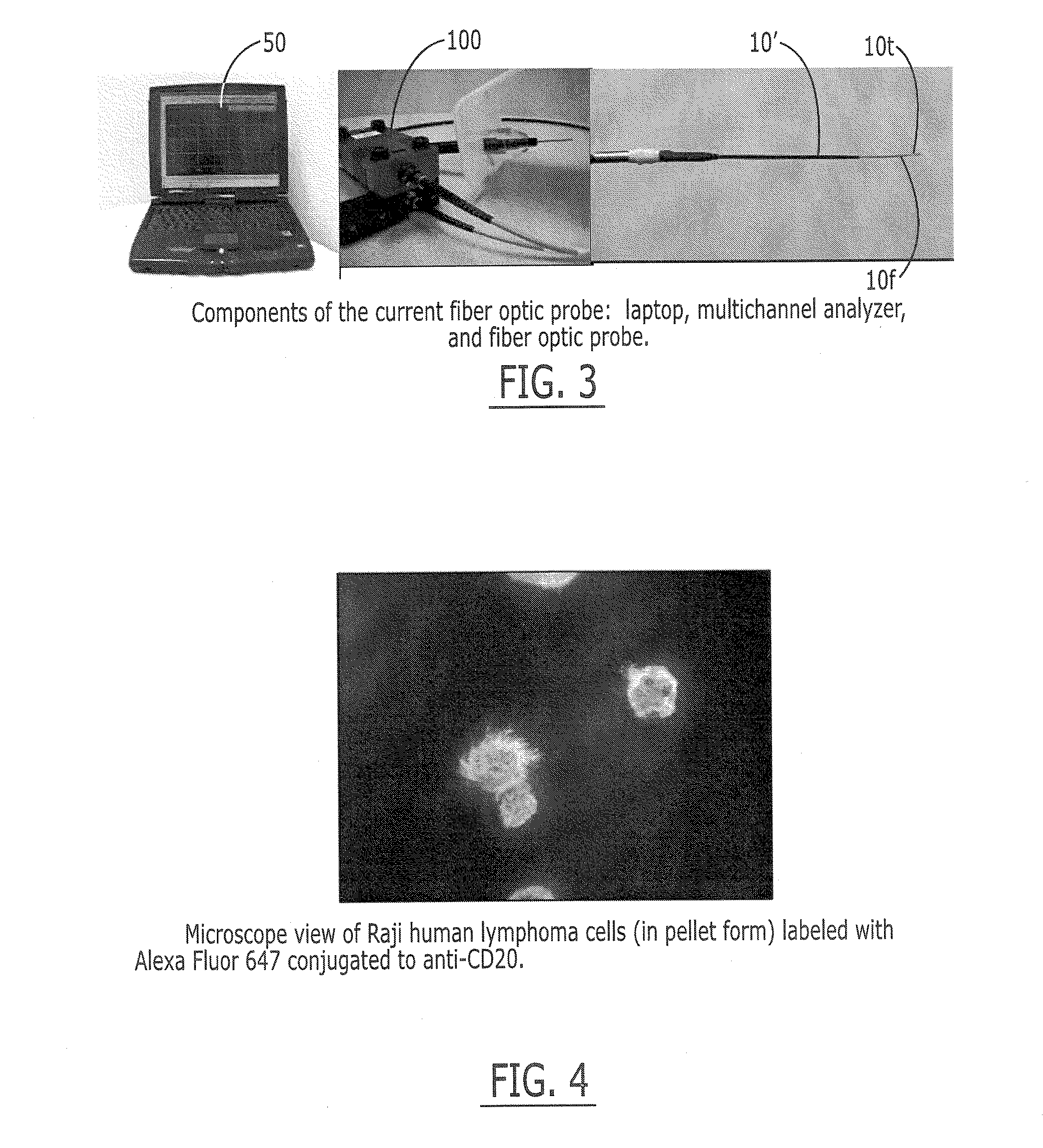
[0149]The fluorescence sensor systems described herein provide real-time, acute and / or chronic, measurement of fluorescently labeled analytes in vivo allowing pharmacokinetics and pharmacodynamics of antibody-based therapies to be assessed on an individualized basis. Initial experiments were successfully completed with a catheter-based version of the probe. The probe uses a laser diode illumination source and an optical multichannel analyzer. The laser diode (650 nm) source effectively penetrates several millimeters of tissue. The antibodies were labeled with Alexa 647 fluorophore (Molecular Probes, Eugene, Oreg.). In vitro tests confirmed this wavelength provides good light transmission through blood, tissue, and serum. Calibration studies with fluor in a colloidal gelatin mixture demonstrated sensitivity in the <10 ng / ml range.

[0150]Experiments were performed using two human cancer cell lines: Raji Burkitt's human lymphoma and BT474 human breast adenocarcinoma. The targeted antige...


PUM

PropertyMeasurementUnit
Timeaaaaaaaaaa
Timeaaaaaaaaaa
Poweraaaaaaaaaa

Abstract

Methods, systems, devices and computer program product include: (i) administering a fluorescent analyte to a subject; (ii) repetitively emitting excitation light from an implanted sensor over a desired monitoring period; (iii) detecting fluorescence intensity in response to the excitation light using the implanted sensor that outputs the excitation light; and (iv) using data associated with the detected fluorescence intensity to perform at least one of: (a) calculate the concentration or dose of the analyte received proximate to the implanted sensor site; (b) evaluate the pharmacodynamic or pharmacokinetic activity of the fluorescent analyte; (c) confirm Ab attachment to a tumor site; (d) monitor a non-target site to confirm it is not unduly affected by a therapy; (e) monitor for changes in cellular properties; (f) use the calculated dose or concentration data to adjust or customize a therapeutic amount of the analyte administered to the subject; (g) confirm micelle concentration at a target site and then stimulate toxin release based on the confirmation; and (h) monitor for the expression of a protein produced from a gene therapy modification.In particular embodiments, the intensity of the excitation signals emitted to the localized tissue can be varied in a predetermined manner to generate optical profiling data of the response of the tissue proximate the sensor.

Description

RELATED APPLICATIONS[0001]The present application is a divisional of application Ser. No. 10 / 779,907, filed Feb. 17, 2004, which claims priority to U.S. Provisional Application Ser. No. 60 / 448,349, filed Feb. 19, 2003 and U.S. Provisional Application Ser. No. 60 / 471,706, filed May 19, 2003, the entire contents of the above-referenced documents are hereby incorporated herein by reference.BACKGROUND[0002]The use of fluorescent reporters to study cells and tissue is known and a body of work has grown up around this technique. Most of this work, however, deals with ex vivo samples and application to living tissue in vivo is difficult given the fluorescence microscopy tools presently available.[0003]The use of therapeutic antibodies (“Ab”) in cancer treatment is a relatively new endeavor. Several compounds are now approved for use in the United States (e.g., HERCEPTIN, RITUXAN, ZEVALIN) and more are in development. These compounds are restricted to treating refractory diseases and are no...

Claims


Application Information

Patent Timeline
IPC IPC(8): A61B10/00A61B5/00A61K49/00
CPCA61B5/0002A61B5/0071A61K49/0058A61B5/417A61K49/0041A61B5/0084
Inventor BLACK, ROBERT D.BOLICK, NATASHA
Owner VTQ IP HLDG
Learn More