
Identification of ligands by selective amplification of cells transfected with receptors

Inactive Publication Date: 2009-06-18
ACADIA PHARMA INC
View PDF0 Cites 3 Cited by
  • Summary
  • Abstract
  • Description
  • Claims
  • Application Information

AI Technical Summary

Benefits of technology

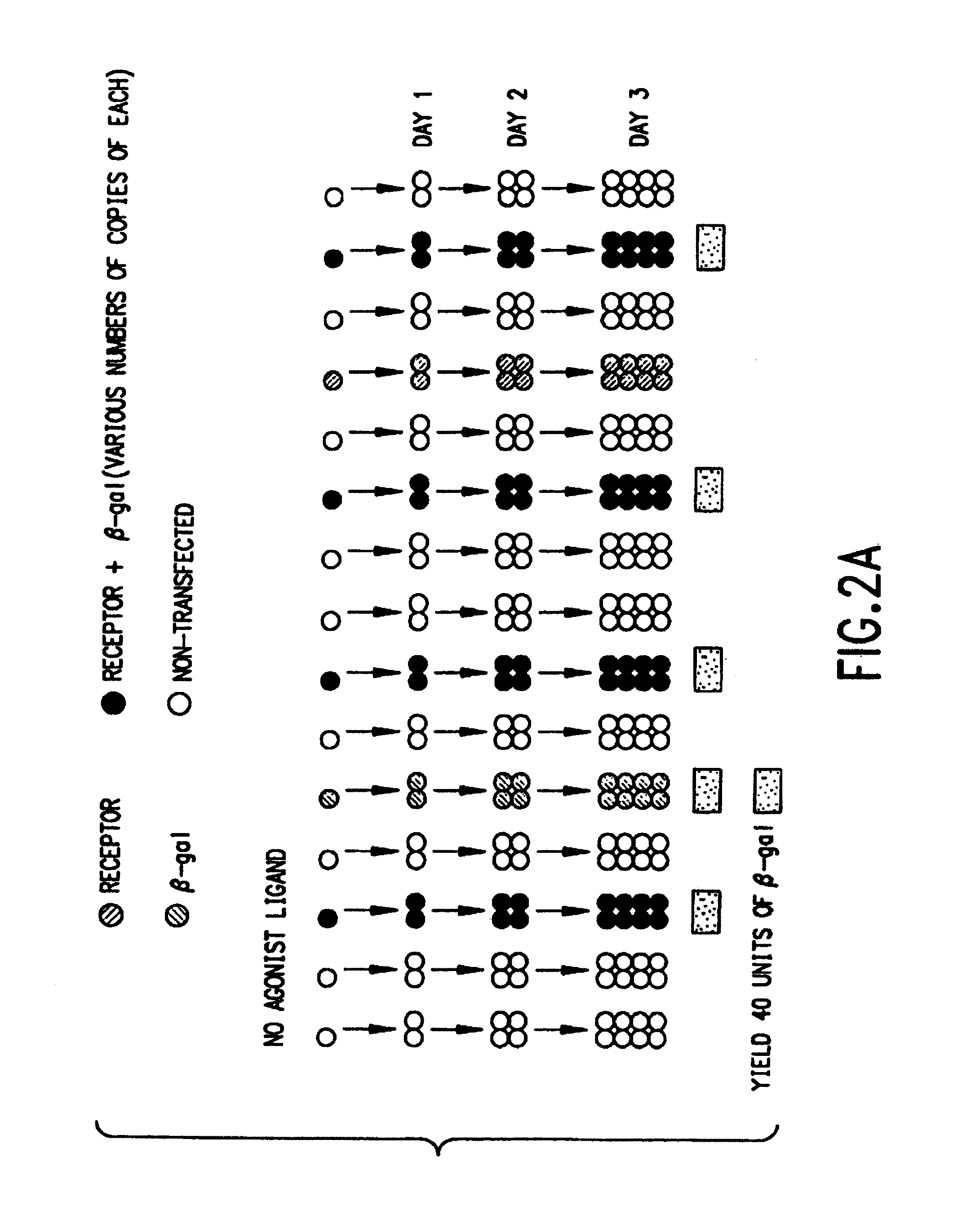
[0035]When the ligand is an antagonist, the action can be determined similarly, but in reverse, i.e., the cells containing the marker will be at a competitive disadvantage relative to the untransfected cells, the population of which will expand at a greater rate than the transfected cells. However, it is preferred that the assay for antagonists be conducted in the presence of an agonist, and the observed effect is a decrease in the amplification response brought about by the presence of the stimulatory ligand alone.
[0063]Furthermore, the present invention relates to a method of decreasing the basal activity level of the 5-HT2A receptor in a subject in need thereof, the method comprising contacting a 5-HT2A receptor in said subject with an inverse agonist of the 5-HT2A receptor in an amount effective to substantially decrease the level of basal activity of said receptor. In a preferred embodiment, the inverse agonist is selective for the 5-HT2A receptor (i.e., has at least about ten times greater affinity for the 5-HT2A receptor than for at least one other neurotransmitter receptor). In another preferred embodiment, the inverse agonist of the 5-HT2A receptor has little or substantially no anti-dopaminergic activity. In a related aspect, the invention relates to a method of decreasing serotonergic neurotransmission through the 5-HT2A receptor, the method comprising contacting a 5-HT2A receptor with an inverse agonist of the 5-HT2A receptor in an amount effective to substantially decrease the level of basal activity of said receptor.
[0101]The Single Receptor Format allows the convenient assay of the interaction of agonist and antagonist ligands with individual receptors. The Multiple Receptor Format allows the convenient assay of the interaction of agonist and antagonist ligands with several receptors at the same time.
[0102]The Single Receptor Format involves very few steps; no expensive reagents; ability to quantitatively discriminate partial agonists, full agonists, and antagonists. Because the assay relies on transfections of recombinant receptor and marker DNA, the assay can be performed with a wide variety of receptors, markers and cell types. In addition to these properties, the Multiple Receptor Format represents the only method known to the inventor which can be applied to screening for ligand activity against large numbers of receptors simultaneously. Thus, the Multiple Receptor Format is particularly suitable for use in a drug screening programme wherein “hits” (that is, substances with ligand activity) may be identified quickly from among a large number of test substances.
[0105]In a further use of the present method, libraries of a given receptor may be prepared by amplifying a specific gene from several individuals, tumors, tissues, or randomly mutated pools. These libraries of cDNAs can then be screened by transfecting pools of DNAs into cells, and growing the cells in the presence or absence of ligand. This strategy is likely to be particularly powerful when applied to identification of constitutively active versions of receptors (e.g. certain oncogenes).

Problems solved by technology

Unfortunately, with available methods and technology, a major limitation in the drug discovery process is the time and expense required to screen these libraries against so many targets.
Binding assays have many limitations: (i) For many technical reasons, binding assays are almost always performed in nonphysiological buffers.
(iv) Since only modest levels of receptor (binding site) expression have been achieved in mammalian cells, propagation of receptors is a major expense in these assays.
(v) The vast majority of labeled ligands are radioisotopes, the purchase, handling and disposal of which are major expenses.
Mol. Endocrinology. 7:12-16 (1993)), these assays have proven to have limited utility when applied to cell surface receptors, presumably because of the more modest transcriptional control that these receptors exert.
The limitations of these assays are that only certain functional types of receptors can be measured.
Also, while the assays are relatively convenient, there are limitations inherent in the endogenous responses and cells that are used.
These pH changes are small in magnitude and require expensive instrumentation for measurement (Cytosensor, Molecular Dynamics Co.).
This device is not compatible with other instruments that are used in mass screening (e.g. use of a 96 well plate format) and because samples must be incubated within the instrument for several minutes, there is limited sample throughput.
A theoretical limitation inherent in all of the above assays is the inability to assay a given ligand against more than a few receptors at the same time.
Because of their limited dynamic range, incompatible assay conditions, and the fact that many receptors cannot be distinguished from one another based on their functional responses, second messenger responses, and most other biochemical effects of receptors, are not at all amenable to multiplexed assay.
Unfortunately, these assays offer few advantages to the approaches described above.
These assays are neither convenient nor inexpensive to perform.
It has been estimated that the total financial cost for the diagnosis, treatment, and lost societal productivity of individuals affected by this disease exceeds 2% of the gross national product (GNP) of the United States.
To date, there exist no definitive diagnostic tests for this disorder.
Antipsychotics are effective in ameliorating positive symptomotology, yet they frequently do not improve negative symptoms, and significant, treatment-limiting side effects are common.
Clinical use of these compounds are limited, however, by their side effect profiles.
Nearly all of the “typical” or older generation compounds have significant adverse effects on human motor function.
Unfortunately, it is likely that antagonism of dopamine D2 receptors also mediates the disabling extrapyramidal side effects.
Antagonists compete against agonists and inverse agonists for access to the receptor, but do not possess the intrinsic ability to inhibit elevated basal or constitutive receptor responses.
233-235) cause reading frame shifts, prematurely terminating translation of these proteins, resulting in non-functioning receptors that subsequently cause color blindness and nephrogenic diabetes insipidus, respectively.
These authors were unable to measure the constitutive activity of the wild type receptor in their assay, and an insufficient number of clinically relevant compounds comprising the various chemical classes of antipsychotics were tested.
This precluded the authors from recognizing the significance of 5-HT2A receptor inverse agonism and efficacy as an antipsychotic.

Method used


Image

  • Identification of ligands by selective amplification of cells transfected with receptors
  • Identification of ligands by selective amplification of cells transfected with receptors
  • Identification of ligands by selective amplification of cells transfected with receptors

Examples


example 1

β-Galactosidase Activity in Cells Transfected with the trk A Receptor

[0232]Nerve growth factor (NGF) is an agonist for the trk A receptor. NGF-stimulated trk A receptors activate tyrosine phosphorylation, and induce foci in NIH 3T3 cells. FIG. 3a illustrates data from an experiment where trk A receptor-transfected cells were grown in the presence or absence of NGF following the general procedure described above. A 10 cm plate of NIH 3T3 cells were transfected with 5 μg of trk A receptor DNA (cloned substantially as described by Kaplan et al., Science 252, 1991, p. 554, and Martin-Zanca et al., Mol. Cell. Biol. 4, 1989, p. 24) and 5 μg of β-gal DNA. The cells were washed after 24 hrs, and after 48 hrs the cells were transferred to 96 wells of a microtiter plate and grown in the presence or absence of NGF for the indicated number of days. β-gal activity was induced by NGF, with a marked induction observed within three days. The data shown in FIG. 3A and FIG. 3B were means of triplicat...

example 2

β-Galactosidase Activity in Cells Transfected with Muscarinic Receptor Subtypes m1, m2, m3, m4 and m5

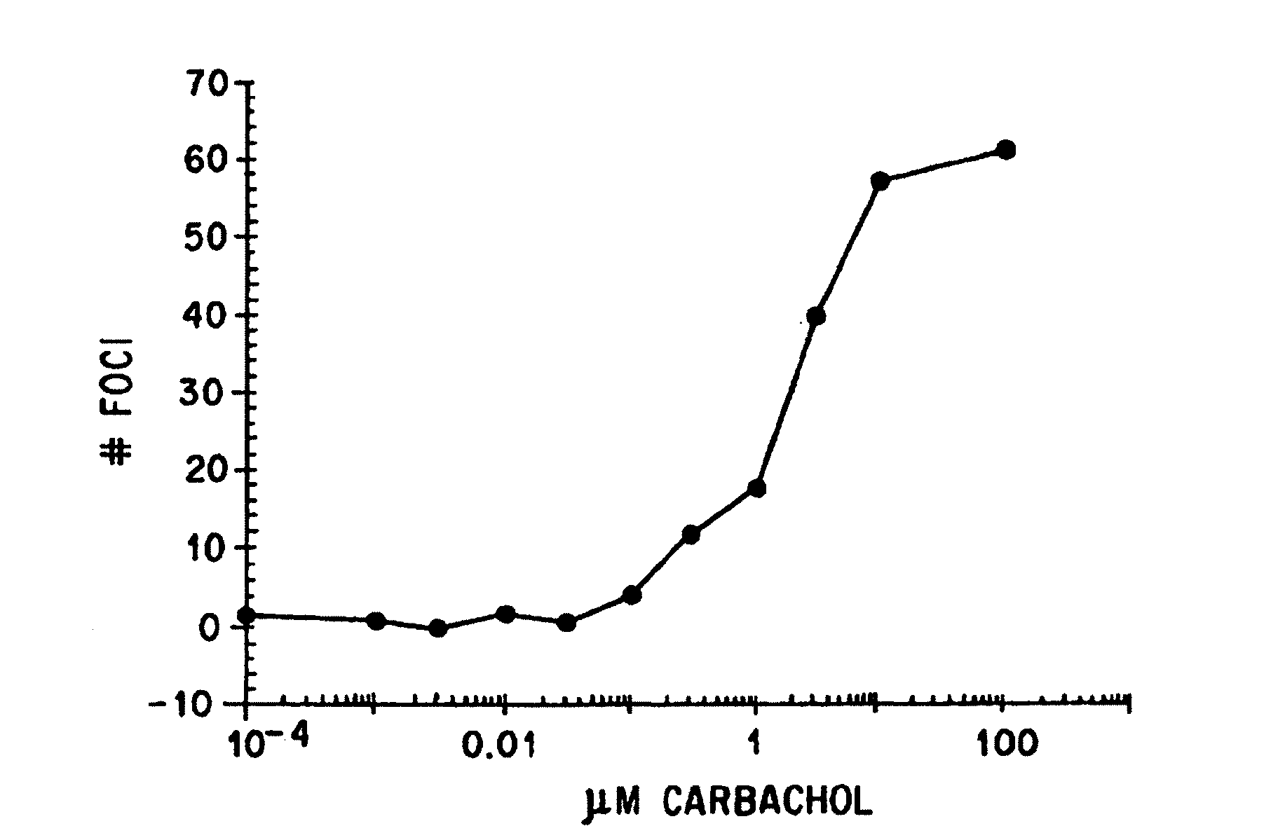
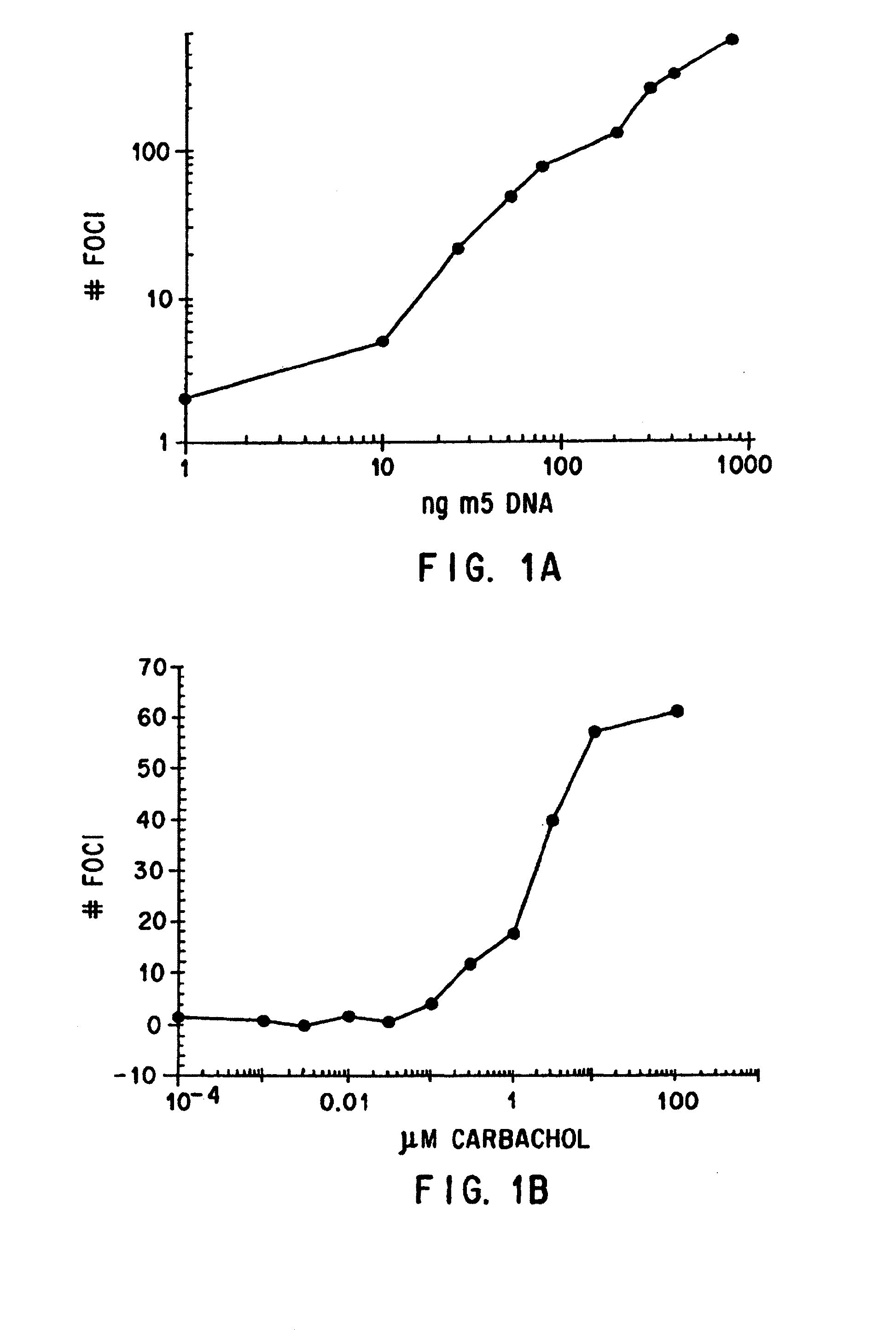
[0234]Muscarinic acetylcholine receptors that stimulate phospholipase c (m1, m3, m5) are able to stimulate cellular growth and induce foci in NIH 3T3 cells, only when the transfected receptors are activated by ligands that have agonist activity. In monoclonal lines isolated from NIH 3T3 cells transfected with these receptors, the agonist dose-response relationships for stimulation of phospholipase c, stimulation of mitogenesis and foci are identical, and these responses are blocked by the muscarinic receptor antagonist atropine. The m2 and m4 muscarinic receptors do not strongly stimulate phospholipase c in NIH 3T3 cells, nor do they induce foci. These data indicate that ligand-induced changes in cellular growth can be used as an assay of the pharmacology of some muscarinic receptor subtypes (Gutkind et al., PNAS 88, 4703 (1991); Stephens et al., Oncogene 8, 19-26 (1993)).

[0235]The d...

example 3

Luciferase Activity in Cells Transfected with the m5 and m2 (Gq-15) Muscarinic Receptors

[0241]Following the general protocol described above, amplification of the m5 muscarinic receptor and the m2 muscarinic receptor (co-transfection with Gq-15) was determined using firefly luciferase (luc, pGL2-control vector, Promega) as a marker instead of β-galactosidase. Receptor, marker, and G-protein DNA concentrations were identical to those described for the β-galactosidase experiments in Example 2. The ED50's of carbachol were 0.22±0.1 μM for m5 and 0.14±0.11 μM for m2 / q-i5 for inducing activity of firefly luciferase. Firefly luciferase was assayed as recommended by the manufacturer (Promega). The data obtained indicate that, like β-galactosidase, firefly luciferase can serve as a sensitive marker of muscarinic receptor activation by a ligand.


PUM

PropertyMeasurementUnit
Volumeaaaaaaaaaa
Volumeaaaaaaaaaa
Volumeaaaaaaaaaa

Abstract

The invention is directed to a method for identifying substances acting as ligands for transfected receptors by using transfected markers to measure receptor / ligand interactions. The present invention also relates to a method of identifying compounds which act as inverse agonists of the 5-HT2A receptor, the method comprising contacting a constitutively active 5-HT2A receptor with at least one test compound and determining any decrease in the amount of basal activity of the receptor so as to identify a test compound which is an inverse agonist of the 5-HT2A receptor. Such inverse agonists may be used in the treatment of schizophrenia and related psychoses.

Description

RELATED APPLICATIONS[0001]This application is a divisional of U.S. application Ser. No. 11 / 417,083, by M. Brann, filed May 3, 2006, and entitled “IDENTIFICATION OF LIGANDS BY SELECTIVE AMPLIFICATION OF CELLS TRANSFECTED WITH RECEPTORS,” which is a continuation of U.S. application Ser. No. 11 / 083,173, by M. Brann, filed Mar. 16, 2005, and entitled “IDENTIFICATION OF LIGANDS BY SELECTIVE AMPLIFICATION OF CELLS TRANSFECTED WITH RECEPTORS,” which claims priority to U.S. application Ser. No. 10 / 130,812, by Weiner et al., filed on Nov. 6, 2002, now abandoned, and entitled “METHOD OF IDENTIFYING INVERSE AGONISTS OF THE SEROTONIN 2A RECEPTOR,” which in turn claims priority under 35 U.S.C. § 371 to the International Application No. PCT / US99 / 21439, by Weiner et al., filed on Oct. 7, 1999, and entitled “METHOD OF IDENTIFYING INVERSE AGONISTS OF THE SEROTONIN 2A RECEPTOR,” which in turn claims priority under 35 U.S.C. § 119 / 365 to U.S. application Ser. No. 09 / 413,626, by Weiner et al., filed on...

Claims


Application Information

Patent Timeline
IPC IPC(8): A61K38/18A61P25/00A61P25/18A61K31/00A61P43/00C12N15/12C12Q1/68G01N33/50G01N33/94
CPCG01N2500/04G01N33/942A61P25/00A61P25/18A61P43/00
Inventor BRANN, MARK R.
Owner ACADIA PHARMA INC
Learn More