
Method for nucleic acid quantitation

Inactive Publication Date: 2009-06-04
HITACHI LTD
View PDF5 Cites 5 Cited by
  • Summary
  • Abstract
  • Description
  • Claims
  • Application Information

AI Technical Summary

Benefits of technology

[0018]In the method of the present invention, the target and standard DNAs may be in a free form. Alternatively, one of them may be immobilized on a solid phase surface. For example, the target DNA sample is immobilized on magnetic beads. As a result, purification or reuse thereof can be performed easily.
[0019]In the present invention, a standard DNA sample can be prepared easily by PCR amplification using a portion of a target DNA sample. A luminescence reaction based on a luciferase reaction is used in quantitative analysis. Unlike dNTPs (particularly, dATP), ddNTPs used in complementary strand synthesis do not serve as a substrate for the luciferase reaction, resulting in no background luminescence. Moreover, two or more of the bases are not incorporated as a nucleic acid substrate. Therefore, precise measurement can be achieved, Furthermore, the amount of luminescence is linear-proportional to the amount of DNA. Therefore, quantitation can be achieved with high accuracy. Moreover, PCR amplification and luminescence reaction measurement can be performed independently. Therefore, in the present invention, a trace amount of sample DNA can be amplified by PCR to a large amount and then analyzed efficiently. The present invention requires only a few minutes from the addition of ddNTPs to the completion of measurement and can achieve quantitative analysis in a much shorter time than that required for real-time PCR. As described later, plural standard DNA samples differing in concentration are prepared. As a result, more precise quantitation can be achieved. The present invention can use, in luminescence measurement, a modification of an inexpensive luminometer or an inexpensive photodetector equipped with a photodiode and does not require expensive reagents such as fluorescent probes. As a result, inexpensive quantitative analysis as a whole can be achieved.
[0020]Specifically, the present invention can achieve all of (i) high quantitative accuracy, (ii) low analysis apparatus cost, (iii) low reagent cost, and (iv) simple analysis procedures, which are hardly achieved by conventional methods.

Problems solved by technology

This method requires low analysis cost but disadvantageously has low quantitative accuracy.
However, the obtained fluorescence intensity ratio is susceptible to the influences of the secondary structures of gene sequences and cross hybridization.
Thus, this method disadvantageously has poor quantitative accuracy and requires huge analysis (apparatus / DNA chip / reagent) cost.
However, the probe is partially degraded during PCR amplification, and the fluorophore released therefrom emits fluorescence.
However, this method presents the problems that: it requires an expensive apparatus for monitoring PCR amplification processes in real time; dilution series (at least five different concentrations) of a standard sample having the same sequences must be prepared prior to analysis and analyzed in-real time simultaneously with a target sample every time; and it requires complicated procedures and much time and labor.

Method used


Image

  • Method for nucleic acid quantitation
  • Method for nucleic acid quantitation
  • Method for nucleic acid quantitation

Examples


example 1

[0056]Example 1 shows the quantitation of an expression level of EFF1G gene in human colon cancer cells (HCT116).

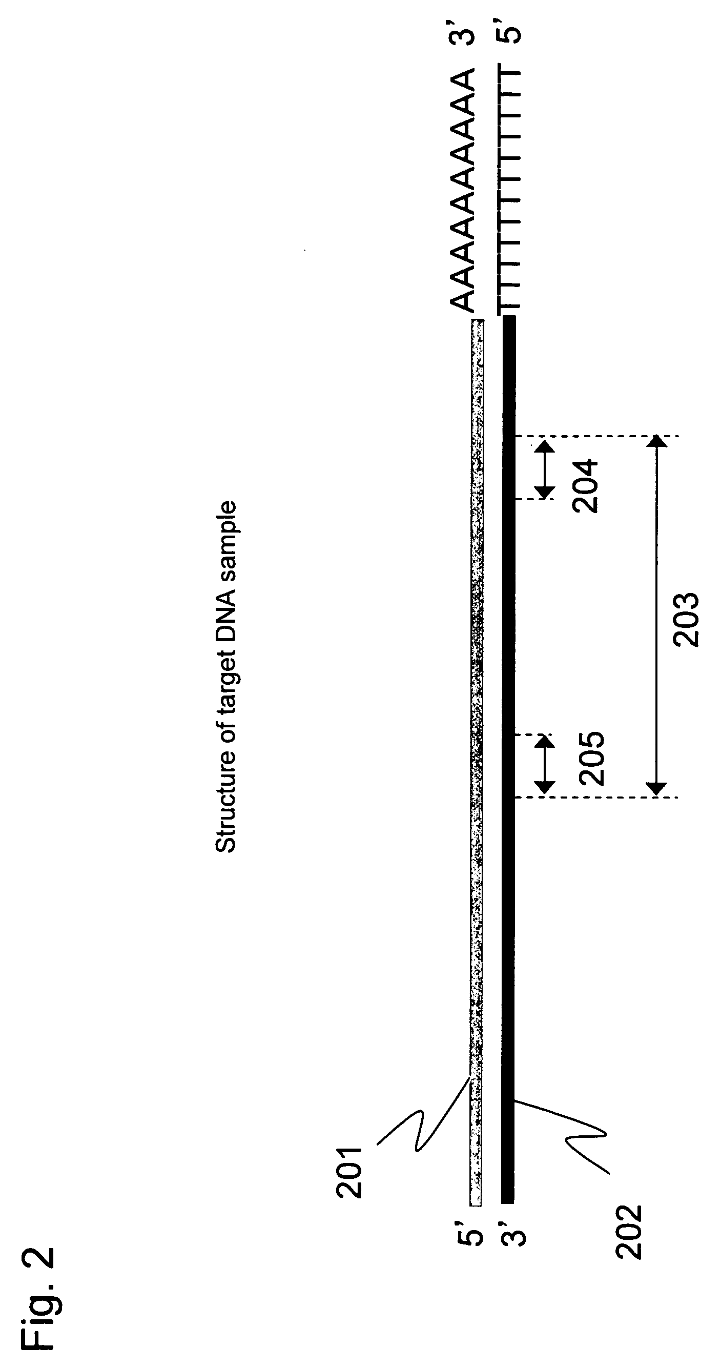
[0057]Full-length cDNA (202) containing a target region was prepared from human colon cancer mRNA (201) according to Step 1 described below. This long cDNA contains a DNA sequence region (203) to be analyzed as a target for quantitative analysis. The target sequence is represented by SEQ ID NO: 1 shown in Sequence Listing. This sequence comprises a priming site of a reverse primer (204), the amplification region to be analyzed (203), and a priming site of a forward primer (205), as schematically shown in FIG. 2.

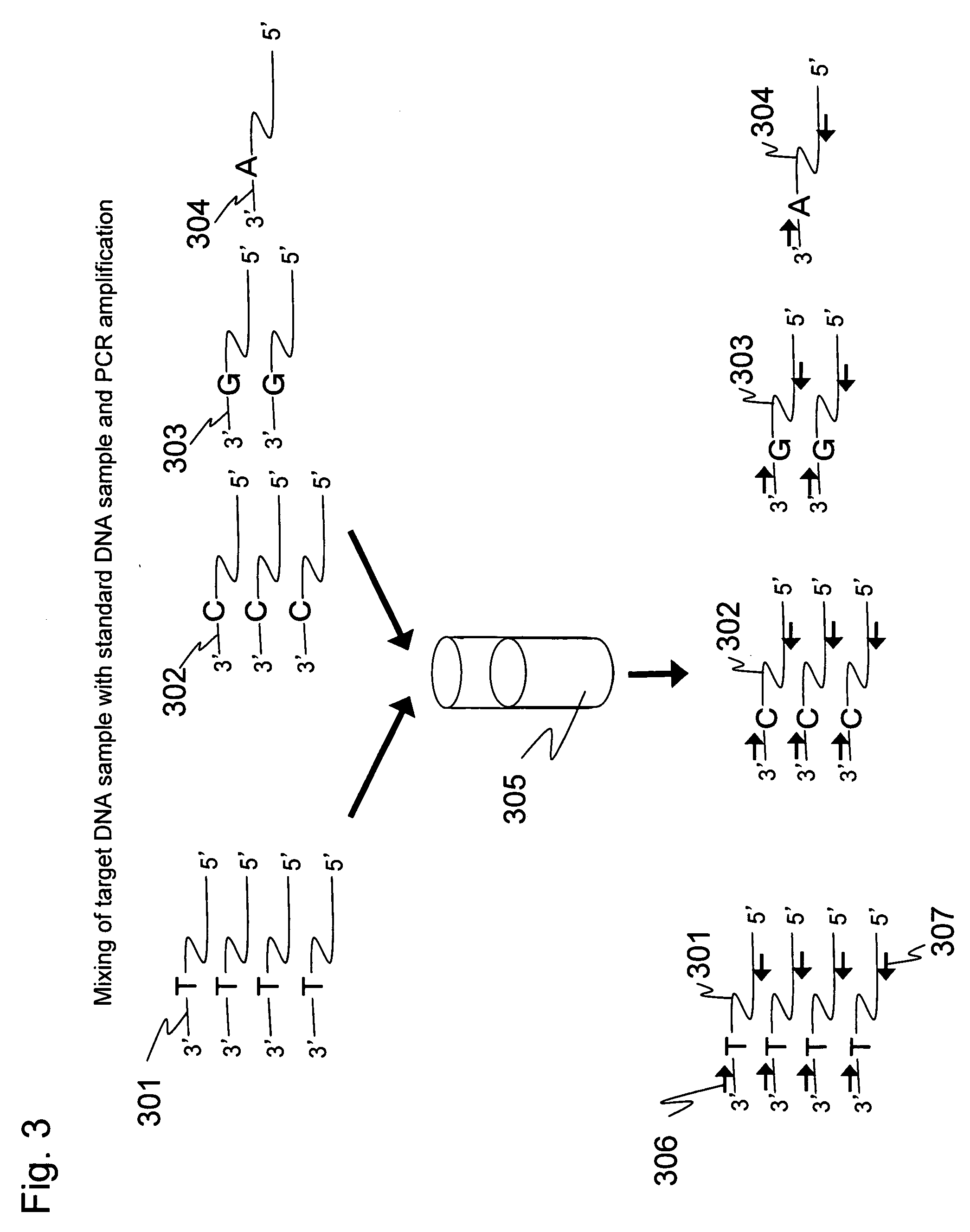
[0058]A portion of the cDNA sample was collected, and a single-stranded DNA fragment having the same sequence as that of the target except for a single-base substitution portion was prepared according to Step 2. In the present Example, the forward primer was designed such that its sequence contained a substitution portion at the 7th base position from the 3′ end. O...

example 2

[0070]Example 2 shows the quantitation of a target DNA sample obtained by reverse transcription of mRNA on oligo(dT)30 magnetic beads.

[0071]An expression level of EFF1G gene in human colon cancer cells (HCT116) was quantitated in the same way as in Example 1. In the present Example, RNA prepared from the colon cancer cells according to the Step 1 is a starting material. cDNA was prepared using oligo(dT)30-immobilized magnetic beads prepared according to Step 6. The cDNA was prepared according to Step 7. Three standard DNA samples were prepared according to the Step 2. These standard DNA samples were mixed at different proportions with the target DNA sample, and the mixed sample was amplified by PCR using the same primers according to Step 8. This amplification product was dissociated into single strands according to the Step 4 of Example 1. Luminescence attributed to single-base extension was measured according to the Step 5 to perform the quantitative analysis of the target DNA sam...

example 3

[0075]Example 3 shows the quantitation of an expression level of EFF1G gene in human colon cancer cells (HCT116) by reusing cDNA immobilized on magnetic beads.

[0076]When a target to be analyzed is obtained in the form of single-stranded cDNA immobilized on magnetic beads as shown in Example 2, the immobilized cDNA is used as a template for 2nd-strand cDNA synthesis. After removal of primers, etc., the synthesized complementary strand is released into a solution and may be used as a target for quantitative analysis. Three standard DNAs (Step 2 of Example 1) are added at different proportions to the synthesized complementary strand sample, and this mixed sample is amplified by PCR using the same primers. The primer used in this PCR amplification is designed to hybridize to the terminal side from the single-base substitution site introduced in the standard DNAs. Therefore, the target DNA sample and the standard DNAs have completely the same application efficiency, as in Examples 1 and ...


PUM

PropertyMeasurementUnit
Concentrationaaaaaaaaaa

Abstract

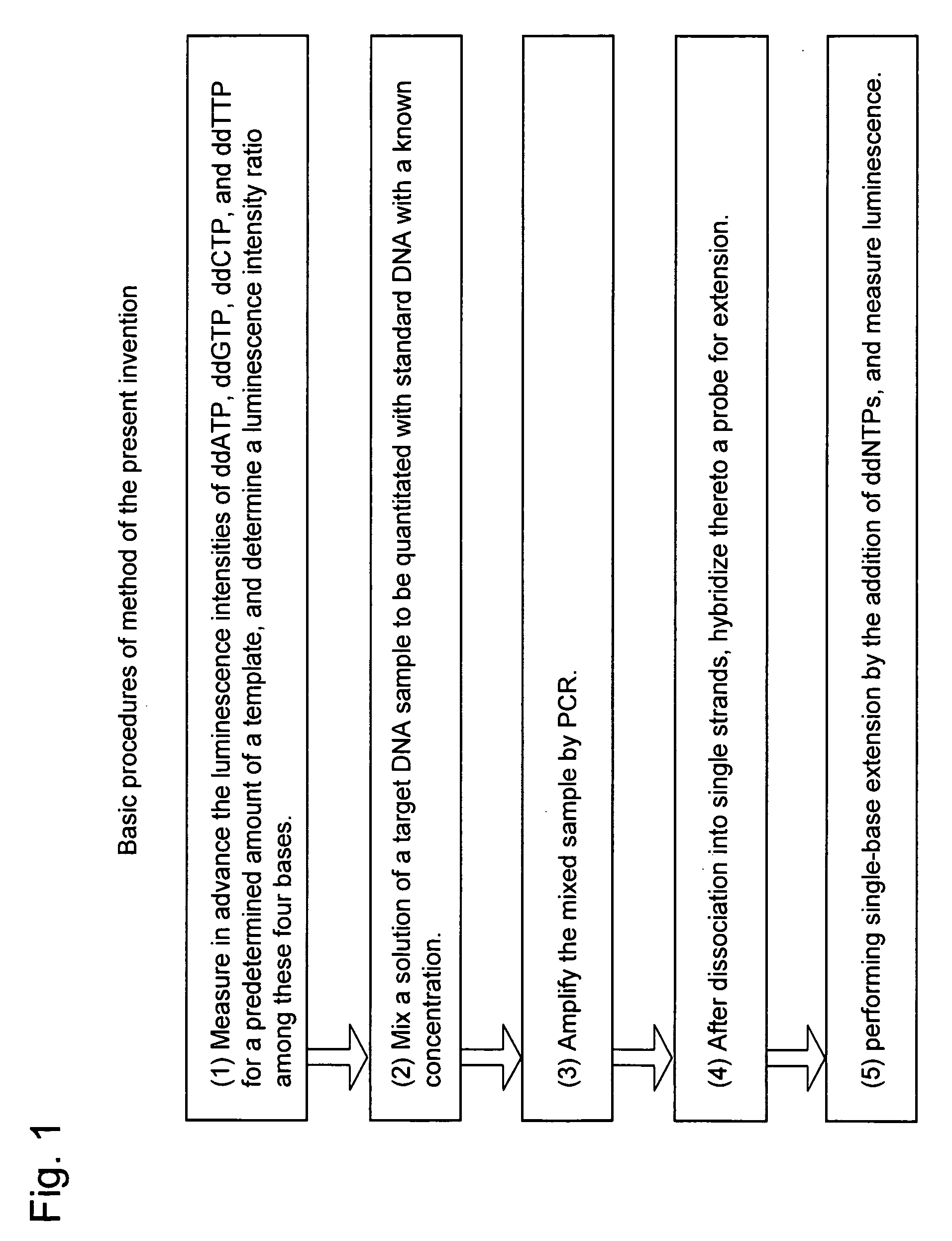
It is intended to provide a novel convenient approach for DNA quantitative analysis that overcomes the disadvantages of conventional formulations. A standard DNA sample is prepared by introducing a single-base substitution into target DNA, and a predetermined amount thereof is mixed with a target DNA sample. The target and standard DNAs are amplified using the same primers designed to amplify a region comprising the single-base substitution site. To a hybridization product of a probe capable of binding to a site immediately before the single-base substitution site, ddATP, ddGTP, ddCTP, and ddTTP are sequentially added one by one to perform a complementary strand synthesis reaction. Luciferase reaction-induced luminescence derived from the formed pyrophosphoric acid is detected. The target DNA is quantitated from the amount of the detected luminescence and the amount of the added standard DNA sample.

Description

CLAIM OF PRIORITY[0001]The present application claims priority from Japanese patent application JP 2007-309089 filed on Nov. 29, 2007, the content of which is hereby incorporated by reference into this application.BACKGROUND OF THE INVENTION[0002]1. Field of the Invention[0003]The present invention relates to a method for DNA or RNA quantitative analysis.[0004]2. Background Art[0005]The study of gene expression levels is important for drug development and the study of disease causation. However, in the biological field centered on DNA, target DNAs are often found in small copy numbers in general and are therefore amplified for measurement. However, it is increasingly demanded to accurately determine the copy numbers.[0006]Examples of typical methods for DNA quantitation used currently include three methods, 1) electrophoresis, 2) DNA microarray (JP Patent Publication (Kokai) No. 2004-33160A (2004)), and 3) real-time PCR (Higuchi R, et al, Bio. Technology, 10: 413-417, (1992)). Of th...

Claims


Application Information

Patent Timeline
IPC IPC(8): C12Q1/68
CPCC12Q1/6851C12Q2545/101C12Q2525/161
Inventor TANIGUCHI, KIYOMIKAMBARA, HIDEKI
Owner HITACHI LTD
Learn More