Tumor Marker For Ovarian Cancer Diagnosis
a tumor marker and ovarian cancer technology, applied in the field of tumor markers for ovarian cancer diagnosis, can solve the problems of not being recommended as a diagnostic tool or target, yielding many false positives, and difficult to detect early ovarian cancer
- Summary
- Abstract
- Description
- Claims
- Application Information
AI Technical Summary
Benefits of technology
Problems solved by technology
Method used
Image
Examples
example 1
Screening of Tumor Markers
[0029]The ovarian tissues collected in the present invention comprised of 36 epithelial ovarian cancers, 10 borderline malignancies and 18 normal ovaries. Clinical and histological characteristics of these 36 ovarian cancer tissue samples are summarized in Table 1.
[0030]The histologic subtypes of ovarian cancer include clear cell, endometrioid, mucinous, serous and others as shown in Table 1. Among these 36 ovarian cancer tissue samples, 10 were of clinical stage I, 6 of clinical stage II, 18 of clinical stage III, and 2 of clinical stage IV.
[0031]Protein extracts from the normal ovarian tissues and the ovarian cancer tissues were separated on SDS-PAGE followed by 2D-polyacrylamide gel electrophoresis.
[0032]2D-polyacrylamide gel electrophoresis was performed on a 130 mm, linear immobilized pH 4-7 Immobiline DryStrip (Amersham Pharmacia Biotech, Piscataway, N.J., USA) using MULTIPHOR II Electrophoresis system. The ovarian tissues were frozen in liquid nitrog...
example 2
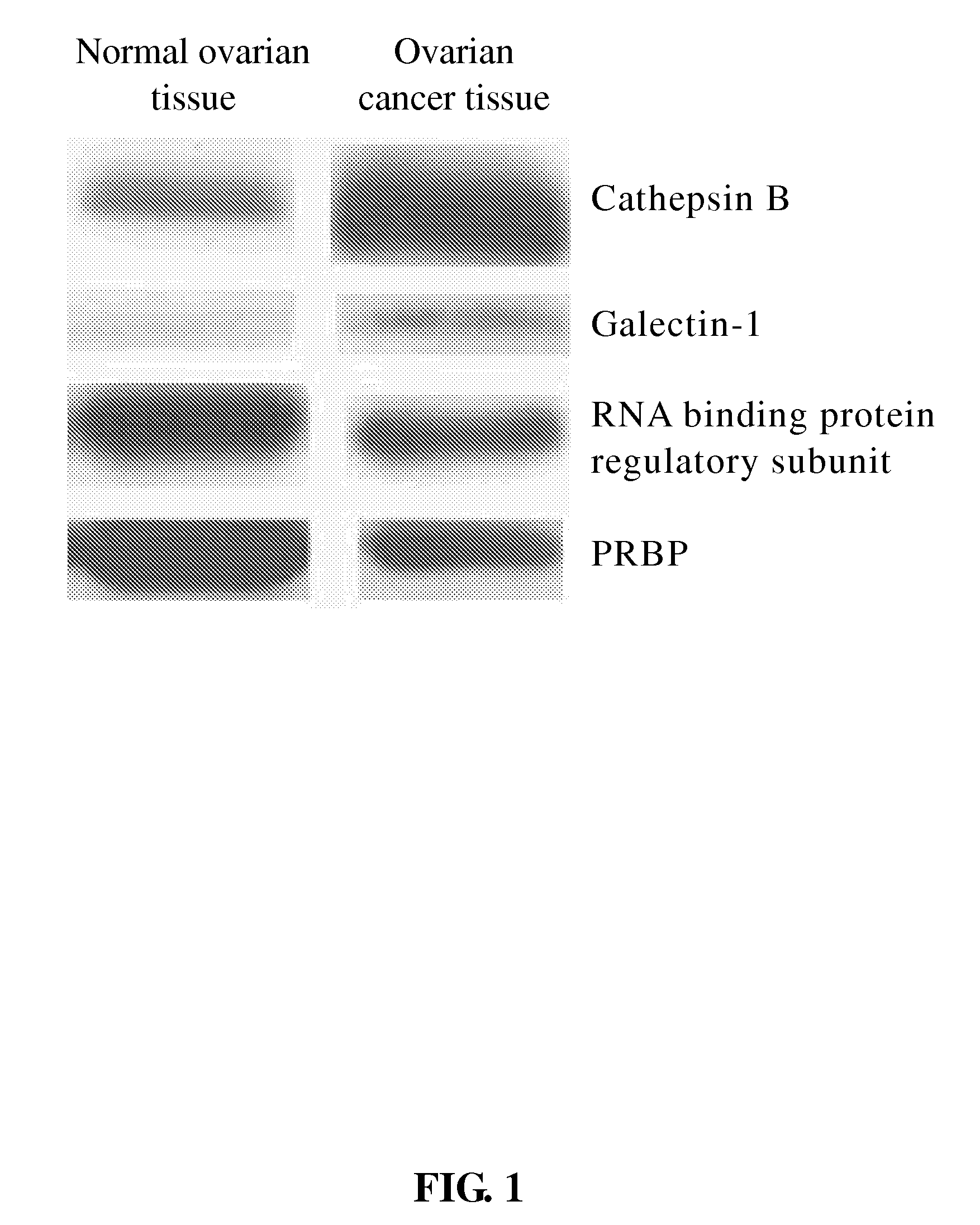
[0040]Four of the protein spots detected in 2D-gel electrophoresis images, which included: cathepsin B, galectin-1, RNA-binding protein regulatory subunit, and plasma retinol-binding protein (PRBP), were further identified through Dot blot, or SDS-PAGE followed by Western blotting analyses.
[0041]The tissues from normal or cancer ovaries were grinded with plastic pestles in ¼ PBS buffer containing protease inhibitor cocktail (Calbiochem). After centrifugation 15,000×g for 10 min at 4° C., the supernatant was transferred to an eppendorf tube and subjected to Dot blot, or SDS-PAGE followed by Western blotting analyses. The protein concentration was determined by the absorption of A280.
[0042]For SDS-PAGE analysis, 30 μg of protein sample was applied to each lane. All samples were heated for 5 min at 95° C. before loading into the 15% polyacrylamide gel. After electrophoresis, proteins were electroblotted onto polyvinylidene difluoride (PVDF) membrane. For Dot blot analysis, 5 μg of prot...
PUM
| Property | Measurement | Unit |
|---|---|---|
| Level | aaaaa | aaaaa |
Abstract
Description
Claims
Application Information
 - R&D
- Intellectual Property
- Life Sciences
- Materials
- Tech Scout
- Unparalleled Data Quality
- Higher Quality Content
- 60% Fewer Hallucinations
Browse by: Latest US Patents, China's latest patents, Technical Efficacy Thesaurus, Application Domain, Technology Topic, Popular Technical Reports.
© 2025 PatSnap. All rights reserved.Legal|Privacy policy|Modern Slavery Act Transparency Statement|Sitemap|About US| Contact US: help@patsnap.com


