
Compositions, Splice Variants and Methods Relating to Cancer Specific Genes and Proteins

a technology of splice variants and cancer genes, applied in the field of newly identified nucleic acids and polypeptides, can solve the problems of increased prostate cancer risk, and increased transactivation ability of gene products

Inactive Publication Date: 2008-10-23
DIADEXUS
View PDF2 Cites 10 Cited by
  • Summary
  • Abstract
  • Description
  • Claims
  • Application Information

AI Technical Summary

Benefits of technology

[0041]In addition, this aspect of the present invention relates to a nucleic acid molecule further comprising one or more expression control sequences controlling the transcription and/or translation of all or a part of a CaSNA or the transcription and/or translation of a nucleic acid molecule that encodes all or a fragment of a CaSP.
[0042]Another aspect of the present invention relates to vectors and/or host cells comprising a nuc

Problems solved by technology

For example, these studies have revealed that as the number of CAG repeats decreases the transactivation ability of the gene product increases, as does the risk of prostate cancer.
Environmental / dietary risk factors which may increase the risk of prostate cancer include intake of saturated fat and calcium.
Despite the need for accurate staging of prostate cancer, current staging methodology is limited.
The wide variety of biological behavior displayed by neoplasms of the prostate has resulted in considerable difficulty in predicting and assessing the course of prostate cancer.
Techniques such as transrectal ultrasound, abdominal and pelvic computerized tomography, and MRI have not been particularly useful in predicting local tumor extension.
While effective at detecting early stage tumors, FOBT is unable to detect adenomatous polyps (premalignant lesions), and, depending on the contents of the fecal sample, is subject to rendering false positives.
Despite the advantages of these procedures, there are accompanying downsides: sigmoidoscopy, by definition, is limited to the sigmoid colon and below, colonoscopy is a relatively expensive procedure, and both share the risk of possible bowel perforation and hemorrhaging.
Double-contrast barium enema (DCBE) enables detection of lesions better than FOBT, and almost as well a colonoscopy, but it may be limited in evaluating the winding rectosigmoid region.
The CEA blood test, which involves screening the blood for carcinoembryonic antigen, shares the downside of FOBT, in that it is of limited utility in detecting colorectal cancer at an early stage.
While computerized tomography and magnetic resonance imaging are useful in staging colorectal cancer in its later stages, both have unacceptably low staging accuracy for identifying early stages of the disease, due to the difficulty that both methods have in (1) revealing the depth of bowel wall tumor infiltration and (2) diagnosing malignant adenopathy.
Rather, techniques such as transrectal ultrasound (TRUS) are preferred in this context, although this technique is inaccurate with respect to detecting small lymph nodes that may contain metastases.
Nonetheless, thirty to forty percent of patients will develop a recurrence of colon cancer following surgical resection, which in many patients is the ultimate cause of death.
All of these lead to genomic instability in colorectal cancers.
Another source of genomic instability in colorectal cancer is the defect of DNA mismatch repair (MMR) genes.
The inactivation of these proteins leads to the accumulation of mutations and causes genetic instability that represents errors in the accurate replication of the repetitive mono-, di-, tri- and tetra-nucleotide repeats, which are scattered throughout the genome (microsatellite regions).
Carcinogen metabolism enzymes such as GST, NAT, CYP and MTHFR are also associated with an increased or decreased colorectal cancer risk.
Moreover, current procedures, while helpful in each of these analyses, are limited by their specificity, sensitivity, invasiveness, and / or their cost.

Method used


Image

  • Compositions, Splice Variants and Methods Relating to Cancer Specific Genes and Proteins
  • Compositions, Splice Variants and Methods Relating to Cancer Specific Genes and Proteins
  • Compositions, Splice Variants and Methods Relating to Cancer Specific Genes and Proteins

Examples


example 1a

Alternative Splice Variants

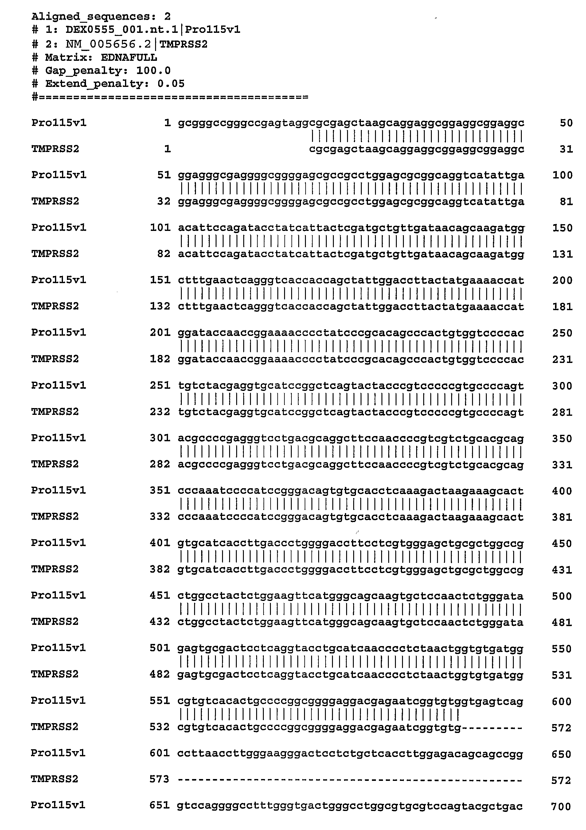
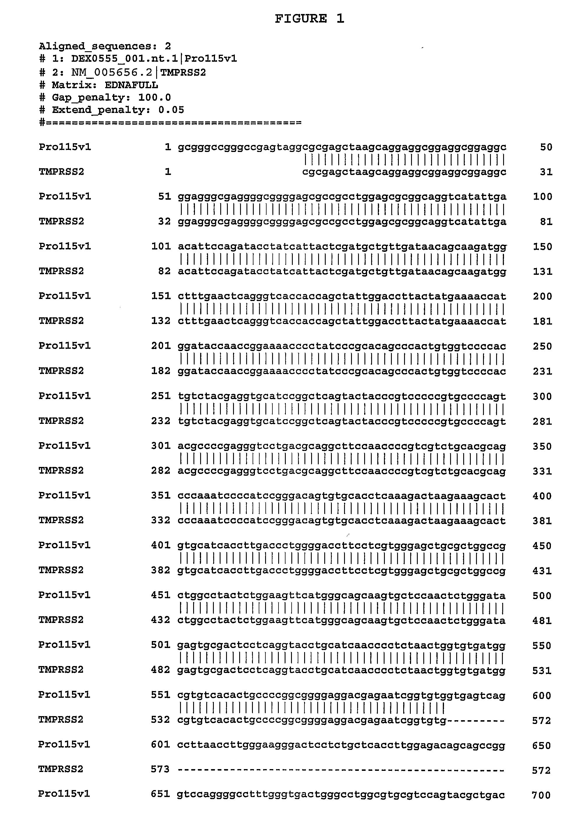
[0483]We identified gene transcripts using the Gencarta™ tools from Compugen Ltd. (Tel Aviv, Israel). Gencarta™ was used to identify splice variant transcripts based on sequences from a variety of public and proprietary databases. These splice variants are either sequences which differ from a previously defined sequence or comprise new uses of known sequences. In general related variants are annotated as DEX0555_XXX.nt.1, DEX0555_XXX.nt.2, DEX0555_XXX.nt.3, etc. The variant DNA sequences encode proteins which differ from a previously defined protein sequence. In relation to the nucleotide sequence naming convention, protein variants are annotated as DEX0555_XXX.aa.1, DEX0555_XXX.aa.2, etc., wherein transcript DEX0555_XXX.nt.1 encodes protein DEX0555_XXX.aa.1. A single transcript may encode a protein from an alternate Open Reading Frame (ORF) which is designated DEX0555_XXX.orf.1. Additionally, multiple transcripts may encode for a single protein. In this c...

example 1b

Sequence Alignment Support

[0503]Alignments between previously identified sequences and splice variant sequences are performed to confirm unique portions of splice variant nucleic acid and amino acid sequences. The alignments are done using the Needle program in the European Molecular Biology Open Software Suite (EMBOSS) version 2.2.0 available at emboss with the extension org of the world wide web from EMBnet (embnet with the extension org of the world wide web). Default settings are used unless otherwise noted. The Needle program in EMBOSS implements the Needleman-Wunsch algorithm. Needleman, S. B., Wunsch, C. D., J. Mol. Biol. 48:443-453 (1970).

[0504]It is well know to those skilled in the art that implication of alignment algorithms by various programs may result in minor changes in the generated output. These changes include but are not limited to: alignment scores (percent identity, similarity, and gap), display of nonaligned flanking sequence regions, and number assignment to ...

example 1c

RT-PCR Analysis

[0505]To detect the presence and tissue distribution of a particular splice variant Reverse Transcription-Polymerase Chain Reaction (RT-PCR) is performed using cDNA generated from a panel of tissue RNAs. See, e.g., Sambrook et al., Molecular Cloning: A Laboratory Manual, 2d ed., Cold Spring Harbor Laboratory Press (1989) and; Kawasaki E S et al., PNAS 85(15):5698 (1988). Total RNA is extracted from a variety of tissues and first strand cDNA is prepared with reverse transcriptase (RT). Each panel includes 23 cDNAs from five cancer types (lung, ovary, breast, colon, and prostate) and normal samples of testis, placenta and fetal brain. Each cancer set is composed of three cancer cDNAs from different donors and one normal pooled sample. Using a standard enzyme kit from BD Bioscience Clontech (Mountain View, Calif.), the target transcript is detected with sequence-specific primers designed to only amplify the particular splice variant. The PCR reaction is run on the GeneAm...


PUM

PropertyMeasurementUnit
Fractionaaaaaaaaaa
Fractionaaaaaaaaaa
Forceaaaaaaaaaa

Abstract

The present invention relates to newly identified nucleic acid molecules and polypeptides present in normal and neoplastic ovarian cells, including fragments, variants and derivatives of the nucleic acids and polypeptides. The present invention also relates to antibodies to the polypeptides of the invention, as well as agonists and antagonists of the polypeptides of the invention. The invention also relates to compositions containing the nucleic acid molecules, polypeptides, antibodies, agonists and antagonists of the invention and methods for the use of these compositions. These uses include identifying, diagnosing, monitoring, staging, imaging and treating ovarian cancer and non-cancerous disease states in ovarian, identifying ovarian tissue, monitoring and identifying and / or designing agonists and antagonists of polypeptides of the invention. The uses also include gene therapy, production of transgenic animals and cells, and production of engineered ovarian tissue for treatment and research.

Description

[0001]This application claims the benefit of priority from U.S. Provisional Application Ser. No. 60 / 616,166 filed Oct. 4, 2004 which is herein incorporated by reference in its entirety.FIELD OF THE INVENTION[0002]The present invention relates to newly identified nucleic acids and polypeptides present in normal and neoplastic cells, including fragments, variants and derivatives of the nucleic acids and polypeptides. The present invention also relates to antibodies to the polypeptides of the invention, as well as agonists and antagonists of the polypeptides of the invention. The invention also relates to compositions comprising the nucleic acids, polypeptides, antibodies, post translational modifications (PTMs), variants, derivatives, agonists and antagonists of the invention and methods for the use of these compositions. These uses include identifying, diagnosing, monitoring, staging, imaging and treating cancer and non-cancerous disease states in breast, intestine & colon, lung, ova...

Claims


Application Information

Patent Timeline
IPC IPC(8): C07K14/435C12N15/11C12N15/12C12Q1/68C12N15/85C12N5/06C12P21/02C07K16/30G01N33/574A61K39/00A61K31/7088A61K38/17C07K14/00
CPCA61K38/00C07K14/4748
Inventor VARTANIAN, STEFFAN F.RODRIGUEZ, MARIA
Owner DIADEXUS
Learn More