
Methods of Light Activated Release of Ligands from Endosomes

a technology of endosomes and ligands, applied in biochemistry apparatus and processes, peptide/protein ingredients, enzymology, etc., can solve the problems of only suitable cells in vitro, invasiveness, and inability to readily apply clinically to in vivo tissue samples, and achieve the effect of enhancing the availability of ligands

Inactive Publication Date: 2008-07-03
LIFE TECH CORP
View PDF27 Cites 9 Cited by
  • Summary
  • Abstract
  • Description
  • Claims
  • Application Information

AI Technical Summary

Benefits of technology

[0011]This invention advances the state of the prior art by providing novel methods of enhancing the availability of ligands inside a cell. Such methods are useful both in vitro and in vivo. In one aspect, the invention...

Problems solved by technology

Further, the leakage rate of oligonucleotides from endosomes to the cytoplasm is extremely slow.
This methodology, however, is invasive and is only appropriate for cells in vitro and cannot be readily applied clinically to in vivo tissue samples.
However, viral peptides are expensive to produce and can be immunogenic.
Liposomes containing fusogenic and pH-sensitive lipids are unsatisfactory because they have a low capacity to entrap oligonucleotides.
At alkaline pH, BPS are predominated by a hydrophobic tail and reside within lipid bilayers due to its limited surface-active properties.
BPS are problematic because if they are not completely degraded before the endosomes mature into lysosomes or are unprotonated until the lysosome stage, then the ionized BPS would disrupt the lysosomes and cause digestive enzyme release that could kill cells.

Method used


Image

  • Methods of Light Activated Release of Ligands from Endosomes
  • Methods of Light Activated Release of Ligands from Endosomes
  • Methods of Light Activated Release of Ligands from Endosomes

Examples


example 1

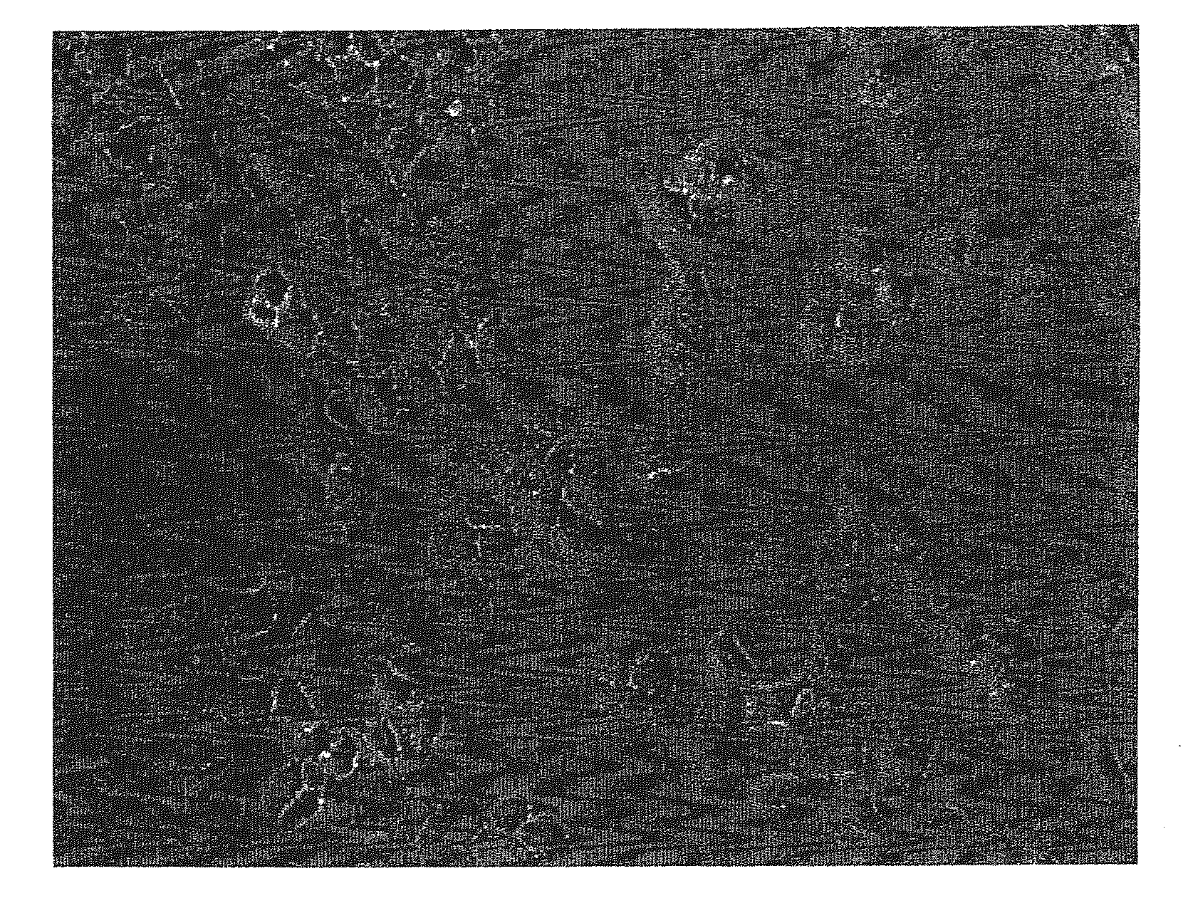
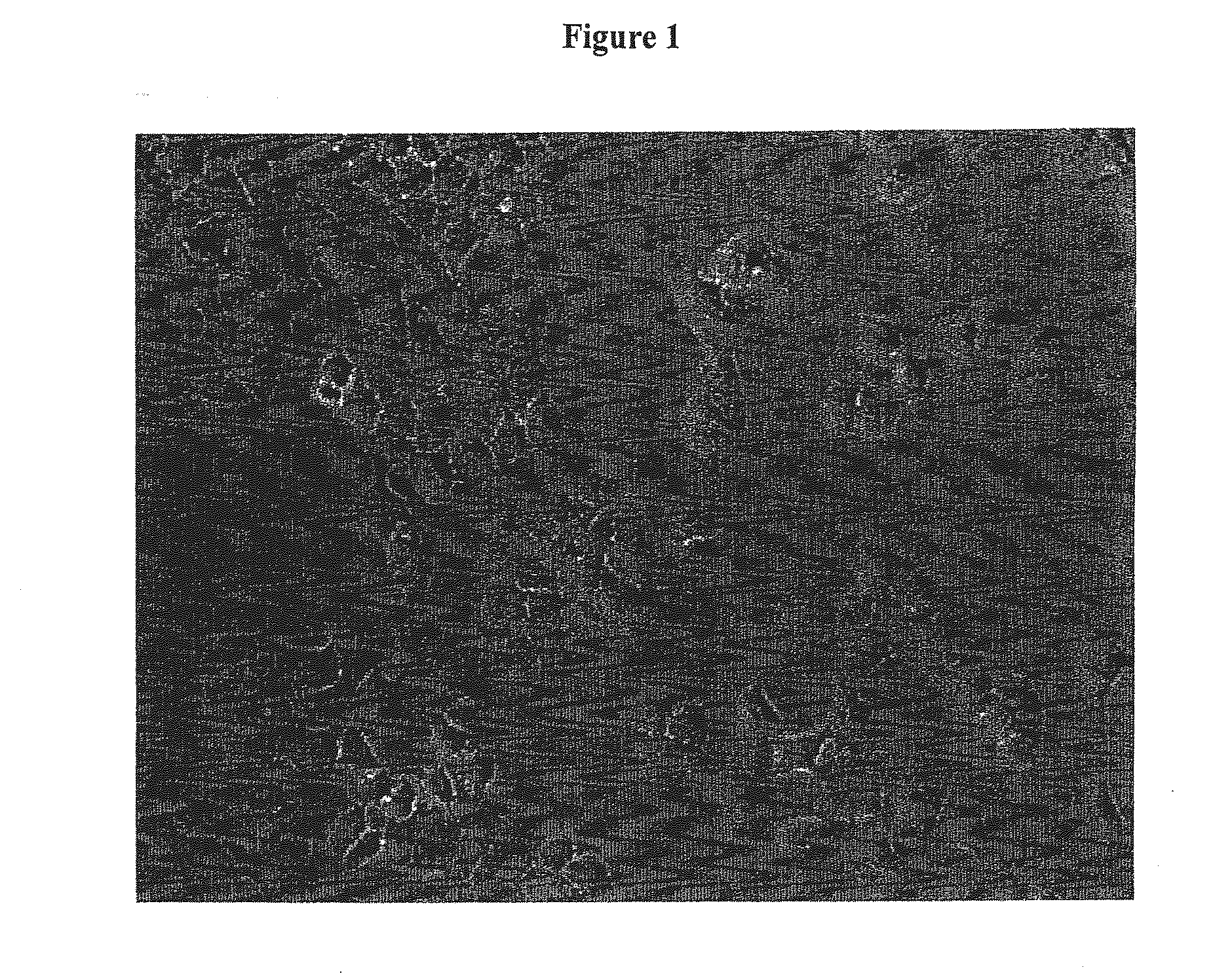
Enhanced Availability of Ligand in A549 Cells

[0172]A549 cells were maintained in high glucose DMEM (Gibco-BRL) supplemented with 10% Fetal Bovine Serum (Gibco-BRL), 2 mM L-Glutamine (Gibco-BRL), and 1× penicillin / streptomycin (Gibco-BRL).

[0173]Seed cell suspensions were prepared by combining 75 μl of 1 mM oligomer (uniformly morpholino modified as taught, e.g., in Summerton, J. et al. Antisense Research and Development, Morpholino and Phosphorothioate Antisense Oligomers Compared in Cell-Free and In-Cell Systems 7:63-70 (1997)) per 425 μl cell suspension.

[0174]24-well plates were seeded with 0.5 ml 10-30K A549 cells per well. The cells are preferably evenly distributed across the plate. Cells were incubated for 18-24 hours at 37° C. in a humidified CO2 incubator. The media was aspirated from the cells and each well rinsed with 0.5 ml of Opti-MEM reduced serum medium (GIBCO-BRL). The media was aspirated and 0.5 ml of Opti-MEM was added to each well a second time.

[0175]The cells were ...

example 2

Enhanced Availability of Ligand in HUVECs (Human Umbilical Vein Endothelial Cells)

[0176]Human umbilical vein endothelial cells (HUVECs) were maintained in HUVEC media: EBM (Clonetics) supplemented with 8% Fetal Bovine Serum (Gibco-BRL), 2 mM L-Glutamine (Gibco-BRL), and 1× penicillin / streptomycin (Gibco-BRL), hEGF, Hydrocortisone, GA-1000, BBE, and 2% FBS (Clonetics).

[0177]On the day before transfection 24-well plates were coated with 0.5 ml / well gelatin for 5 minutes. Gelatin was aspirated and plates seeded with 0.5 ml 10-30K HUVECs per well. (Cells are preferably about 70% confluent at the start of transfection, and should be evenly distributed across the plate.) The cells are incubated at 37° C. in a humidified CO2 incubator.

[0178]On the day of transfection, a 300 μM stock solution of uniformly morpholino modified oligomer was prepared by diluting 300 μl of oligomer per 700 μl of HUVEC media. The media was aspirated from the cells, and 0.5 ml of the oligomer / HUVEC media was added...


PUM

PropertyMeasurementUnit
Wavelengthaaaaaaaaaa
Flexibilityaaaaaaaaaa

Abstract

Methods for delivering ligands to a cell by using light to activate fluorescent ligands causing their release from endosomes. The instant methods thus increasing the efficiency of ligands, e.g. in vitro or at localized sites within a subject. The invention provides for the release of ligands by shining a light source on a cell to promote release of ligands into the cell where they can effect their function.

Description

RELATED APPLICATIONS[0001]This application claims the priority of U.S. Provisional Patent Application No. 60 / 267,272, filed Feb. 8, 2001.BACKGROUND OF THE INVENTION[0002]For antisense oligonucleotides to regulate gene expression, they must penetrate the cell membrane. Because oligonucleotides are anionic, they cannot passively diffuse through cell membranes. Oligonucleotides are believed to enter cells through two different active transport processes: adsorbtive endocytosis and fluid phase endocytosis (pinocytosis).[0003]Adsorbtive endocytosis requires that the oligonucleotide adsorb to the surface of the cell. Charged oligonucleotides adsorb better to the surface of the cell than uncharged oligonucleotides and, therefore, are internalized better. Cell surface heparin-binding proteins facilitate the adsorbtion process.[0004]Pinocytosis is the process where cells constitutively engulf water and dissolved solutes from the fluid phase. Pinocytosis is especially important for the intern...

Claims


Application Information

Patent Timeline
IPC IPC(8): C12N13/00A61K38/00C12N15/113C12N15/87
CPCA61K38/00C12N15/113C12N2310/3517C12N2310/3233C12N2310/3513C12N15/87
Inventor WOOLF, TOD M.
Owner LIFE TECH CORP
Learn More