
Ion-selective quantum dots

Active Publication Date: 2008-06-05
CHARLES STARK DRAPER LABORATORY
View PDF5 Cites 16 Cited by
  • Summary
  • Abstract
  • Description
  • Claims
  • Application Information

AI Technical Summary

Benefits of technology

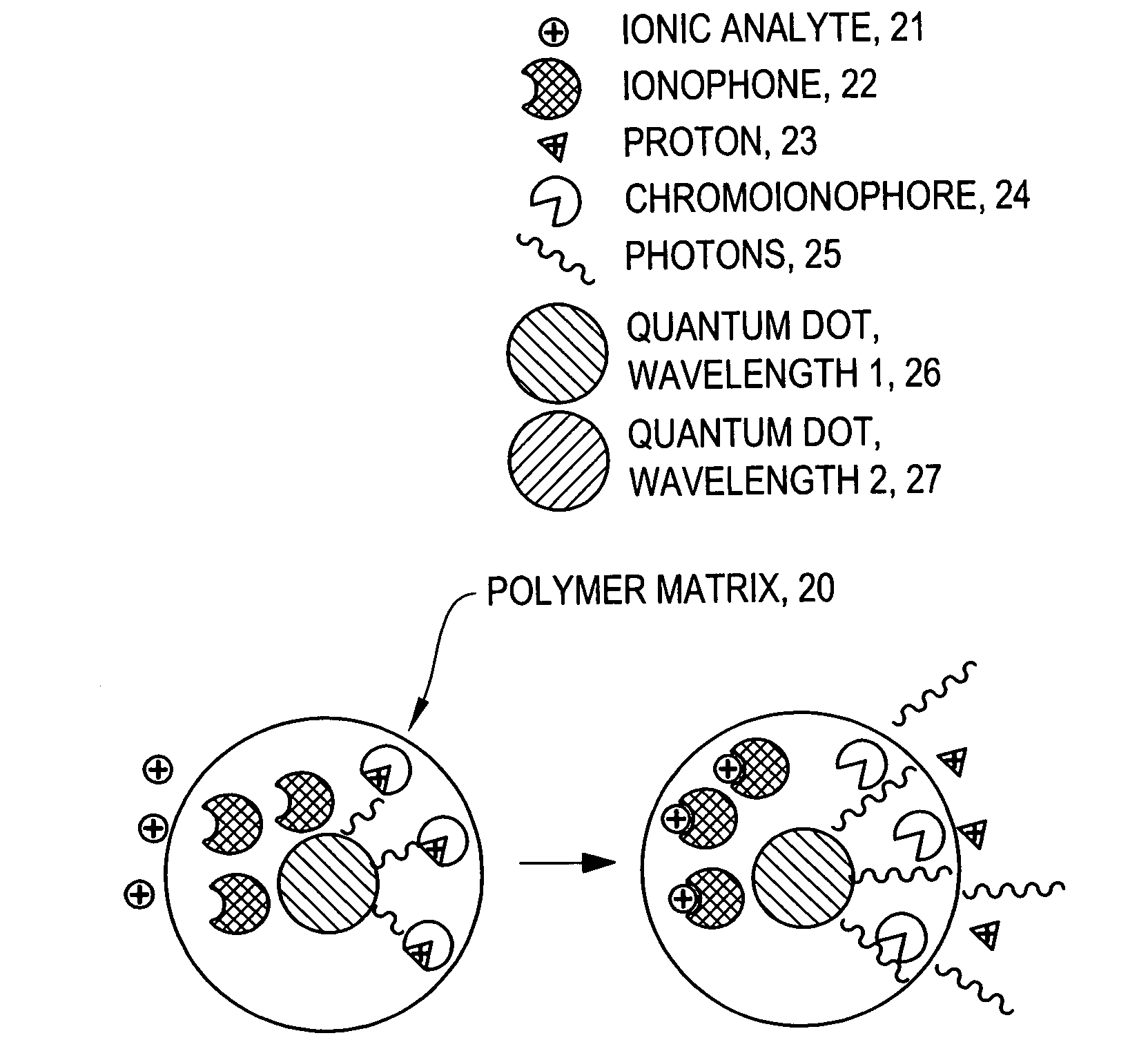
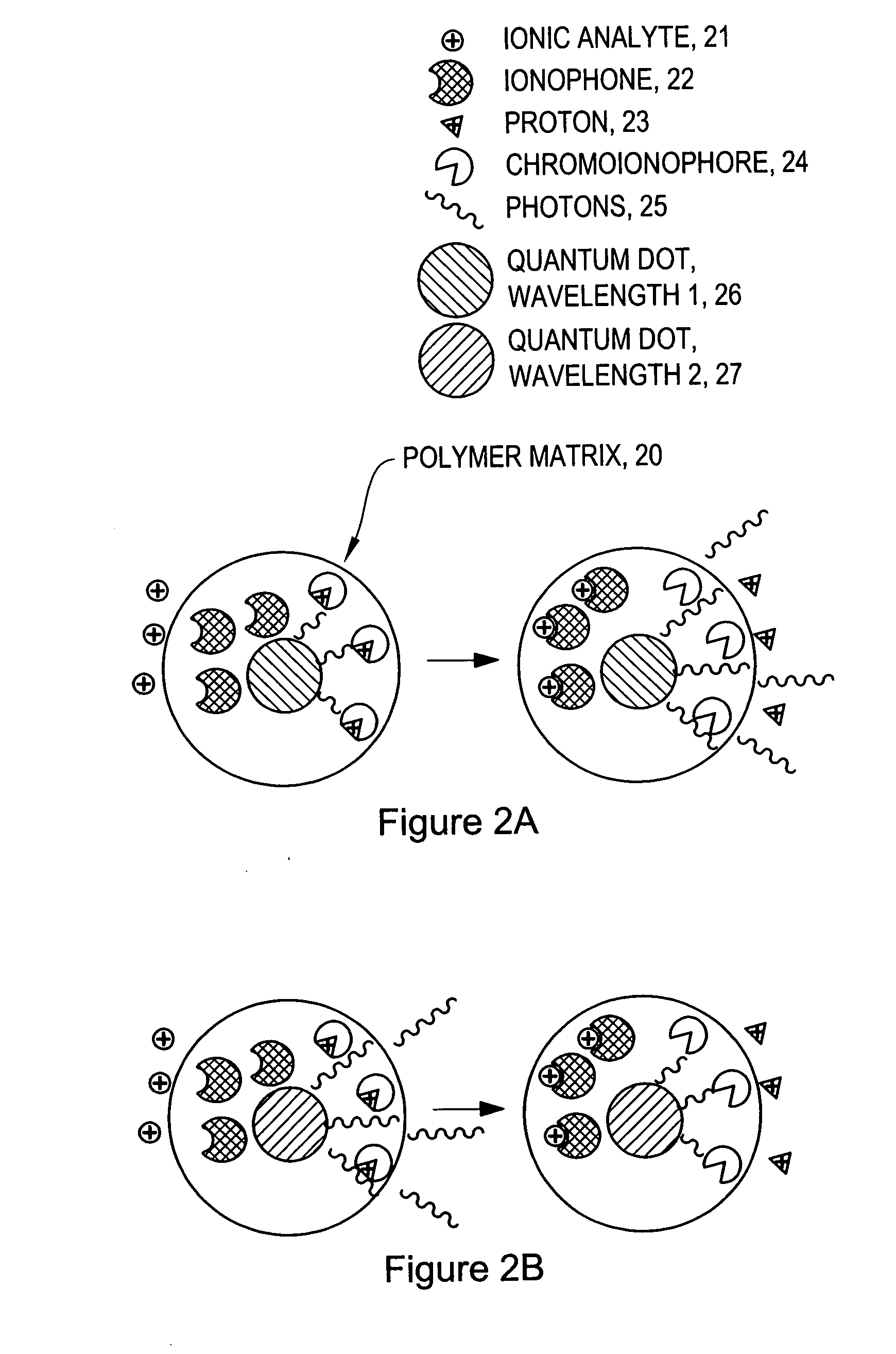
[0005]The invention provides ion-selective sensors comprising quantum dots capable of selectively measuring ions, e.g., Na+, K+, Cl−, etc., in the cytosol of a single living cell. Quantum dots are attractive probes for microscopy due to their photophysical advantages over fluorescent dyes, including prolonged photostability, brightness and quantum efficiency. In certain embodiments, a sensor comprises one or more quantum dots, a pH-sensitive dye, and optionally an ion-selective component such as an ionophore. These elements may, for example, be disposed in a polymer matrix. In certain embodiments, the sensors may detect ionic analytes by selective ion extraction by the polymer, thereby inducing a pH change within the sensor which in turn changes the absorbance of the pH-sensitive dye. The change of absorbance may in turn attenuate the intensity of detectable emissions, e.g., fluorescence, from the quantum dot by directly absorbing its fluorescence emission.

Problems solved by technology

While it is generally believed that sodium is essential to the function of excitable cells, its role is not as well understood.
There are, however, disadvantages to the available methods of ion monitoring.
For example, whole-cell patch clamp monitoring which involves contacting cells with a pulled glass capillary to electrically monitor ion channels, can be performed on single cells but not reliably in a high-throughput fashion.
Fluorescent ion-indicator dyes while effective for monitoring certain ions, lack selectivity for other physiological ions and are easily photobleached.
Ion-selective nanosensors (e.g., probes encapsulated by biologically localized embedding (PEBBLEs)) which comprise a fluorescent indicator dye display fast response time to ions as they diffuse into the polymer matrix, however, the components of the nanosensors, such as the fluorescent dye, are also prone to photobleaching.
Dye-loaded liposomes are highly biocompatible due to their lipid construction but are limited in their range of detectable analytes particularly to gases.

Method used


Image

  • Ion-selective quantum dots
  • Ion-selective quantum dots
  • Ion-selective quantum dots

Examples


examples

[0075]Ion-Selective Polymer Solution. The ion-selective polymer solution was made from the following components: 30 mg High Molecular Weight Polyvinyl Chloride, 60 mg Bis-2-Sebacate, 0.1 mg Sodium Ionophore X, 0.1 mg Sodium tetrakis[3,5-bis(trifluoromethyl)phenyl]borate, and 0.1 mg Chromoionophore I. The combined reagents were stirred in 500 μL of tetrahydrofuran (THF) to afford a homogenous solution.[0076]Ion-Selective Sensor Fabrication. Quantum dots (ITK organic 655, Invitrogen) were flocculated in a methanol / isopropanol mixture with the addition of toluene in a 1:1 (v:v) ratio of toluene:quantum dot solution. The supernatant was removed and the quantum dots were resuspended in THF containing 3.3 mM 1-decanethiol. To the quantum dot solution (0.2 nMoles) was added the ion-selective polymer solution (17.2 nMoles Chromoionophore I, 50 μl) and the mixture was stirred.[0077]Immobilized Polymer Matrix of Sensors. To form an immobilized polymer matrix of sensors, 1 μl of the polymer / qu...


PUM


Abstract

The invention provides ion-selective sensors comprising quantum dots capable of selectively measuring ions, e.g., Na+, K+, Cl−, etc., in various environments, including in the cytosol of a living cell. Quantum dots are attractive probes for microscopy due to their photophysical advantages over fluorescent dyes, including prolonged photostability, brightness and quantum efficiency. In certain embodiments, a sensor comprises one or more quantum dots, a pH-sensitive dye, and optionally an ion-selective component such as an ionophore. These elements may, for example, be disposed in a polymer matrix. In certain embodiments, the sensors may detect ionic analytes by selective ion extraction by the polymer, thereby inducing a pH change within the sensor which in turn changes the absorbance of the pH-sensitive dye. The change of absorbance may in turn attenuate the intensity of detectable emissions, e.g., fluorescence, from the quantum dot by directly absorbing its fluorescence emission.

Description

CROSS-REFERENCE TO RELATED CASES[0001]This application claims the benefit of U.S. Provisional Patent Application No. 60 / 834,973, entitled “Ion-Selective Quantum Dots,” filed on Jul. 31, 2006, the entire disclosure of which is hereby incorporated by reference as if set forth herein in its entirety.BACKGROUND OF THE INVENTION[0002]Ion channels lie at the heart of the dynamics of excitable cells and control the action potential responsible for the beat of a cardiac cell or the firing of a synapse. Traditionally, calcium has been the most studied ion in the intracellular space, and as a result a good deal is understood about calcium ion dynamics, regulation and impact on the cell. While it is generally believed that sodium is essential to the function of excitable cells, its role is not as well understood. Sodium channels occur at sites of action potential generation in neurons and are responsible for the upstroke of the action potential in cardiac cells. Sodium channelopathies are know...

Claims


Application Information

Patent Timeline
IPC IPC(8): G01N33/53C12Q1/20C12M1/34
CPCB82Y5/00B82Y15/00G01N33/5005G01N2333/726G01N33/588G01N33/6872G01N33/84G01N33/54313Y10S977/774
Inventor CLARK, HEATHER A.HARJES, DANIEL I.DUBACH, JOHN M.
Owner CHARLES STARK DRAPER LABORATORY
Learn More