
In Vivo Device for Assisting and Improving Diastolic Ventricular Function

a diastolic ventricular function and in vivo technology, applied in the field of in vivo devices for improving diastolic ventricular function, can solve the problems of increased modulus of chamber stiffness, affecting diastolic function, and congestive heart failur

Inactive Publication Date: 2008-03-20
CORASSIST CARDIOVASCULAR LTD
View PDF8 Cites 24 Cited by
  • Summary
  • Abstract
  • Description
  • Claims
  • Application Information

AI Technical Summary

Benefits of technology

[0044] The term “anatomically compatible” as used hereinbefore refers to the fact that the structure of the device of the invention is such that it may readily be adapted in situ to the precise shape and size of the heart to be treated.
[0045] The term “physiologically compatible” as used hereinbefore refers to the fact that the structure of the device of the invention is such that it may readily be adapted in situ to the precise movement vectors of the heart to be treated.
[0054] In another aspect, the present invention also encompasses a method for improving diastolic function of either the left or right ventricle of the heart, comprising attaching an anatomically-compatible and physiologically-compatible in vivo device as disclosed hereinabove to the external wall of said ventricle(s) by means of connecting elements,
[0064] e) Increased ease with which the device may be delivered to the desired region of the left ventricle by minimally-invasive endoscopic means.
[0069] j) Significantly greater ease of construction of the device; and
[0070] k) Significantly lower cost of construction of the device.

Problems solved by technology

In these patients, diastolic dysfunction is implicated as a major contributor of congestive heart failure.
Thus, diastolic dysfunction increases the modulus of chamber stiffness.
The fundamental problem in diastolic heart failure (DHF) is the inability of the left ventricle to accommodate blood volume during diastole at low filling pressures, as described by Mandinov, L., Eberli, F. R., Seiler, C., and Hess, M. O., in “Diastolic Heart Failure”, Cardiovascular Res. 2000, 45: 813-825.
More severe resistance to LV filling may cause inadequate filling even in enhanced diastolic pressure with an additional leftward shift of the diastolic pressure-volume relation, resulting in a decreased end diastolic volume and depressed stroke volume, as described by Mandinov, L., et al.
The management of diastolic heart failure is difficult.
However, they must be used with care because the volume sensitivity of patients with diastolic dysfunction bears the risk that excessive diuresis may result in a sudden drop in stroke volume.
Because of the steep pressure-volume relationship, a small decrease in diastolic volume will cause a large decrease of the filling pressure, and will result in a drop in stroke volume, and thus, in cardiac output.
Nonetheless, vasodilators have not been useful in the management of isolated diastolic heart failure and are more effective in combined heart failure, as indicated in the previously cited Braunwald, E. text.
However, currently, use of Ca2+ channel blockers is limited due to their negative inotropic effects (negative influence on the systolic function of the heart), and clinical trials have not clearly proven them to be beneficial.
Biventricular pacing, however, has not been clinically proven effective in the treatment of patients with diastolic heart failure.
Moreover, there is a need for such a device which is biocompatible and is specially configured for compact and long-term reliable use in humans.

Method used


Image

  • In Vivo Device for Assisting and Improving Diastolic Ventricular Function
  • In Vivo Device for Assisting and Improving Diastolic Ventricular Function
  • In Vivo Device for Assisting and Improving Diastolic Ventricular Function

Examples


example

In Vivo Demonstration of the Implantation and Use of the Device of the Present Invention in a Mammalian Subject

Methods

[0123] Note: All animals in the study received humane care in compliance with the Public Health Service Policy on Humane Care and Use of Laboratory Animals, prepared by the office of laboratory animal welfare—National Institute of Health, amended August 2002.

Anesthesia and Instrumentation:

[0124] A healthy sheep, (12 month, 40 Kg) was anesthetized as follows: [0125] 1. Induction: I. M Ketamine 5 mg / kg & Xlyazine 0.25 mg / kg. [0126] 2. Intubation and artificial ventilation. [0127] 3. Maintenance of anesthesia by Isoflurane(0.5%-2.5%) [0128] 4. Following intra-tracheal intubation, positive pressure mechanical ventilation was instituted using the above inhalants. [0129] 5. A peripheral vein was cannulated for crystalloid solution infusion to assist the maintenance of stable hemodynamics.

Test Procedure:

[0130] The following test procedure was then performed: [0131]...


PUM


Abstract

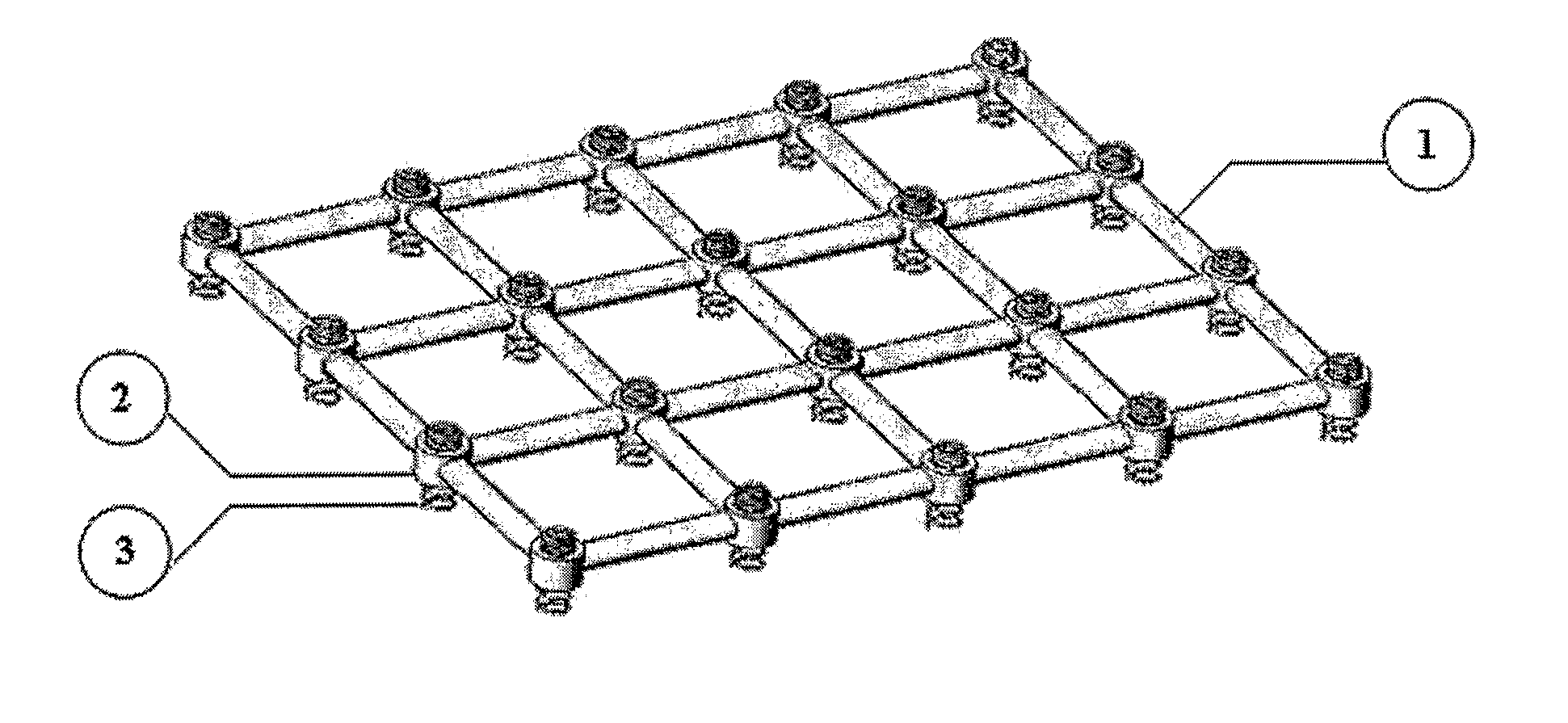
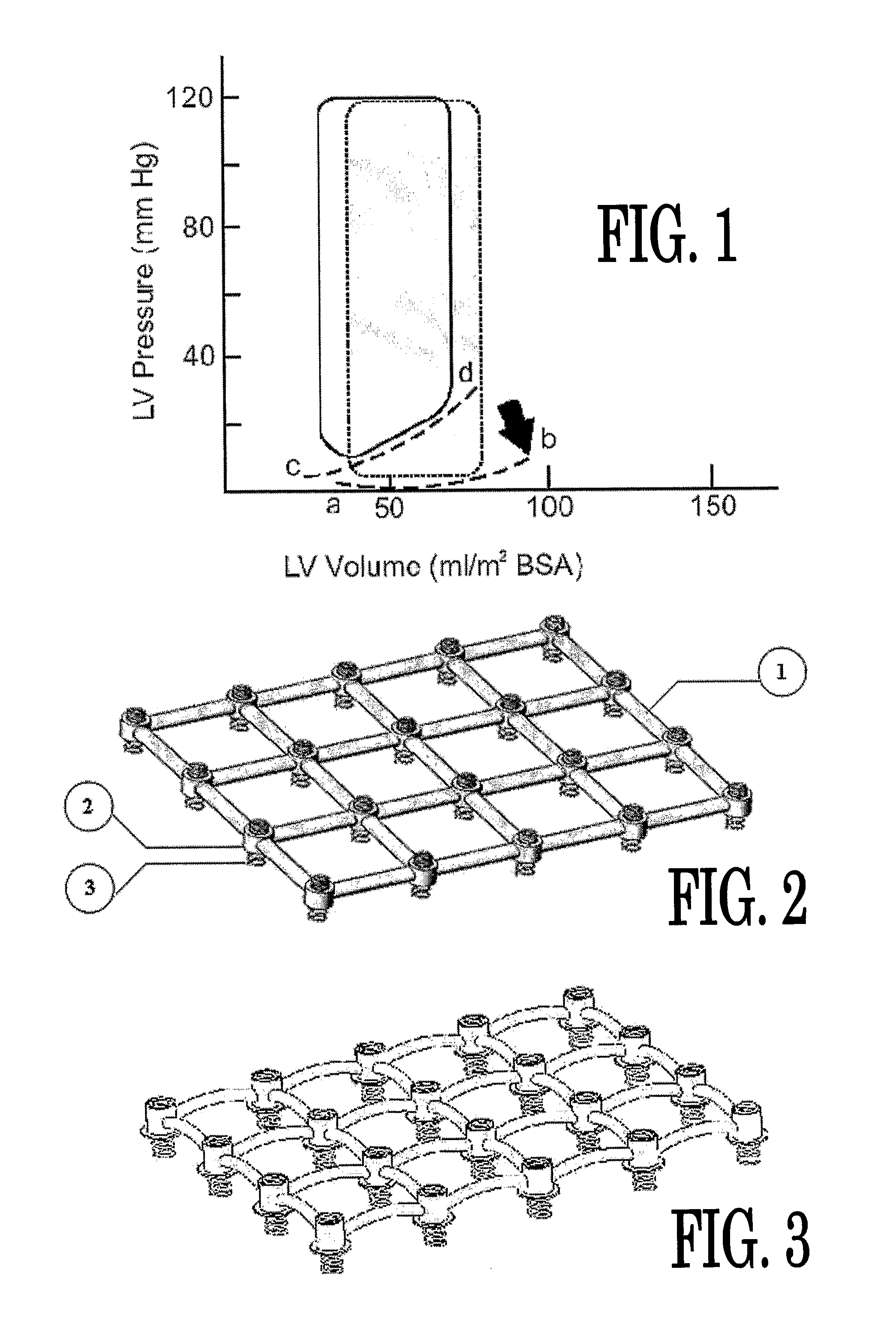
The present invention is primarily directed towards an anatomically-compatible and physiologically-compatible in vivo device for improving diastolic function of either the left or right ventricle of the heart, wherein said device comprises at least one elastic component in the form of a lattice capable of being arranged in a curved conformation such that one surface of said lattice may be adapted to the curvature of the external ventricular surface of the heart, or a portion thereof, and wherein said at least one elastic component is capable of being operatively connected to the external ventricular surface of the heart by means of one or more connecting elements.

Description

FIELD OF THE INVENTION [0001] The present invention relates to a device for improving ventricular function of the heart and, more particularly, to a modified in vivo device for improving diastolic function of the left ventricle of the heart. BACKGROUND OF THE INVENTION [0002] Heart failure is commonly defined as the inability of the left ventricle, herein, also referred to as LV, to generate an adequate cardiac output at rest or during exertion, while operating at a normal or enhanced LV filling pressure. Congestive heart failure (CHF) is a clinical syndrome in which heart failure is accompanied by the symptoms and signs of pulmonary and / or peripheral congestion. Heart failure is most commonly associated with impaired LV systolic function. A widely used index for quantifying systolic function is ‘ejection fraction’ (EF), defined as the ratio of stroke volume to end-diastolic volume, which can be estimated using techniques such as radiocontrast, radionuclide angiography, and / or, echo...

Claims


Application Information

Patent Timeline
Patent Type & Authority Applications(United States)
IPC IPC(8): A61M1/10
CPCA61F2/2481
Inventor DUBI, SHAYHIRSZOWICZ, ERAN
Owner CORASSIST CARDIOVASCULAR LTD
Learn More