
Diagnostics And Therapeutics For Transmissible Spongiform Encephalopathy And Methods For The Manufacture Of Non-Infective Blood Products And Tissued Derived Products

a technology of transmissible spongiform encephalopathy and non-infective body fluid, which is applied in the direction of antibody medical ingredients, animal cells, peptide/protein ingredients, etc., can solve the problems of epidemic proportions, difficulty in diagnosing and treating them effectively, and dairy cattle in particular at the highest measurable risk

Inactive Publication Date: 2008-03-13
ABBOTT LAB INC
View PDF4 Cites 13 Cited by
  • Summary
  • Abstract
  • Description
  • Claims
  • Application Information

AI Technical Summary

Benefits of technology

The present invention provides a medicament for the treatment of pathologies where the depletion of B-cells or T-cells is therapeutically effective. The medicament can be made into a drug or a combination of drugs for this purpose. The invention also provides a method for manufacturing a body fluid or tissue derived product that is depleted from B-cells or T-cells, and an antibody directed against tse-infected B-cells or T-cells for use in diagnostic assays. The technical effects of the invention include the development of a new treatment for transmissible spongiform encephalopathy (TSE) in humans or animals, the use of B-cell depletants or T-cell depletants for the treatment of TSE, and the development of a method for manufacturing a body fluid or tissue derived product that is depleted from B-cells or T-cells.

Problems solved by technology

However, BSE reached epidemic proportions in England and was spread by the use of rendered materials in cattle-feed.
Dairy cattle in particular are at the highest measurable risk.
These examples point out the potential danger of these new diseases and the difficulties in diagnosing and treating them effectively.
Since there is no reliable marker of transmissible spongiform encephalopathy infectivity, the kinetics of replication of the infectious agent cannot be studied specifically since the physical carriers of prions are not known.
The extremely rapid establishment of a plateau of the infectious titer in the spleen at a relatively early time point during the latency time suggests that the availability of prion replication sites is rate-limiting in the LRS.
It is not known however, whether this plateau is due to a limited number of spleen cells supporting prion replication, or rather to limited availability of prion replication sites within each cell.

Method used


Image

  • Diagnostics And Therapeutics For Transmissible Spongiform Encephalopathy And Methods For The Manufacture Of Non-Infective Blood Products And Tissued Derived Products
  • Diagnostics And Therapeutics For Transmissible Spongiform Encephalopathy And Methods For The Manufacture Of Non-Infective Blood Products And Tissued Derived Products
  • Diagnostics And Therapeutics For Transmissible Spongiform Encephalopathy And Methods For The Manufacture Of Non-Infective Blood Products And Tissued Derived Products

Examples


example 1

Generation of t11 μMT Mice

[0193] The V-gene segment of the immunoglobulin heavy chain of the B-cell hybridoma VI41 (ref. 27) secreting a VSV-neutralizing antibody was cloned into an expression vector encoding the mouse β-chain of allotype a. Transgenic mice were generated and backcrossed to μMT mice. t11 μMT mice exclusively expressed the transgenic β-chain of the allotype a; endogenous IgM of the allotype b and immunoglobulins of other subclasses were not detected in their serum (not shown).

example 2

2.1. Scrapie Inoculation

[0194] Mice were inoculated with a 1% homogenate of heat and sarcosyl-treated brain prepared from mice infected with the Rocky Mountain laboratory (RML) scrapie strain. Thirty microliters were used for intra-cranial (i.c.) injection, whereas 100 μl were administered by intra-peritoneal (i.p.) route. Mice were monitored every second day, and scrapie was diagnosed according to standard clinical criteria.

2.2. Western-Blot Analysis

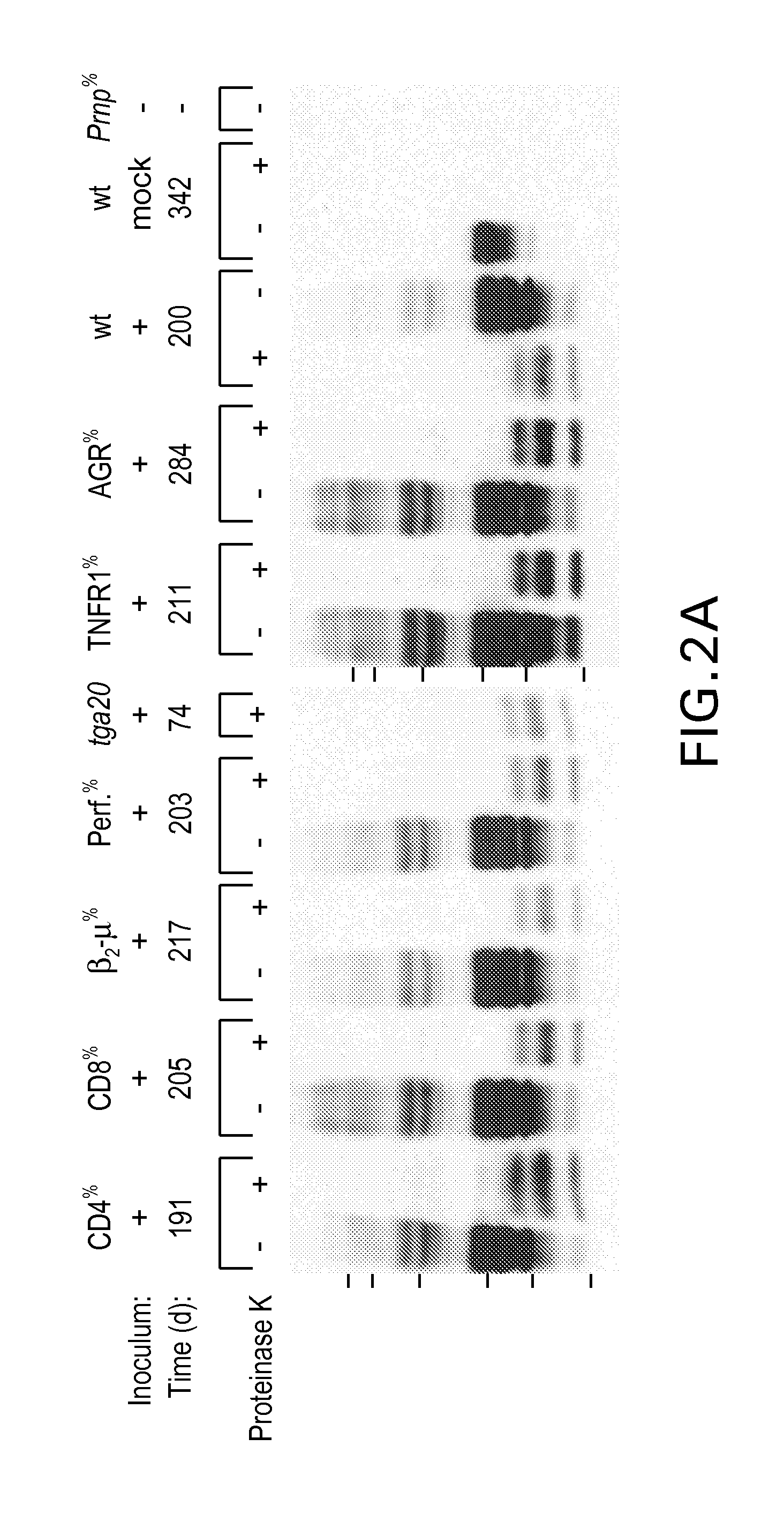
[0195] Ten percent brain homogenates were prepared as described 1 and, where indicated, digested with 20 μg / ml of proteinase K for 30 minutes at 37° C. Eighty μg of total protein were then electrophoresed through 12% SDS-polyacrylamide gel, transferred to nitrocellulose membranes, probed with monoclonal antibody 6H4. (Prionics AG, Zurich) or polyclonal antiserum IB3 (reference 26) against mouse PrP, and developed by enhanced chemiluminescence.

2.3. Detection of PrP Antibodies

[0196] Brain Lysates from wild-type and Prnpo / o mice, as w...

example 3

FACS Analysis Shown in FIG. 2D

[0210] Peripheral blood cells were incubated with serum from t11 μMT mice, washed, incubated with anti-mouse IgM-FITC conjugate followed by anti-CD3-PE (Pharmingen), and analyzed with a Becton-Dickinson FAScan instrument after erythrocyte lysis and fixation. For analysis, cells were gated on CD3-positive T-cells. EL4 cells infected with vesicular stomatitis virus (VSV) were stained with 5 μg VSV-specific monoclonal antibody VI24 (ref. 27) and with FTC-labelled antibody to mouse IgG2a (Southern Biotechnology), or with serum of t11 μMT mice, and with FITC-labelled F(ab′)2 antibody to mouse IgM (anti-IgM-FITC, Tago), or with serum of C57BL / 6 mice and anti-IgM-FITC. All data acquisition and analysis were performed with CellQuest software (Becton Dickinson).


PUM

PropertyMeasurementUnit
resistanceaaaaaaaaaa
temperaturesaaaaaaaaaa
Resistanceaaaaaaaaaa

Abstract

B-cells have been identified as being the crucial carriers of infectivity in the spread of transmissible spongiform encephalopathy within an infected organism. In a second step, B-cells may infect further components_of the immune system, e.g. T-cells. Accordingly, the present invention provides B-cell and T-cell specific ligands for the use in diagnostics and therapeutics for transmissible spongiform encephalopathy and provides methods for the manufacture of non-infective blood products and tissue derived products. Thus, the present invention provides medicaments comprising B-cell and / or T-cell depletants, for the treatment of pathologies where the depletion of B-cells and / or T-cells, and more particularly of tse-infected B-cells and / or T-cells is therapeutically effective.

Description

RELATED APPLICATIONS [0001] This application is a continuation of U.S. Ser. No. 09 / 554,567, filed May 16, 2000, which claims priority to Patent Cooperation Treaty Application No. PCT / EP98 / 08271, filed Dec. 16, 1998, now expired, and European Patent Application No. EP97122186.6, filed Dec. 16, 1997, currently pending, of which all are incorporated herein by reference in their entireties.FIELD OF THE INVENTION [0002] The present invention relates to diagnostics of and therapeutics for transmissible spongiform encephalopathy (tse). Further, the invention relates to non-infective body fluid products and to non-infective tissue derived products and to-suitable methods for the manufacture thereof. BACKGROUND ART [0003] Transmissible spongiform encephalopathies (TSE's) comprise a group of slow degenerative diseases of the CNS such as Creutzfeldt-Jakob disease (CJD), new variant CJD (termed nvCJD)1,2, Gerstmann-Sträussler-Scheinker disease (GSS) and kuru in man and scrapie in sheep or BSE (...

Claims


Application Information

Patent Timeline
Patent Type & Authority Applications(United States)
IPC IPC(8): A61K31/56A61K38/00A61K39/395A61K49/00A61P43/00C07K16/00C12N5/06G01N33/53A61K31/415A61K31/4166A61K31/4427A61K31/57A61K31/675A61K35/12A61K35/14A61K35/16A61K38/13A61K39/00A61K45/00A61P25/00A61P31/00C07K14/435C07K16/28C12P21/08G01N33/543G01N33/569G01N33/68
CPCA61K31/4166A61K31/4427A61K31/675A61K2039/505C07K16/28G01N2800/2828G01N33/56972G01N33/6896A61K2300/00A61P25/00A61P31/00A61P43/00
Inventor AGUZZI, ADRIANO P.KLEIN, MICHAEL A.RAEBER, ALEX J.WEISSMANN, CHARLESZINKERNAGEL, ROLF M.
Owner ABBOTT LAB INC
Learn More