Monocyte cell
a monocyte and cell technology, applied in combinational chemistry, blood/immune system cells, chemical libraries, etc., can solve the problems of excessive angiogenesis, tissue death risk, and damage to normal tissue, and achieve the effect of inhibiting angiogenesis and inhibiting angiogenesis
- Summary
- Abstract
- Description
- Claims
- Application Information
AI Technical Summary
Benefits of technology
Problems solved by technology
Method used
Image
Examples
example 1
Methods
Cell Purification and Cell Sorting
Human Cells:
[0143] PB was obtained from healthy volunteers following informed consent, according to the Declaration of Helsinki and a protocol approved by the H. San Raffaele Bioethical Committee. Total leukocytes were analysed after lysis of erythrocytes using ammonium chloride. PBMCs were isolated using Ficoll-Hypaque gradient. Granulocytes, T and B lymphocytes were positively selected by magnetic sorting (using CD15, CD3 or CD19 MicroBeads, respectively; Miltenyi). T cell-depleted PBMCs were negatively selected by CD3 MicroBeads. Resident monocytes (CD16+CD14low) were enriched from PBMCs by negative selection of T, B and NK cells (using a cocktail of CD3, CD19 and CD56 MicroBeads), followed by positive selection by CD16 MicroBeads. Inflammatory monocytes (CD16-CD14+) were enriched by negative selection of CD16+ cells, followed by positive selection by CD14 MicroBeads. For cell sorting, we used a Becton Dickinson FACS Vantage SE-FACSDi...
example 2
Tie2 Expression in Human Peripheral Blood Identifies a Subset of Noninflammatory Monocytes
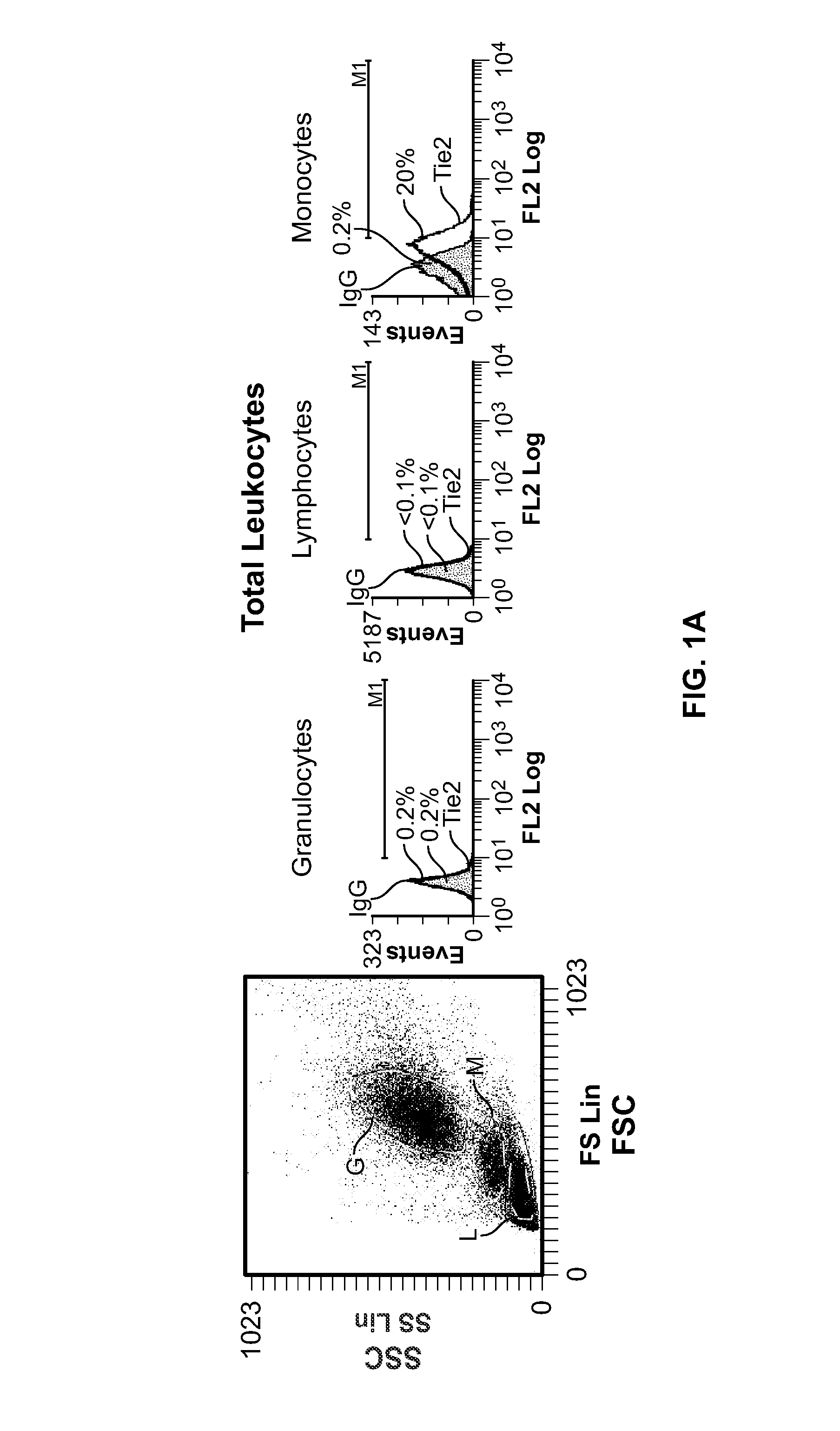
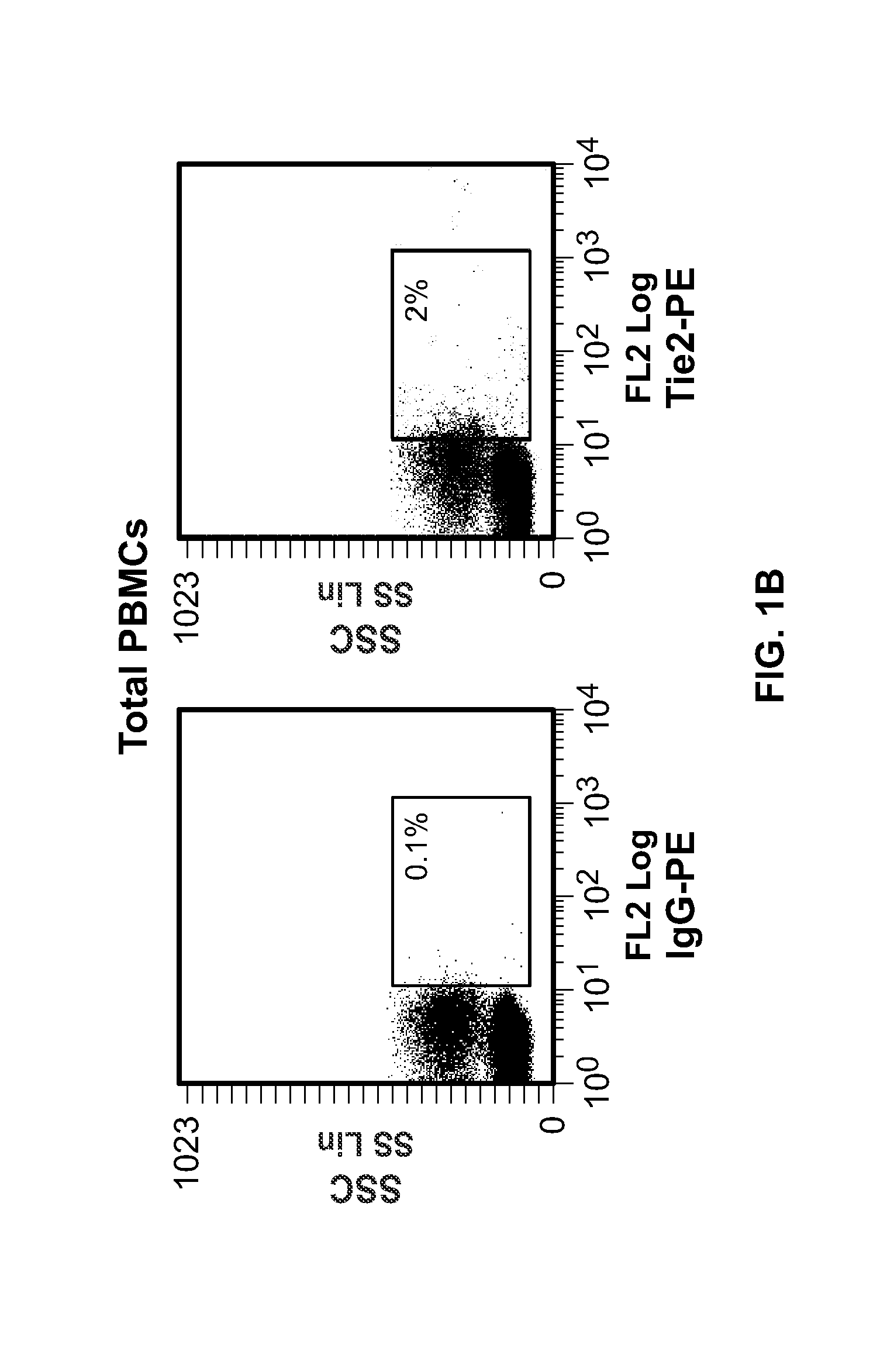
[0159] In order to investigate Tie2 expression by human haematopoietic cells, we stained peripheral blood (PB) obtained from healthy donors with a mouse anti-human Tie2 monoclonal antibody (clone 83715 from R&D systems; or clone 9 from RELIAtech. See methods above) and analysed cells by flow cytometry after lysis of erythrocytes.
[0160] The main haematopoietic populations in PB are granulocytes (50-70%), lymphocytes (25-40%) and monocytes (5-10%). We found that only a small fraction (0.1-0.5%; n=7) of these leukocytes expressed Tie2 to detectable levels (FIG. 1A), implying that the wide majority of granulocytes were Tie2−, a finding in contrast with a previous report that showed that human neutrophils expressed Tie2 (Lemieux et al., 2005). However, we noted that Tie2+ cells co-purified with peripheral blood mononuclear cells (PBMCs; FIG. 1B)—the haematopoietic cell fraction containing lymphocy...
example 3
Tie2-Expressing Monocytes are Recruited to Human Tumours
[0166] We showed that TEMs infiltrate murine tumours, including spontaneously and orthotopically growing neoplasms (De Palma et al., 2005). To study whether human Tie2+ monocytes are present in human solid tumours, we analysed the haematopoietic infiltrate of 28 human carcinoma specimens, including kidney, colorectal, breast, gastric, pancreatic and lung cancers, by four-color FACS analysis, immunohistochemistry and immunofluoresce triple staining (Table 2).
TABLE 2Cancer specimens and normal organs analyzed in this studyTie2 expressionin mononuclearMethod ofcellsanalysisCancer specimensColon adenocarcinoma (5)5 / 5C, F,Gastric adenocarcinoma (2)2 / 2C, IPancreatic adenocarcinoma (1)1 / 1C, ILiver metastasis (1)0 / 1IBreast carcinoma (1)1 / 1C, F,Renal clear cell carcinoma (2)2 / 2F,Non small cell lung cancer (2)2 / 2C, F,Brain glioblastoma (1)0 / 1IPapillary cell carcinoma of thyroid (1)1 / 1F,Soft tissue tumours (5)4 / 5C, INormal organsColon ...
PUM
| Property | Measurement | Unit |
|---|---|---|
| concentration | aaaaa | aaaaa |
| concentration | aaaaa | aaaaa |
| concentration | aaaaa | aaaaa |
Abstract
Description
Claims
Application Information
 - R&D
- Intellectual Property
- Life Sciences
- Materials
- Tech Scout
- Unparalleled Data Quality
- Higher Quality Content
- 60% Fewer Hallucinations
Browse by: Latest US Patents, China's latest patents, Technical Efficacy Thesaurus, Application Domain, Technology Topic, Popular Technical Reports.
© 2025 PatSnap. All rights reserved.Legal|Privacy policy|Modern Slavery Act Transparency Statement|Sitemap|About US| Contact US: help@patsnap.com


