
Systems and methods for monitoring temperature during electrosurgery or laser therapy

a technology applied in the field of electrosurgical and laser therapy, can solve the problems of liver tumor recurrence, incomplete necrosis of tissue surrounding the vessel, and the failure of conventional electrosurgical devices and procedures

Inactive Publication Date: 2008-02-07
LUMASENSE TECH HLDG
View PDF14 Cites 75 Cited by
  • Summary
  • Abstract
  • Description
  • Claims
  • Application Information

AI Technical Summary

Benefits of technology

[0022]In some embodiments, the tissue or organ (e.g. tissue site or organ site) is a site of a tissue disease, such as a liver anomaly, stomach cancer, bowel cancer, pancreatic cancer, kidney cancer, or lung cancer. In some embodiments, the tissue or organ is ablated during to a controlled depth by plasma-induced volumetric removal of the tissue. In some embodiments, the tissue or organ is exposed to a temperature in the range of 40° C. to 90° C. during such ablation. In some embodiments, the tissue is skin. In some embodiments, the organ is heart, bladder, lung, liver, muscle, salivary gland, colon, spleen, pancreas, gallbladder, liver, kidney, stomach, tongue, thyroid gland, gallbladder, brain, large intestine, or small intestine.

Problems solved by technology

Conventional electrosurgical devices and procedures, however, suffer from a number of disadvantages.
For example, in certain situations, complete ablation of target tissue that is adjacent a vessel may be difficult or impossible to perform, since significant bloodflow may draw the produced heat away from the vessel wall, resulting in incomplete necrosis of the tissue surrounding the vessel.
Due to the increased vascularity of the liver, the heat sink effect may cause recurrence of liver tumors after a radio frequency ablation.
Another drawback is that conventional electrosurgery devices are not suitable for the precise removal (ablation) of tissue.
At the point of contact of the electric arcs with tissue, rapid tissue heating occurs due to high current density between the electrode and tissue.
The tissue is parted along the pathway of vaporized cellular fluid, inducing undesirable collateral tissue damage in regions surrounding the target tissue site.
Most of the thermal injury is thought to result from conduction of heat from the surface layer.
While Eick and Bierbaum demonstrate the importance of tissue temperature controlled radiofrequency delivery in ablation procedures, the setup used by Erick and Bierbaum is wholly unsatisfactory for use in treatment of patients because it requires the injection of a thermocouple deep into the tissue being ablated.

Method used


Image

  • Systems and methods for monitoring temperature during electrosurgery or laser therapy
  • Systems and methods for monitoring temperature during electrosurgery or laser therapy
  • Systems and methods for monitoring temperature during electrosurgery or laser therapy

Examples


Embodiment Construction

[0030]Radiofrequency ablation (RFA) is used for local tissue ablation. See Decadt and Siriwardena, 2004, “Radiofrequency ablation of liver tumors: systematic review,” Lancet Oncol 5, 550-560, which is hereby incorporated by reference. In RFA, a needle electrode (e.g., 14-17.5 G) with an insulated shaft and a non-insulated distal tip is inserted into or over a lesion, often with imaging-guidance. For example, the physician may be guided in the placement of the needle by images from an imaging provided by ultrasound, a CT scanner, or magnetic resonance. In some procedures, once the needle is in place, tines are deployed from the hollow core of the needle. These tines penetrate the tissue. The patient is made into an electrical circuit by placing grounding pads in appropriate places (e.g. on the thighs or back muscles). RFA energy is then sent through the needle and tines, destroying the tissue.

[0031]Radiofrequency ablation is an attractive tool for cancer patients, especially for live...


PUM


Abstract

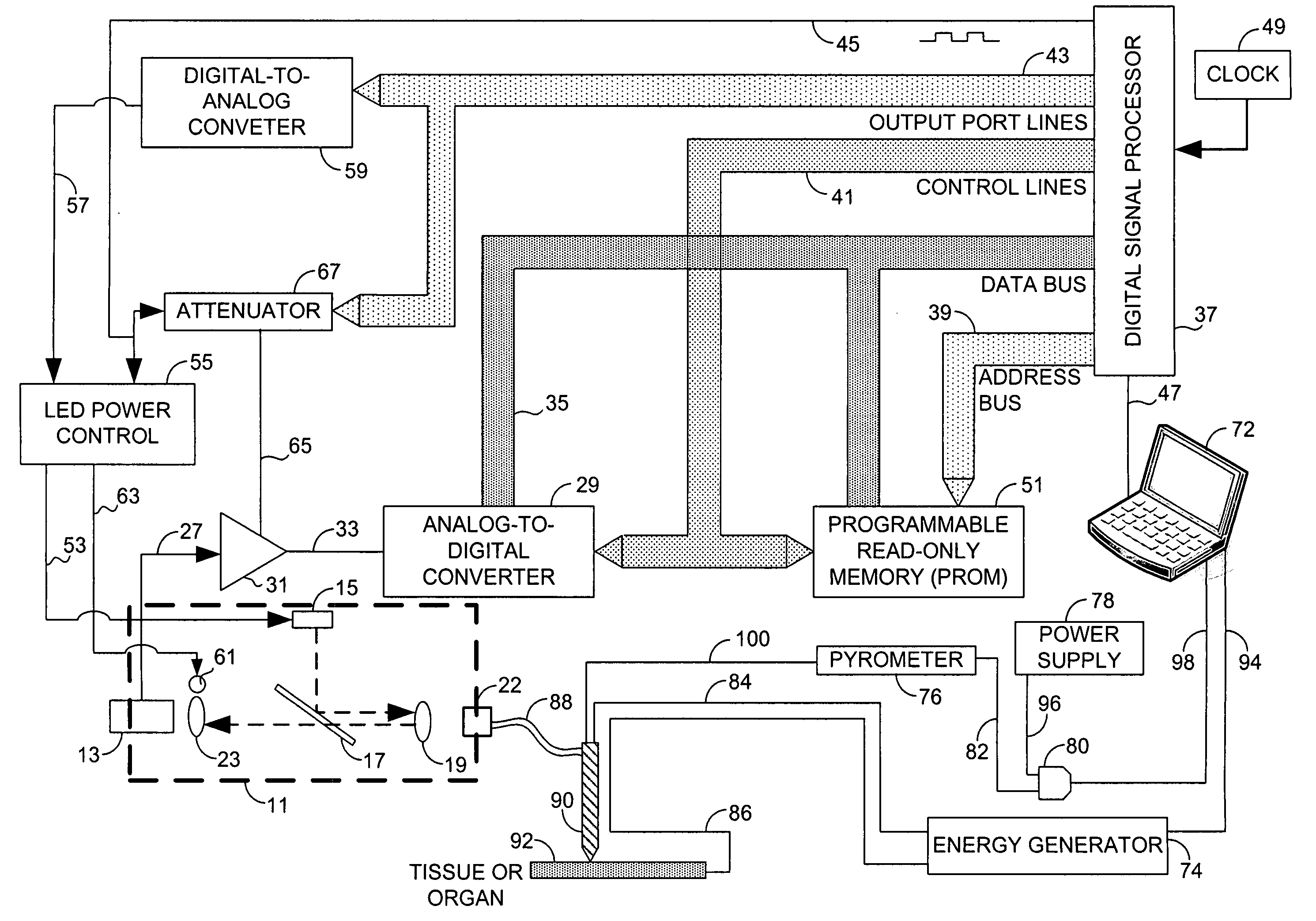
Systems that measure temperatures of tissue during electrosurgery or laser therapy of the tissue or organs is provided. One system provides a pyrometer that measures infrared electromagnetic energy emitted by a surface of a tissue or organ thereby determining a sub-surface temperature of the tissue or organ. The system further has an energy generator and an ablation electrode or laser probe that delivers energy from the energy generator to a tissue or organ, responsive to a sub-surface temperature determined by the pyrometer. The pyrometer can be calibrated using a luminescent material having known optical properties as a function of temperature. The luminescent material can be positioned on the surface of the tissue or organ or inserted directly into the tissue or organ using a catheter. Methods in which the surface or sub-surface temperature of the tissue is measured during therapy are also provided.

Description

FIELD OF THE INVENTION[0001]The present invention relates generally to the fields of electrosurgery and laser therapy, and more particularly to surgical and cosmetic devices and methods which employ energy (e.g., RF ablation energy, laser energy, etc.) to resect, coagulate, or ablate tissue or organs. The present invention also relates to apparatus and methods for removing tissue at a target site by a procedure that is capable of measuring tissue or organ surface temperature and / or temperatures below the surface of the tissue or organ.BACKGROUND OF THE INVENTION[0002]Tissue may be destroyed, ablated, or otherwise treated using thermal energy during various therapeutic procedures. Many forms of thermal energy may be imparted to tissue, such as radio frequency electrical energy, microwave electromagnetic energy, laser energy, acoustic energy, or thermal conduction. In particular, radio frequency ablation (RFA) may be used to treat patients with tissue anomalies, such as liver anomalie...

Claims


Application Information

Patent Timeline
Patent Type & Authority Applications(United States)
IPC IPC(8): A61B6/00A61B18/18
CPCA61B18/14A61B2017/0007A61B18/20
Inventor HOANG, ANHHU, JI-DIH
Owner LUMASENSE TECH HLDG
Learn More