
Apparatus and method for cell migration assays

a cell migration and apparatus technology, applied in the field of apparatus and method for detecting cell migration, can solve the problems of inability to examine individual cell migration, suffer from several limitations, and suffer from immune suppression, nausea and kidney damag

Inactive Publication Date: 2008-02-07
TRUSTEES OF TUFTS COLLEGETHE
View PDF29 Cites 42 Cited by
  • Summary
  • Abstract
  • Description
  • Claims
  • Application Information

AI Technical Summary

Benefits of technology

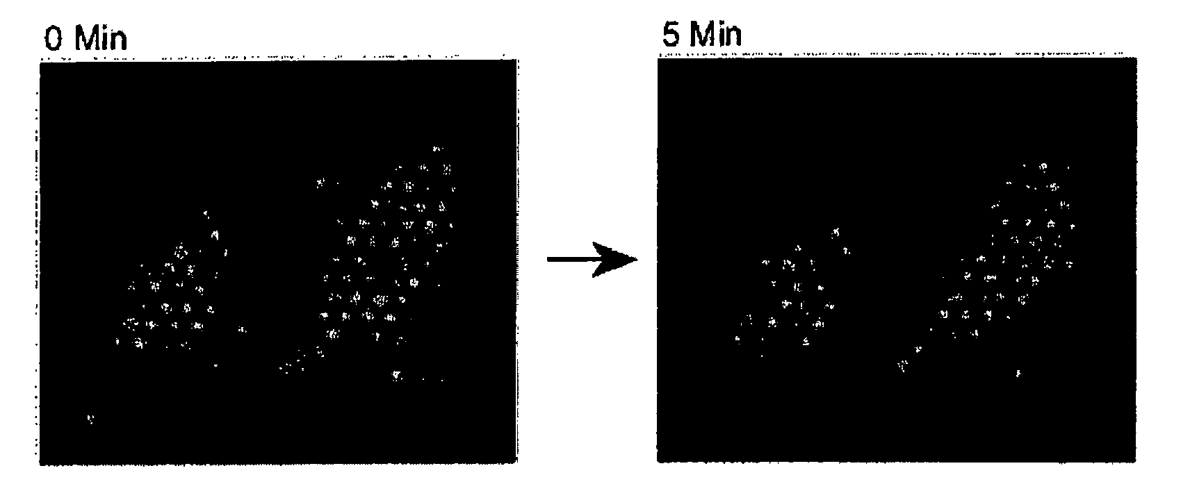
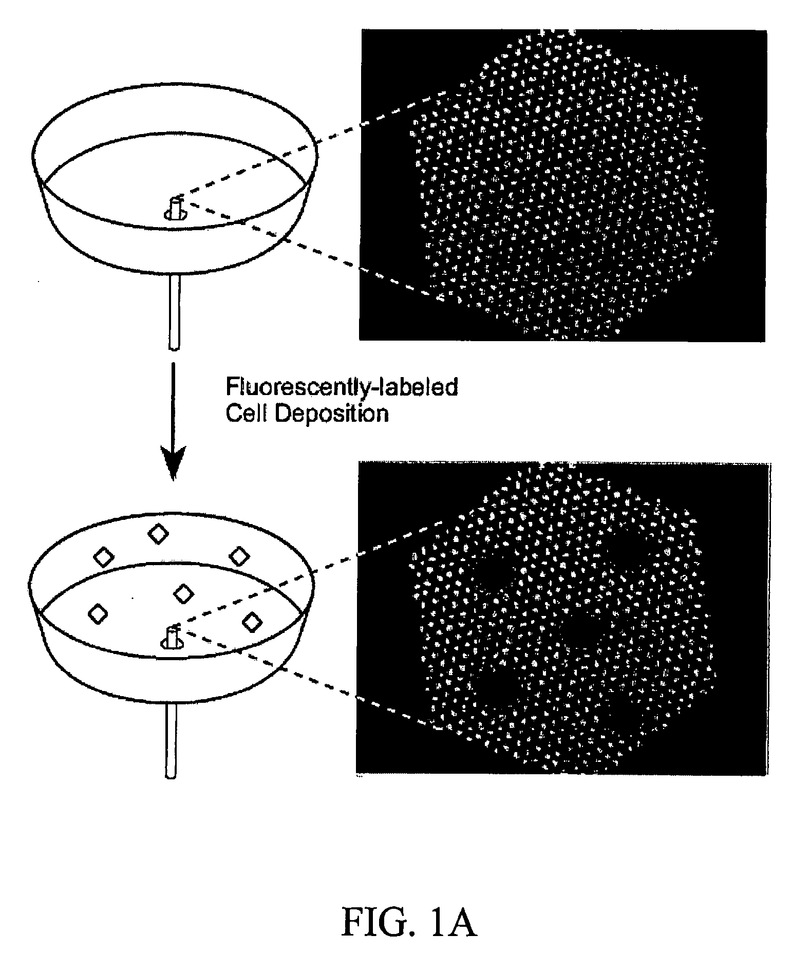
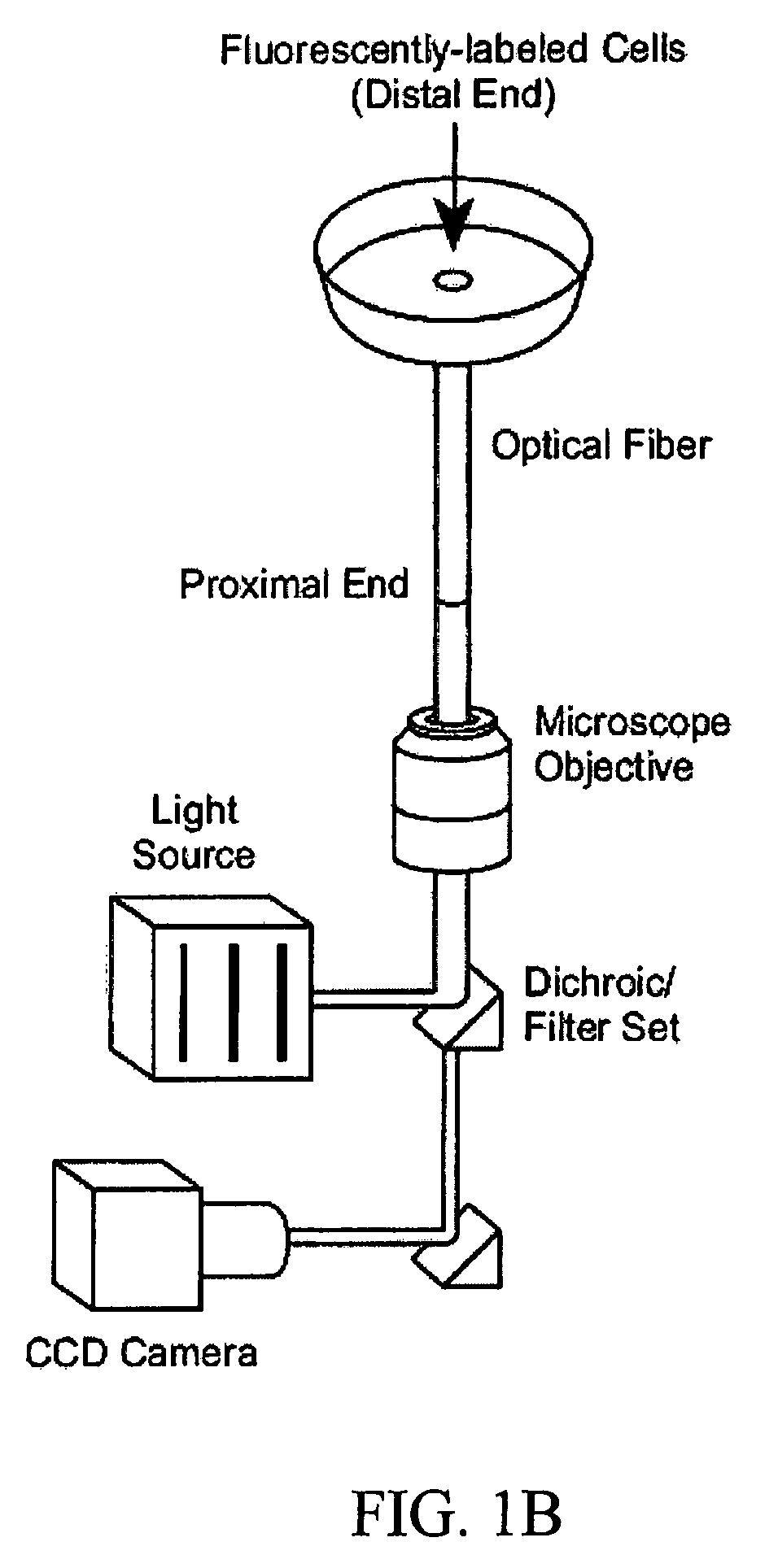
[0007] The present invention, in one embodiment, is directed to a method of detecting cell migration. The method includes providing a plurality of cells on a substrate having a plurality of detection units, wherein each of the cells is disposed over at least one of the detection units. The method further includes detecting cellular movement of at least one of said cells. In one alternative aspect of the invention, each of the cells is disposed over more than one of the detect...

Problems solved by technology

Antiproliferative drugs are harmful to most dividing cells, causing side effects such as immune suppression, nausea, and kidney damage.
Techniques such as the Boyden chamber and the wound assay are among the most widespread techniques used to study cell migration, but suffer from several limitations.
The Boyden chamber relies on a chemical gradient to produce directed cell movement, and cannot be used for examining individual cell migration.
In addition, characteristics of the platform itself can cause variations in migration, such as pore size and gradient dissipation.
The nature of the wound assay also makes it difficult to examine individual cells, as the entire group is a dynamic monolayer of cells.
Although these two methods are very useful, they are population-based techniques that examine a collective response from many cells, and consequently miss important details such as cell-substrate adhesion and intracellular motion.
More recently developed techniques, such as cell patterning and software tracking have the ability to record the movement of individual cells, but these techniques involve complex procedures and / or computer software, and generally take from hours to days to determine drug efficacy.

Method used


Image

  • Apparatus and method for cell migration assays
  • Apparatus and method for cell migration assays
  • Apparatus and method for cell migration assays

Examples


examples

Methods

[0110] NIH / 3T3 fibroblasts were obtained from American Type Tissue Collection (ATCC No. CRL-1658). Dulbecco's modified eagle medium, penicillin-streptomycin, L-glutamine, calf serum, trypsin-EDTA were obtained from Invitrogen (Carlsbad, Calif.). Fibronectin was obtained from Calbiochem-Novabiochem Corporation (La Jolla, Calif.). Live / Dead Viability / Cytotoxicity kit for animal cells and Vybrant DiO cell-labeling solution were obtained from Molecular Probes (Eugene, Oreg.). Nocodazole and sodium bicarbonate were obtained from Sigma Chemical Co. (St. Louis, Mo.). Fiber-optic imaging bundles were obtained from the Schott Corporation (Southbridge, Mass.) and Illumina, Inc. (San Diego, Calif.). Delta T4 culture dish system, and delta T culture dishes were obtained from Bioptechs (Butler, Pa.). Aluminum oxide lapping films were obtained from Mark V Laboratory (East Granby, Conn.).

[0111] After labeling, the cells were allowed to settle and adhere to a fibronectin-coated fiber bundl...


PUM


Abstract

The present invention is directed to a method and apparatus for detecting and analyzing cell migration. More specifically, the present invention is directed to novel technology for analyzing cellular movement, including whole cell migration and subcellular component movement. Cells are distributed onto a substrate and monitored for migration or movement. According to one embodiment, when a labelled cell or portion of a cell passes over one of the delineations between detection units, such as individual fibers in a fiber optic bundle, the label causes a large intensity increase, which stays for a given “residence time” until the cell departs from the detection unit.

Description

FIELD OF THE INVENTION [0001] The present invention relates to an apparatus and method for detecting cell migration. In addition the invention relates to a method of detecting migration of intracellular organelles in living cells. BACKGROUND OF THE INVENTION [0002] Cells from a malignant tumor are able to leave the original site and establish metastases throughout the body through a combination of adhesion, proteolysis, and migration. The importance of studying cell migration becomes apparent when it is realized that the prevention of cellular migration can stop cancer proliferation. Until recently, chemotherapy regimens consisted entirely of antiproliferative compounds, which typically operate by inhibiting nucleic acid function. Antiproliferative drugs are harmful to most dividing cells, causing side effects such as immune suppression, nausea, and kidney damage. The side effects of these compounds have driven the development of alternative strategies to preventing cancer prolifera...

Claims


Application Information

Patent Timeline
IPC IPC(8): C12N1/02C12M1/34
CPCG01N15/1475G01N2015/0065G01N21/6458G01N21/6428G01N15/01G01N15/1433
Inventor WALT, DAVID R.DI CESARE, CHRISTOPHER J.
Owner TRUSTEES OF TUFTS COLLEGETHE
Learn More