
Radiotherapy device

a radiation therapy and device technology, applied in the field of radiation therapy devices, can solve the problems of increasing the discomfort of patients, affecting the patient's comfort, so as to achieve the effect of reliable identification, precise localization of metabolic anomalies, and good patient accessibility

Inactive Publication Date: 2008-01-10
SIEMENS AG
View PDF18 Cites 31 Cited by
  • Summary
  • Abstract
  • Description
  • Claims
  • Application Information

AI Technical Summary

Benefits of technology

[0008]An object particularly underlying the invention is to specify a radiotherapy device, the images from which enable reliable identification and precise localization of metabolic anomalies, in particular of malignant tissues affected by tumors, and which offers good patient accessibility, so that operative and / or minimally-invasive interventions can be carried out on the patient and examined contemporaneously with the radiation treatment and / or image recording and without moving the patient.
[0009]This object is achieved by a radiotherapy device of the type mentioned in the introduction, with which in accordance with the invention the image recording apparatus comprises an angiography CT. There is no need for a gantry and one of the elements of the image recording apparatus, together with its housing, can be moved about the patient. In this way, anatomical and functional imaging can be combined with good patient accessibility.
[0010]The angiography CT can be a conventional angiography CT. A further development of an angiography CT, which supplies good 3D soft tissue recordings, has been known for a short time under the name of “Angiographische Computertomographie” (ACT) [Computed Tomography Angiography, CTA] or also as “DynaCT” and is described for instance in DE 10 2004 057 308 A1, which is incorporated by reference herein in its entirety. This further development relates on the one hand to improved detector technology and on the other hand to improved image processing methods, which revert back to correction algorithms which have been specially adjusted to the soft tissue display during the processing and visual conversion of the detector signals. CTA allows both the vascular system and also the surrounding soft tissue or any deposits on the vessel wall to be displayed with a high resolution, up to 5 HU. If complications occur during radiation therapy, an operation or a catheter intervention, there is no need to reposition the patient in an MRI or CT device in order to produce a control image, as a result of which valuable time can be gained for the introduction of further therapy steps.

Problems solved by technology

This alignment of the image data sets, also referred to as registration, generally only exhibits limited accuracy and frequently requires time-consuming user interaction despite large-scale computer-aided automation.
An essential disadvantage of this concept lies in the poor accessibility of the patient during the examination, due to one or two closed detector tubes which are arranged adjacent to one another.
An arrangement of this type may not only increase the patient's discomfort, but also generally precludes the performing of minimally-invasive or surgical interventions on the patient during the examination procedure for instance.

Method used


Image

  • Radiotherapy device
  • Radiotherapy device
  • Radiotherapy device

Examples


Embodiment Construction

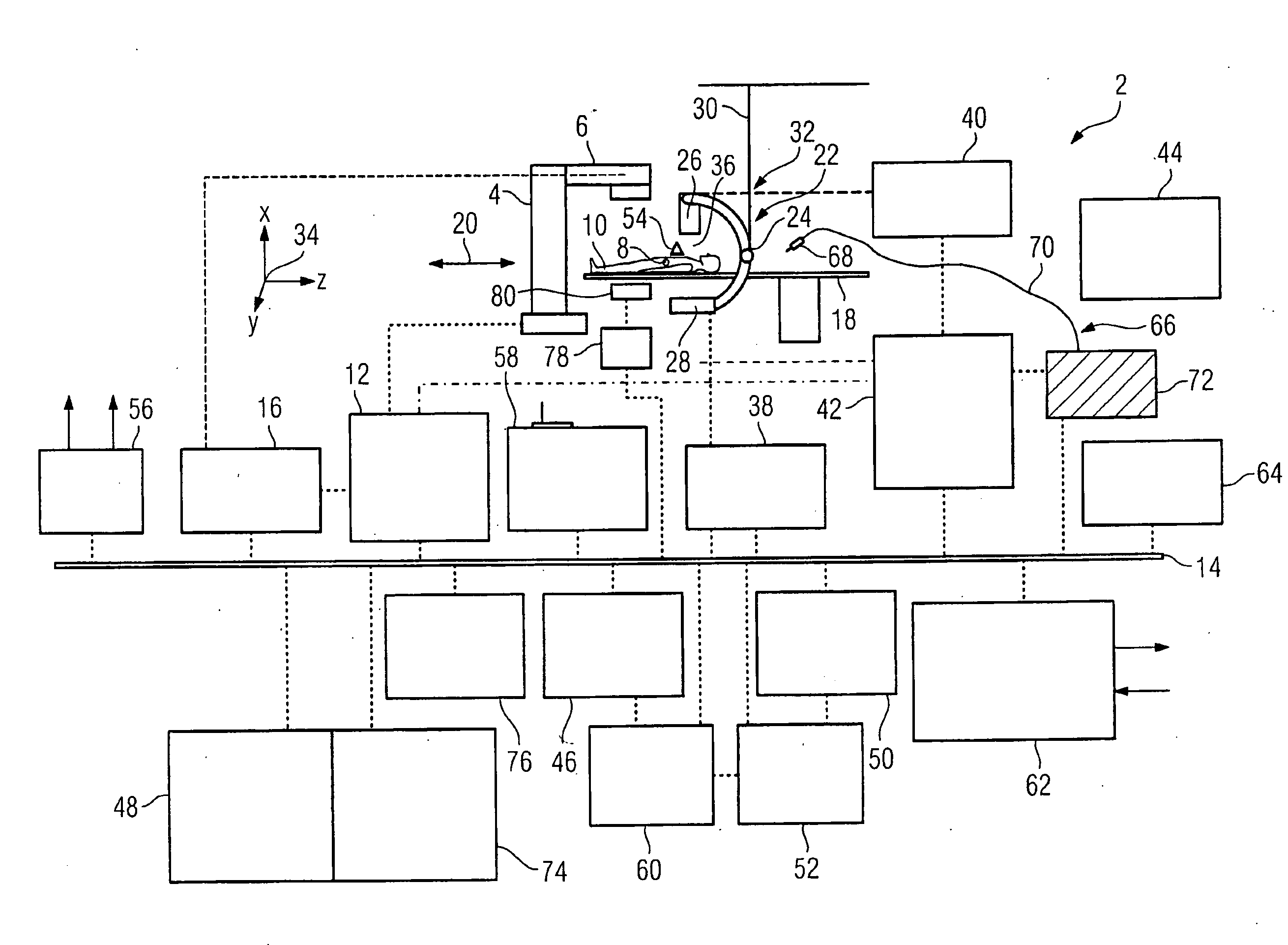
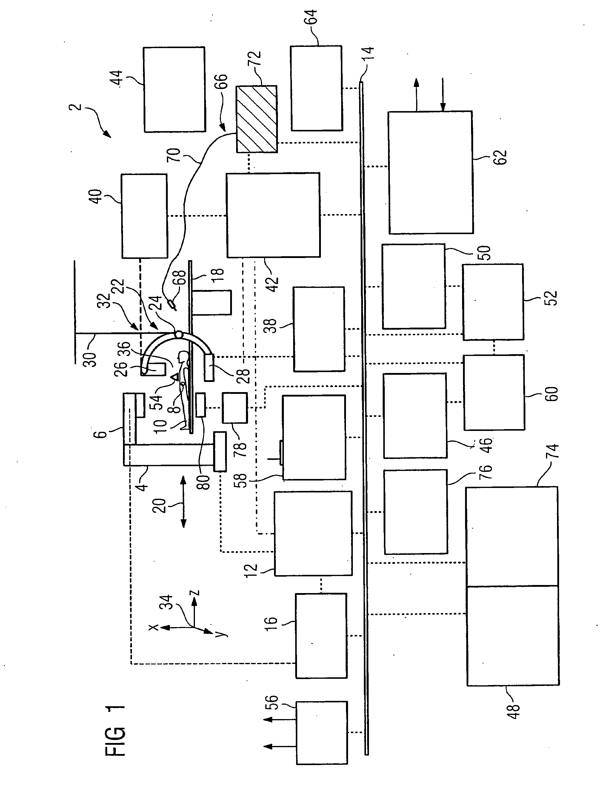
[0031]FIG. 1 shows a schematic overview of a medical radiotherapy device 2, which comprises a therapeutic radiation unit 4 having a linear accelerator with a radiation source 6 for generating a high energy electron or ion beam. The radiation unit 4 is designed such that the radiation source 6 rotates about an isocenter, which corresponds to an area of the body 8 and / or radiation region to be radiated, and thus the beams strike the radiation region successively from different directions. The radiation source 6 can be mounted on a stand or on a 3D robot. The stand and the 3D robot can be secured in each instance to the floor, to the wall or to a ceiling. By rotating about an isocenter, a very high intensity is achieved in the radiation region and / or in the isocenter, in which a tumor of a patient 10 is located, while the intensity is considerably lower in the surrounding tissue. The movement of the radiation source 6 is controlled by a motion controller 12, which is connected to a dat...


PUM


Abstract

There is described a radiotherapy device having an integrated unit composed of a therapeutic radiation unit for treating an area of a body of a patient and an image recording apparatus for recording images of the area of the body using an element configured as a radiation source and an element configured as a radiation detector. To generate images for a reliable identification and precise localization of a tumor and consequently achieve good patient accessibility the image recording apparatus comprises an angiography CT.

Description

CROSS REFERENCE TO RELATED APPLICATIONS[0001]This application claims priority of German application No. 10 2006 026 490.8 DE filed Jun. 7, 2006, which is incorporated by reference herein in its entirety.FIELD OF INVENTION[0002]The invention relates to a radiotherapy device having an integrated unit composed of a therapeutic radiation unit for treating an area of the body of a patient and an image recording apparatus for recording images of the area of the body having an element which is configured as a radiation source and as a radiation detector.BACKGROUND OF INVENTION[0003]With tumor diseases or suchlike, a rapid and reliable diagnosis and immediately introduced therapy are of particular importance for the recovery process. The diagnosis of such diseases is supported by imaging methods. The radiation of tumors and metastases has been established within the field of cancer treatment for decades. Linear accelerators have been used for years for semi-deep and deep radiation therapy, ...

Claims


Application Information

Patent Timeline
Patent Type & Authority Applications(United States)
IPC IPC(8): A61B8/00A61N5/00
CPCA61B5/6833A61B6/5247A61B6/12A61B6/466A61B6/467A61B6/504A61B6/5235A61B6/527A61B8/0833A61B8/0841A61B8/5238A61B8/5276A61N5/1049A61N2005/1061A61B5/721
Inventor MASCHKE, MICHAEL
Owner SIEMENS AG
Learn More