Method of deriving pluripotent stem cells from a single blastomere
a single blastomere, stem cell technology, applied in the direction of artificial cell constructs, genetically modified cells, skeletal/connective tissue cells, etc., can solve the problems of destroying the donor embryo, destroying the developing embryo, and the known methods of excising and culturing single blastomeres have failed to produce stem cells that demonstrated pluripotency, etc., to achieve the effect of reducing the expression of genes
- Summary
- Abstract
- Description
- Claims
- Application Information
AI Technical Summary
Benefits of technology
Problems solved by technology
Method used
Image


Examples
example 1
[0045]Female C57BL / 6 mice were superovulated by injection of 5 units of pregnant mare's serum gonadotropin (Sigma-Aldrich, St. Louis, Mo.-Aldrich, St. Louis, Mo.) followed 48 hours later by injection of 5 units of human chorionic gonadotropin (Sigma-Aldrich, St. Louis, Mo.). The superovulated females were then paired overnight with C57BL / 6 males for mating. The following morning, females with vaginal plugs were selected for embryo collection. Typically, by this time post coitus (p.c.), most embryos could be expected to be at the 2 cell or 4 cell stages of development. Embryos at the 8 cell stage of development could be expected to have formed by 2.5 days p.c. Embryos were collected by flushing the oviduct with M2 medium (Sigma-Aldrich, St. Louis, Mo.). After briefly washing the embryos in M2 medium, the embryos were transferred into 35 mM non-adherent tissue culture plates that contained KSOM culture medium (Specialty Media, Phillipsburg, N.J.). The tissue culture pla...
example 2
Isolation and Culture of Single Blastomeres of Preimplantation Embryos
[0046]The zona pellucida of each 2 cell, 4 cell, or 8 cell embryo was removed by placing embryos into acidified (pH 2.5) Tyrode's medium (MediCult, Denmark) for 2-3 minutes at 37° C. followed by two brief washings in M2 medium (Sigma-Aldrich, St. Louis, Mo.). Zona-free embryos were then incubated in Ca2+ and Mg2+ free biopsy medium (MediCult, Denmark) for 10 minutes at 37° C. Individual blastomeres were isolated by repeatedly pipetting the zona-free embryos with a flame-polished glass micropipette. Subsequently, following a brief recovery in KSOM medium, isolated blastomeres were each transferred to 20 μl droplets of KSOM medium and incubated at 37° C. and 5% CO2. Generally, within about 2.5 days, the blastomeres would rise to morula-like cell clusters, which were then cultured in one of three different culture conditions, designated conditions A, B, and C, respectively as depicted in FIG. 1. Culture condition A w...
example 3
Oct-4 Expression by Blastomere Derivatives
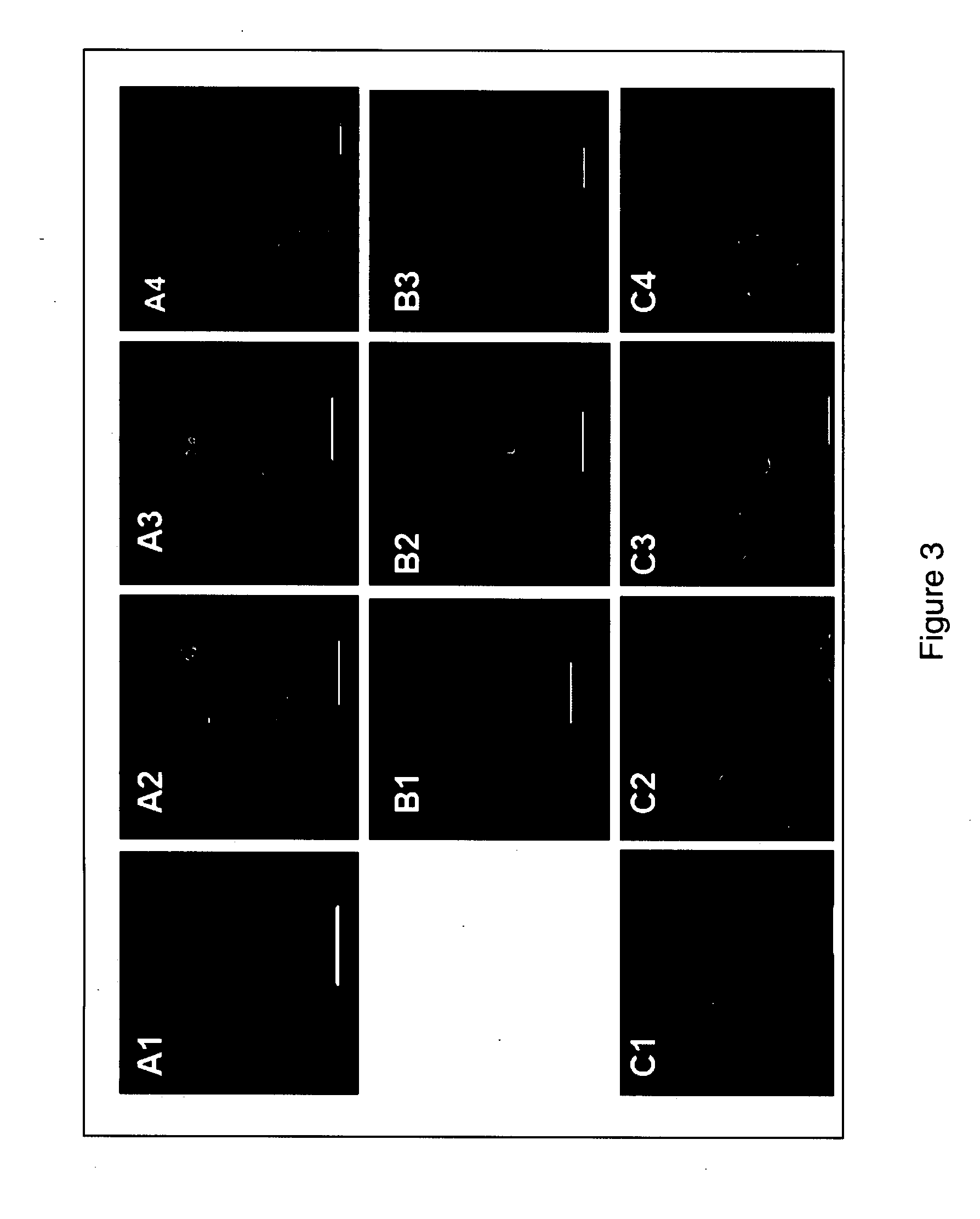
[0050]Immunofluorescence analysis was used to assess expression of Oct-4 expression by blastomere derivatives. The expression of Oct-4 by a cell was considered to correlate with pluripotency. The protocol for immunofluorescence staining was performed as follows (Kuo H C et al. (2003) Biol Reprod 68:1727-35).
[0051]Blastomeres or their derivatives were plated onto cover slips coated with Matrigel (Invitrogen, Carlsbad, Calif.) and fixed with 4% paraformaldehyde for 20 minutes at room temperature. Blocking solution, which included PBS (Invitrogen, Carlsbad, Calif.), 0.1% BSA (Sigma-Aldrich, St. Louis, Mo.), 10% normal goat serum (Sigma-Aldrich, St. Louis, Mo.), and 0.2% Triton X-100 (Sigma-Aldrich, St. Louis, Mo.) was then added to the fixed cells. The fixed cells were incubated overnight with primary antibodies (Table 2) in PBS containing 0.1% BSA (Sigma-Aldrich, St. Louis, Mo.) and 10% normal goat serum (Sigma-Aldrich, St. Louis, Mo.) at 4° C...
PUM
 Abstract
Description
Claims
Application Information
 - R&D
- Intellectual Property
- Life Sciences
- Materials
- Tech Scout
- Unparalleled Data Quality
- Higher Quality Content
- 60% Fewer Hallucinations
Browse by: Latest US Patents, China's latest patents, Technical Efficacy Thesaurus, Application Domain, Technology Topic, Popular Technical Reports.
© 2025 PatSnap. All rights reserved.Legal|Privacy policy|Modern Slavery Act Transparency Statement|Sitemap|About US| Contact US: help@patsnap.com