
Method and System for Non-Contact Fluorescence Optical Tomography with Patterned Illumination

a fluorescence optical tomography and patterned illumination technology, applied in the field of molecular imaging, can solve the problems of inability to accurately resolve inability to accurately solve image reconstruction problems, and inability to provide patterned illumination, etc., to achieve the effect of significantly increasing the cost and improving the accuracy of determining fluorescent targets at varying depths

Active Publication Date: 2007-12-13
BAYLOR COLLEGE OF MEDICINE
View PDF1 Cites 28 Cited by
  • Summary
  • Abstract
  • Description
  • Claims
  • Application Information

AI Technical Summary

Benefits of technology

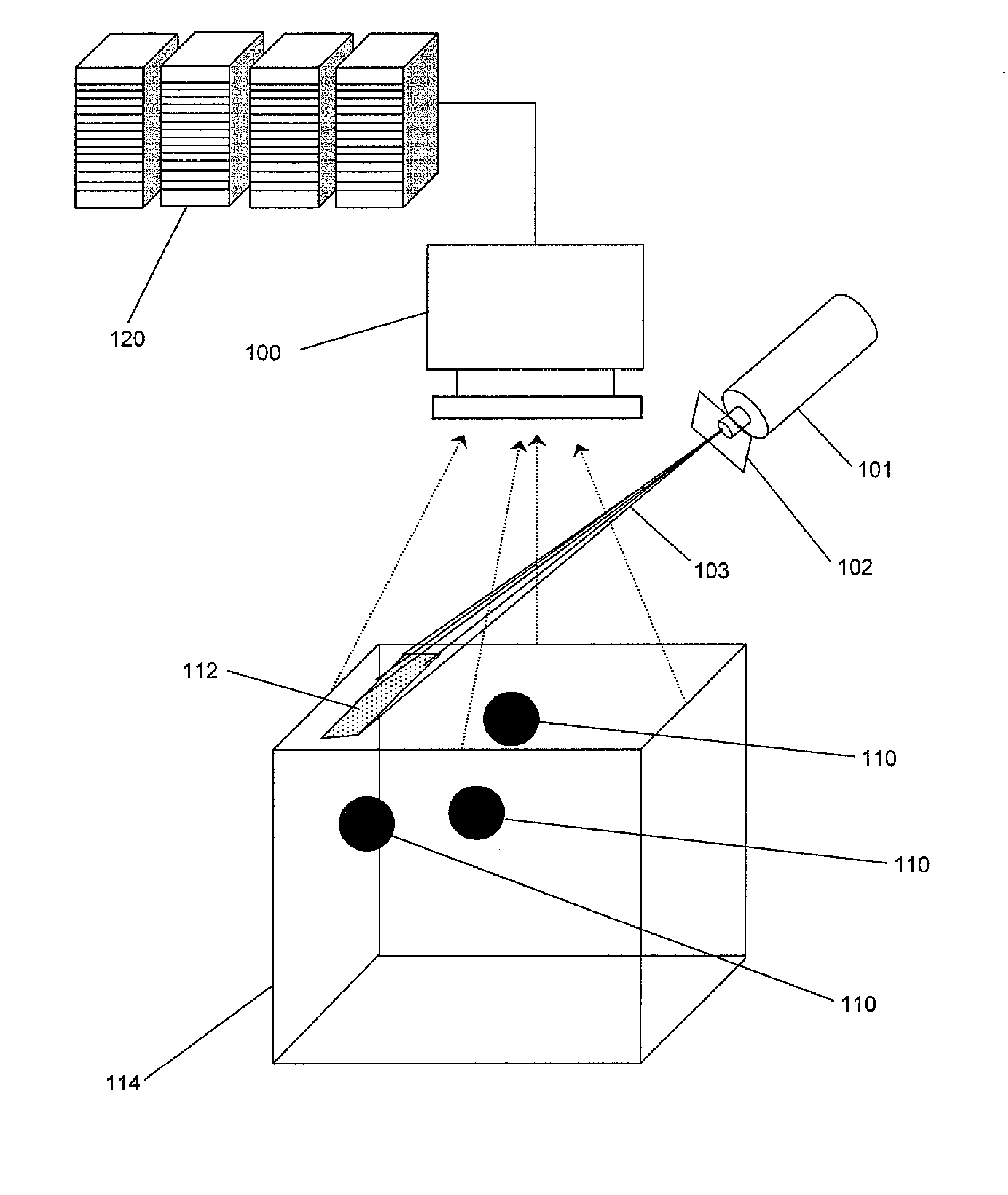
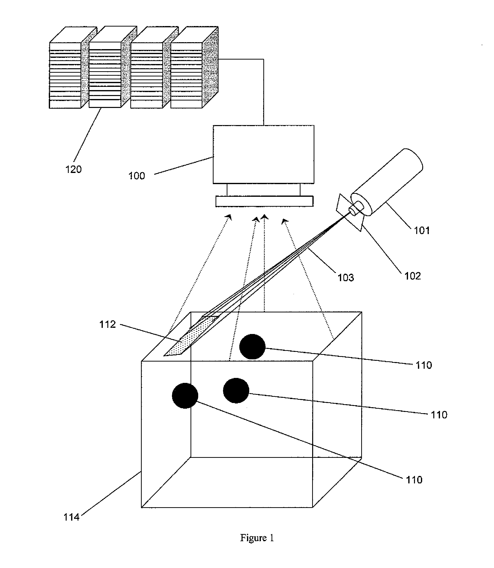
The patent describes a new method and system for getting 3D images of multiple fluorescent targets embedded in a medium. The method involves illuminating the surface of the medium with light and measuring the reflected and emitted fluorescence. An iterative algorithm is used to create a 3D image that minimizes the difference between a predicted data set and the measured data sets. The system includes an excitation light source, a sensor, and a computer capable of executing multiple calculations in parallel. The invention is simple and cost-effective and can resolve fluorescent targets at varying depths more accurately than previous techniques.

Problems solved by technology

Because photon propagation in a biological medium is diffuse, the image reconstruction problem can be ill-posed.
However, fiber optics based tomography systems suffer from sparse measurement data, and inadequate excitation light penetration into the tissue interior.
While area-illumination provides enhanced excitation light penetration, the tomography analysis is complex because of the increased ill-posedness introduced by the availability of only the reflectance measurements.

Method used


Image

  • Method and System for Non-Contact Fluorescence Optical Tomography with Patterned Illumination
  • Method and System for Non-Contact Fluorescence Optical Tomography with Patterned Illumination
  • Method and System for Non-Contact Fluorescence Optical Tomography with Patterned Illumination

Examples


example 1

[0086] Synthetic measurements were generated on a 512 ml cubical tissue phantom with optical properties of 1% Liposyn. In a right handed coordinate system with the origin at the vertex of the phantom, z=8 plane was set as the illumination and detection plane. Excitation light modulated at 100 MHz was delivered on the illumination plane via a multiple illumination scheme employing (i) four line sources, (ii) four Gaussian sources, and (iii) a combination of diffractive optics patterns. These source patterns are depicted in FIG. 5. The simulated fluorescence amplitude and phase was collected over the illumination plane. On a workstation cluster, the maximum number of sources which can be simulated is only limited by the number of compute nodes. The fluorescent targets were simulated to be 5 mm diameter spheres filled with 1 μM Indocyanine Green solution in 1% liposyn. The phantom background was assumed to be devoid of fluorophores.

[0087] A. Single Target Reconstruction

[0088] The sim...

example 2

Animal Model

[0092] Yorkshire swine were chosen as imaging subjects because the swine dermis and lymphatic network is considered to be similar to human. The animal was anesthetized, intubated, and maintained with isoflurane to prevent movement. A two month old 60 lb female swine was injected with 100 μL of Hyaluronan-I IRDye783 conjugate near the mammary chains. The fluorescence activity of the injected agent was equivalent to 32 μM indocyanine green solution. The dye injection was performed while imaging with a dynamic fluorescence imaging system which showed drainage of the injected contrast agent into the lymph nodes in swine groin. Hyaluronan binds with LYVE-1 receptor which is expressed only in lymphatic channels. After the excess agent was flushed away by the lymphatic pulsing, the vessels and the nodes were observed to be stained with the fluorescence agents and an almost steady state fluorescence signal was observed. Datasets for tomographic imaging were acquired 4 hours aft...


PUM


Abstract

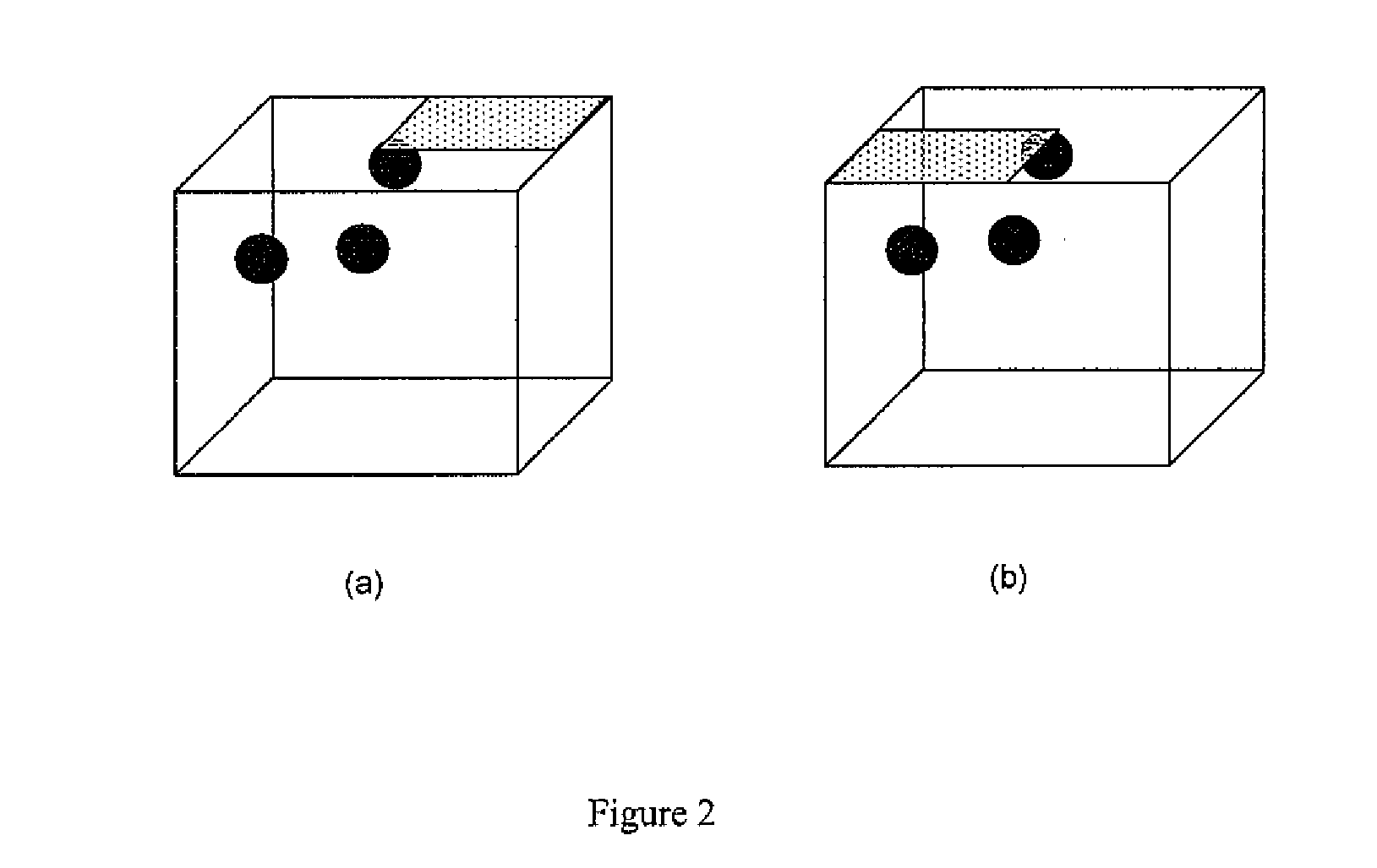
A method and system for non-contact fluorescent optical tomography using patterned illumination is disclosed. The method comprises illuminating a surface of a medium with light from at least one excitation light source to project at least two patterns. Each pattern comprises at least one motif, wherein the medium comprises at least one fluorescent target. The method further comprises for each pattern, measuring excitation light reflected from the medium to generate an excitation data set. In addition, the method comprises, for each pattern, measuring fluorescence emitted from the at least one fluorescent target to generate a fluorescence data set. The method also comprises generating a 3D image of the at least one fluorescent target in the medium by applying iterative algorithm. The iterative algorithm minimizes the difference between a predicted data set based on a mathematical model, and each excitation data set and each fluorescence data set.

Description

CROSS-REFERENCE TO RELATED APPLICATIONS [0001] This application claims priority to U.S. provisional application Ser. No. 60 / 743,577, filed on Mar. 20, 2006 and incorporated herein by reference in its entirety for all purposes.BACKGROUND [0002] 1. Field of the Invention [0003] This invention relates generally to the field of molecular imaging. More specifically, the invention relates to fluorescence optical tomography using patterned illumination. [0004] 2. Background of the Invention [0005] Molecular imaging is a rapidly advancing research area with the potential of providing early diagnosis and identification of the human diseases. Optical fluorescence tomography is a novel molecular imaging modality that attempts to recover the spatial distribution of light emitting fluorophores inside a highly scattering medium, such as biological tissue, from measurements made on the surface of the medium. This technique offers many advantages including non-invasiveness and the ability to constr...

Claims


Application Information

Patent Timeline
Patent Type & Authority Applications(United States)
IPC IPC(8): G06K9/00
CPCA61B5/0073A61B5/415G01N21/6456G01N21/4795A61B5/418
Inventor JOSHI, AMITSEVICK-MURACA, EVA
Owner BAYLOR COLLEGE OF MEDICINE
Learn More