
Fluorescent detector systems for the detection of chemical perturbations in sterile storage devices

a detector system and detector technology, applied in the direction of chemical methods analysis, instruments, analysis using chemical indicators, etc., can solve the problems of limiting the sensitivity and specificity of this method, difficult to store pcs with high platelet counts, and easy use of indicators

Active Publication Date: 2007-11-01
BLOOD CELL STORAGE
View PDF86 Cites 37 Cited by
  • Summary
  • Abstract
  • Description
  • Claims
  • Application Information

AI Technical Summary

Benefits of technology

[0019] In one embodiment, the method further includes writing parameter data to the vessel containing the sample, the vessel having a means for receiving the parameter data that further allows the data to be read from the vessel at a later time.
[0029] In one embodiment, the system further includes a means for writing parameter data to the vessel containing the sample, the vessel having a means for receiving the parameter data that further allows the data to be read from the vessel at a later time.
[0045] In one embodiment, the system further includes a means for writing pH data to the vessel containing the sample, the vessel having a means for receiving the pH data that further allows the data to be read from the vessel at a later time.

Problems solved by technology

The growth of bacteria in platelet concentrates (PCs) can be monitored by utilizing reagent dipsticks for pH and glucose however the time of detection following inoculation with a low dose of organisms (e.g. 50 colony forming units / mL) varies from organism to organism, limiting this method's sensitivity and specificity given the shelf life of the product (Brecher, Hogan et al.
The main disadvantage of this method is the requirement for sampling of a clinical specimen and introduction of the sample into a separate culture vessel at one point in time.
The sampling is disadvantageous because the contaminating organisms may not be included in the sample volume, resulting in a false negative test.
The BTHC plasticized PVC material also has high permeability for CO2, excess of which in some cases leads to difficulties storing PCs which have high platelet counts.
Certain aromatic organic compounds (like phenolphthalein) change color with pH and can be immobilized on solid supports to form “pH paper.” These visual indicators are easy to use, but do not provide a quantitative reading.
The color changes can be difficult to distinguish accurately, and can be masked by colored analyte.

Method used


Image

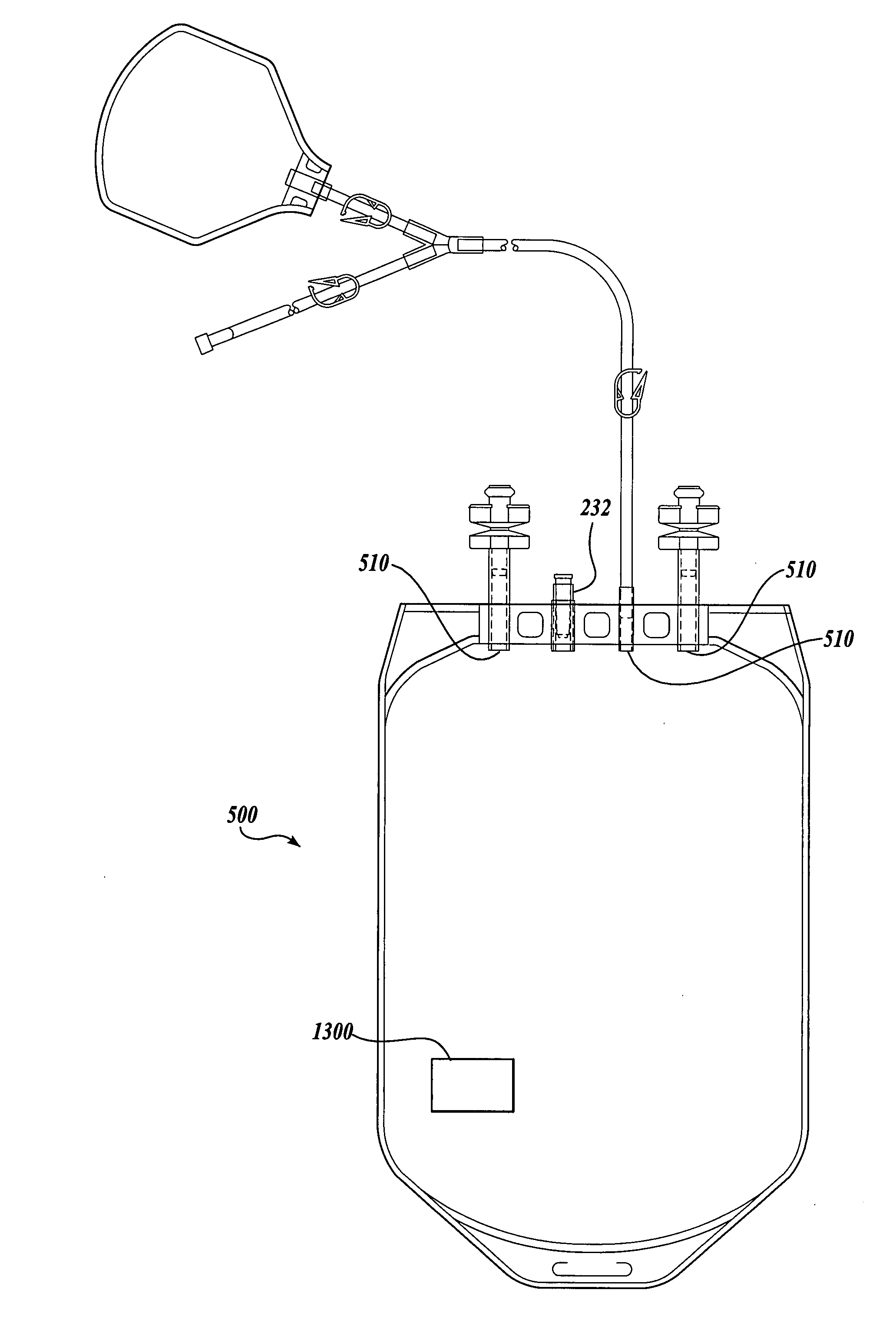
  • Fluorescent detector systems for the detection of chemical perturbations in sterile storage devices
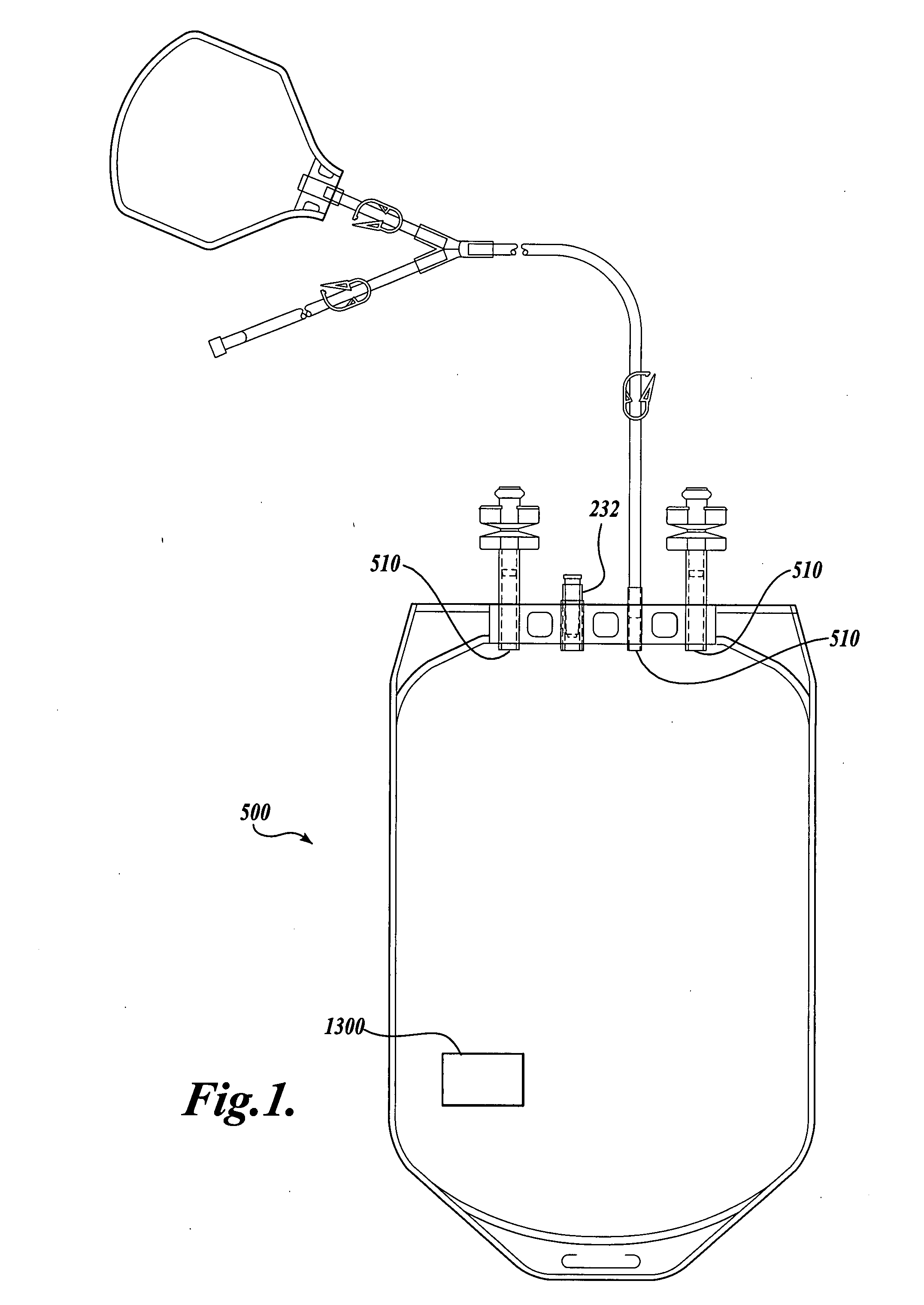
  • Fluorescent detector systems for the detection of chemical perturbations in sterile storage devices
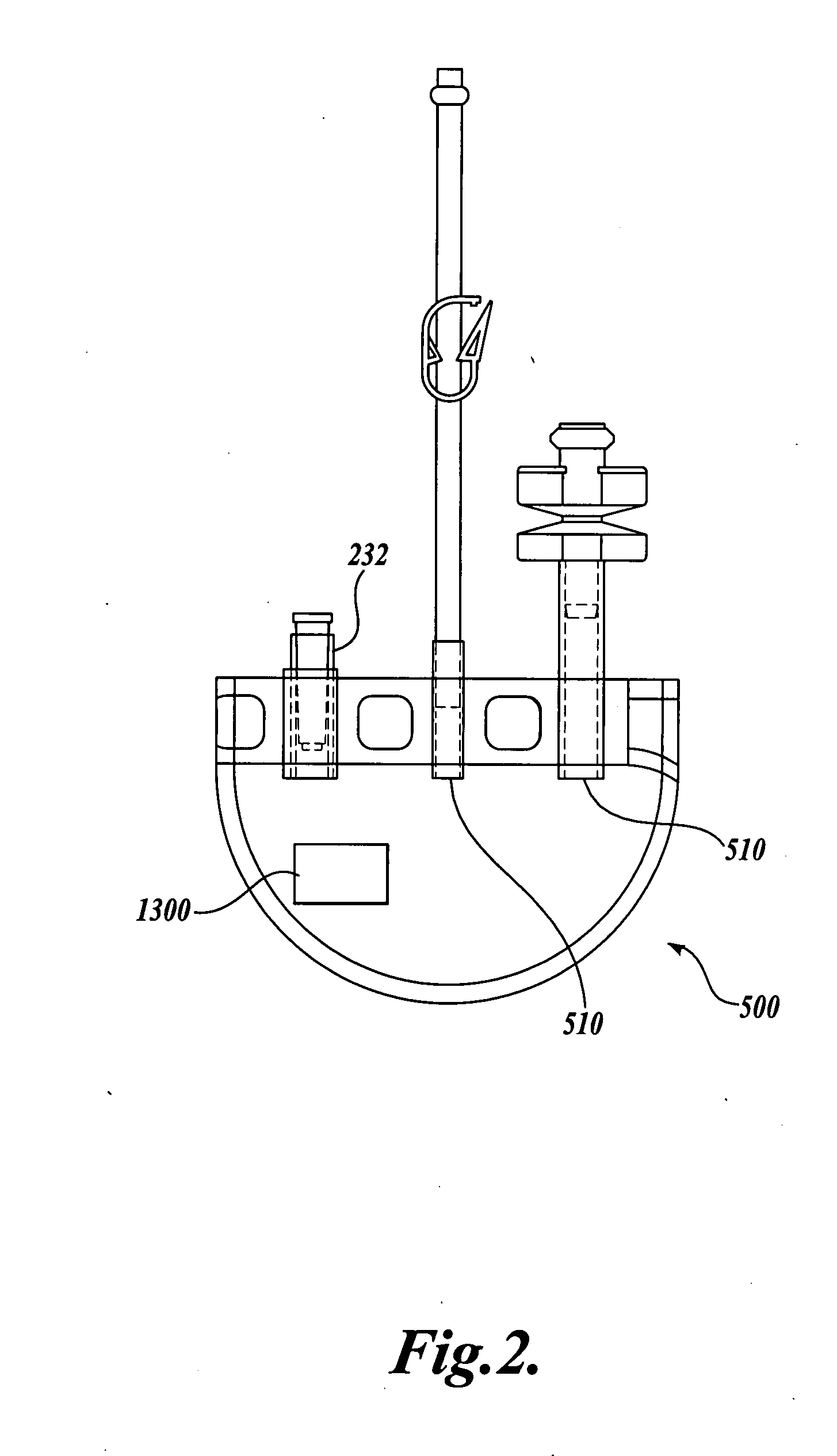
  • Fluorescent detector systems for the detection of chemical perturbations in sterile storage devices

Examples


example 1

The Preparation of Representative Fluorophore-Protein Conjugates

EBIO-3 / HSA

[0258] Method A. A 0.1 M stock solution of EDC (Sigma / Aldrich Chemical Co., St. Louis Mo.) was prepared by dissolving 6.2 mg of EDC in 0.2 mL of DMF and 0.123 mL of 50 mM phosphate buffer (pH 5.8). 1.0 mg of EBIO-3 acid (Nanogen, Bothell, Wash.) was dissolved in 0.102 mL of DMF to give a 20 mM solution. 3.0 mg (0.045 micromoles) of HSA (Sigma / Aldrich Chemical Co., St. Louis, Mo.) was dissolved in 0.3 mL of pH 8.5 sodium bicarbonate in each of two 1.7 mL Eppendorf tubes. 0.1M EDC (0.045 mL) was added to 20 mM EBIO-3 (0.045 mL, 0.9 micromoles) in a separate Eppendorf tube and this was added to one of the HSA tubes to give an EBIO-3:HSA offering ratio of 20:1. An offering ratio of 5:1 was used in the other HSA tube by adding a premixed solution of 0.0225 mL of EDC (0.1 mM) and 0.0225 mL of EBIO-3 (20 mM). The homogeneous dark red HSA conjugate solutions were incubated at room temperature in the dark. After 21 h...

example 2

Immobilization of Representative Fluorophore-Protein Conjugates

EBIO-3 / HSA

[0260] Spotting Immobilization Method. EBIO-3 / HSA conjugate was prepared as described in Example 1 at a ratio of 2:1. Nitrocellulose membranes were obtained from Schleicher and Schuell under the trade name PROTRAN. The discs were treated in the same way as the general immobilization method described in Example 14 using a 4 mg / mL solution of EBIO-3 / HSA.

[0261] Soaking Immobilization Method. EBIO-3 / HSA conjugate was prepared as described in Example 1 at a ratio of 2:1. Mixed ester nitrocellulose and cellulose acetate membranes were obtained from Millipore under the product series TF. The EBIO-3 / HSA conjugate is diluted to 0.2 mg / mL and 45 mL is added to a 9 cm disc of the membrane. The disc is agitated overnight at room temperature and protected from light. The unbound conjugate is removed and the disc is washed with two 1 hour washes and one overnight wash all with agitation. The disc is then desiccated and st...

example 3

The Manufacture of a Vessel Incorporating a Representative Substrate-Immobilized Fluorescent Species

[0262] PVC material is compounded with a number of additives, for example, plasticizers, stabilizers, and lubricants. The formulation is used for making bags and tubes. The compounded PVC is extruded through a die or calendared in a press for converting the plasticized material into sheet form. The extruded sheet, after slitting, is cut into the desired size and sent to the welding section. The donor and transfer tubings are made by extrusion of similar PVC compounds. The tubes are then cut to the appropriate length and sent to the welding section. The components, such as transfusion ports, needle covers, and clamp, are produced by injection molding. The components are ultrasonically cleaned and dried in a drying oven.

[0263] Welding. The blood bags are fabricated by a high frequency welding technique. Sized PVC sheets are placed between electrodes and high frequency at high voltage ...


PUM

PropertyMeasurementUnit
Timeaaaaaaaaaa
Timeaaaaaaaaaa
Timeaaaaaaaaaa

Abstract

System and method for detecting and measuring chemical perturbations in a sample. The system and method are useful for non-invasive pH monitoring of blood or blood products sealed in storage bags.

Description

CROSS-REFERENCE TO RELATED APPLICATIONS [0001] This application claims the benefit of U.S. Patent Application No. 60 / 794,193, filed Apr. 21, 2006; and is a continuation-in-part of U.S. patent application Ser. No. 11 / 207,580, filed Aug. 19, 2005, which claims the benefit of U.S. Provisional Application No. 60 / 602,684, filed Aug. 19, 2004, and U.S. Provisional Application No. 60 / 674,393, filed Apr. 22, 2005, each of which is expressly incorporated herein by reference in its entirety.BACKGROUND OF THE INVENTION [0002] Platelets are a component of blood comprised of anucleate megakaryocyte fragments that circulate in the blood for about 10 days (van der Meer, Pietersz et al. 2001; Dijkstra-Tiekstra 2004). As they age in the circulatory system platelets are known to undergo biochemical changes that eventually leads to their clearance in the spleen and liver. When separated as a component of whole blood, platelets are routinely concentrated, resuspended in plasma and / or platelet additive ...

Claims


Application Information

Patent Timeline
IPC IPC(8): G01N33/00
CPCG01N21/6428G01N21/77G01N21/80G01N33/492G01N33/542G01N33/582C12Q1/54G01N2021/6421G01N2021/6484G01N2021/7786G01N2201/084C12Q1/04C12Q1/22G01N33/84G01N33/66G01N2021/6434G01N2201/062G01N2201/068G01N2201/0826G01N2400/00
Inventor REED, MICHAEL W.GEELHOOD, STEVEN J.HARRIS, PAUL C.BARKER, LYNN M.HARGRAVE, PERRYNANASSY, OLIVER
Owner BLOOD CELL STORAGE
Learn More