Methods for treating bone tumors
- Summary
- Abstract
- Description
- Claims
- Application Information
AI Technical Summary
Benefits of technology
Problems solved by technology
Method used
Image
Examples
example 1
Cell Culture and Cell Morphology Study
[0132] Three human tumor cells lines were used as models: osteosarcoma cell lines SaOS-2 and MG-63 and lung carcinoma cell line A549. Cell lines were obtained from American Tissue Culture Collection (ATCC) and were grown and sustained in the appropriate media using standard techniques. Morphology of the cultured cells was monitored with an Olympus CK2 inverted microscope equipped with a CCD camera. Images were captured using phase contrast with 100× magnification.
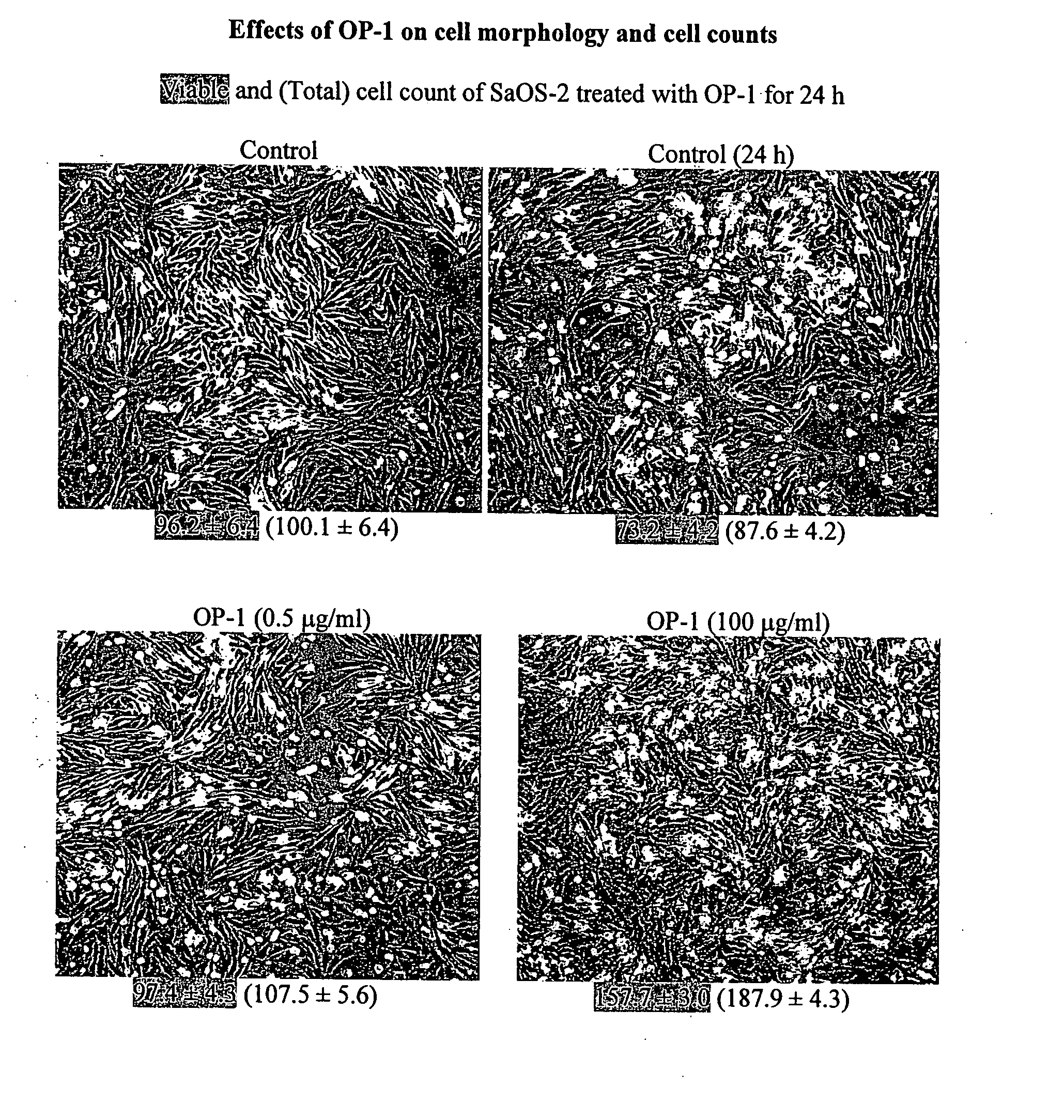
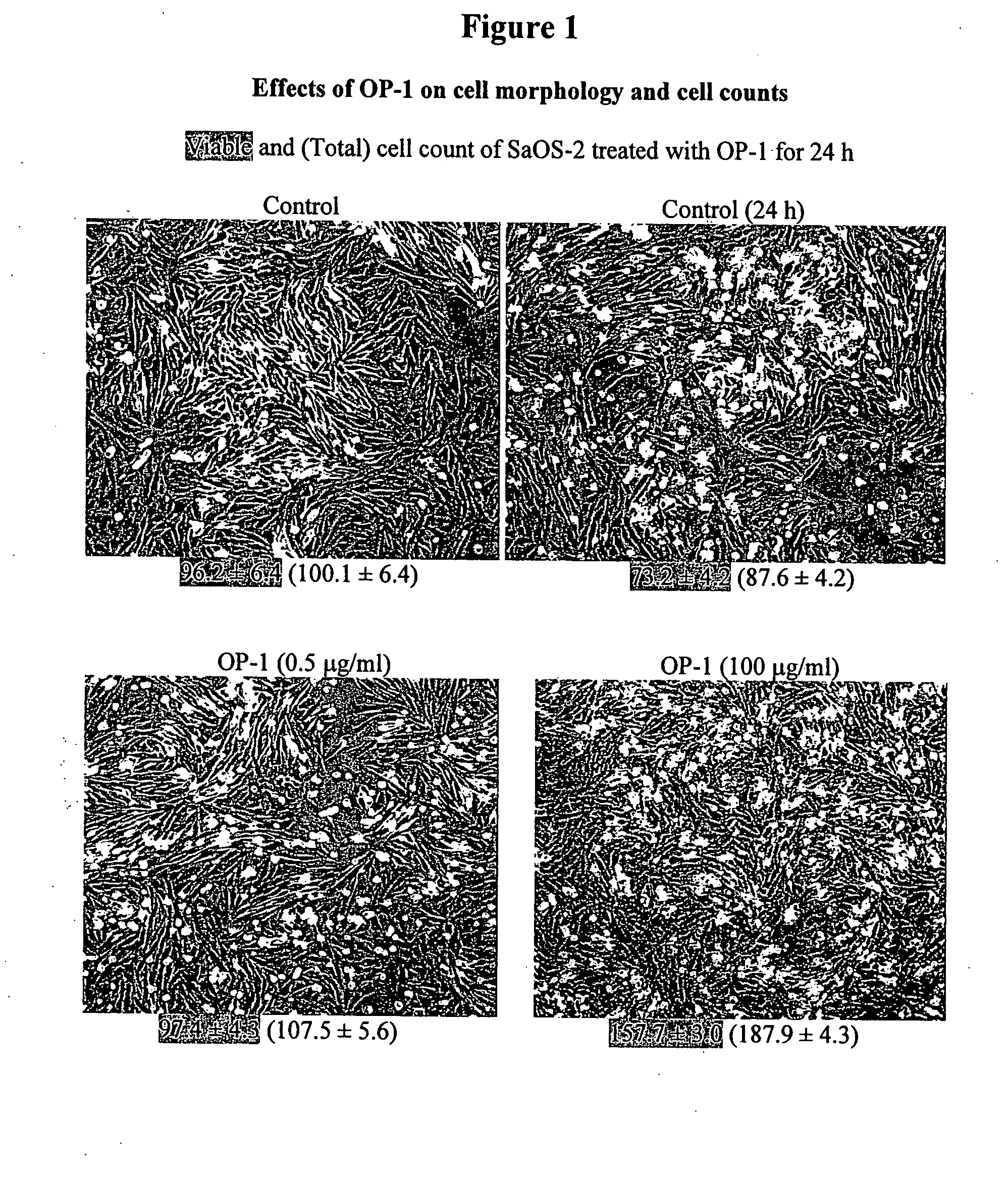
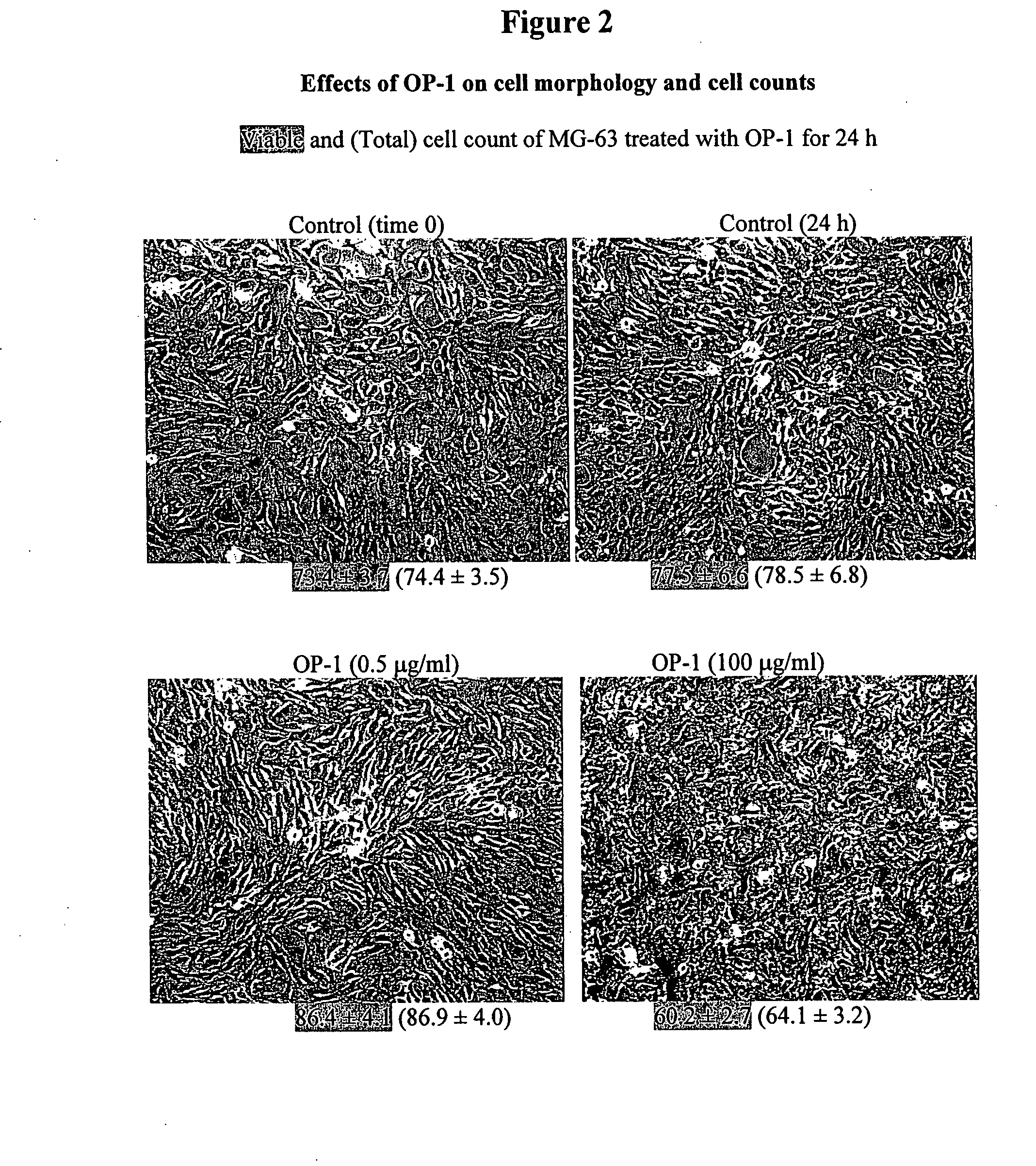
[0133] To examine the effects of OP-1 on cell morphology and cell counts, cells were treated with different concentrations of OP-1 (0.5 μg / ml and 100 μg / ml) for 24 h. The viable and total cell counts following OP-1 treatment were measured using the trypan blue exclusion assay. Treatment of SaOS-2 cells with OP-1 increased both viable and total cell counts in a dose-dependent manner as compared with untreated control cells (see FIG. 1). OP-1 treatment of MG-63 cells did not increase vi...
example 2
[0134] Total RNA was isolated using TRI reagent (Molecular Research Center, Inc., Cincinnati, Ohio) following the manufacturer's recommendation. The cDNA probes for ActR-I, BMPR-IA, BMPR-IB, and BMPR-II were obtained by digestion of the corresponding plasmids with the appropriate restriction endonucleases as reported previously (Yeh et al., J Cell Physiol 185:87-97 (2000). Specifically, the 580-bp ActR-I insert was obtained by digestion of the parent plasmid containing the ActR-I insert with EcoRI / Aval. The 530-bp BMPR-IA insert was obtained by digestion with HindIII / PvuII. The 660-bp BMPR-IB insert was obtained by digestion with HpaI / SacI. The 800-bp BMPR-II insert was obtained by PstI digestion of hBMPR-II cloned in pCMV5. The resultant cDNA fragments were purified by agarose gel electrophoresis and were labeled with [α-32P]dATP using the Strip-EZ DNA labeling system (Ambion Co, Austin, Tex.). The labeled cDNA probes were purified through a Midi-SELECT...
example 3
Effects of OP-1 on Cell Proliferation In Vitro
[0137] To examine the effects of OP-1 on cell proliferation, cells were subcultured at a cell density of 2×10⁴ / ml in a 48-well plate and grown in the appropriate medium with serum until mid-log phase. The specific day at which the culture reached mid-log (the doubling time) varied according to the individual cell line. Cells were then treated with various concentrations of OP-1 (0, 0.1, 0.5, 1.0, 5.0, 10, 50, and 100 μg / ml; Stryker Biotech, Hopkinton, Mass.) in serum free-medium containing 0.1% BSA for 18 h. Cells were pulsed with [3H] thymidine for 6 h after treatment (with the exception of the SaOS-2 cell line in which cell proliferation was evaluated by a tetrazolium calorimetric assay; see discussion below). The extent of incorporation of [3H]thymidine (5 μCi / ml) into DNA and the number of cells were determined as previously described (Yeh et al., Endocrinology 138:4181-4190 (1997)). After removal of the medium containing the uninco...
PUM
 Abstract
Description
Claims
Application Information
 - R&D
- Intellectual Property
- Life Sciences
- Materials
- Tech Scout
- Unparalleled Data Quality
- Higher Quality Content
- 60% Fewer Hallucinations
Browse by: Latest US Patents, China's latest patents, Technical Efficacy Thesaurus, Application Domain, Technology Topic, Popular Technical Reports.
© 2025 PatSnap. All rights reserved.Legal|Privacy policy|Modern Slavery Act Transparency Statement|Sitemap|About US| Contact US: help@patsnap.com


