
Simultaneous quantification of nucleic acids in diseased cells

a nucleic acid and nucleic acid technology, applied in the field of viral infection detection and analysis, can solve the problems of trivial sample preparation, laborious sample preparation, and reduced pcr efficiency, so as to assess potential side effects and eliminate variability

Inactive Publication Date: 2007-08-23
GILEAD PHARMASSET LLC
View PDF10 Cites 15 Cited by
  • Summary
  • Abstract
  • Description
  • Claims
  • Application Information

AI Technical Summary

Benefits of technology

"The patent text describes a method for simultaneously quantifying the nucleic acid of a host cell and a virus in a sample using real-time PCR. This technique is reliable and accurate, and can be used to evaluate the effectiveness of anti-viral agents and toxicity of compounds. The method also allows for the assessment of the specificity of a compound's inhibitory effect on the host and viral nucleic acids. The host and viral nucleic acid levels can be compared to a standard or an internal standard to monitor changes in infection. The method is economic, non-radioactive, rapid, and amenable to large through-put."

Problems solved by technology

PCR itself is quite simple, but sample preparation can be laborious.
Inhibitors of PCR are naturally occurring substances which reduce the efficiency of PCR, and which are often present in clinical samples.
When the specimen contains a large amount of target nucleic acid, sample preparation is trivial.
But sample preparation is more difficult in most clinical specimens, particularly when a large volume specimen must be processed and only a few pathogens are present.
Complex protocols are often required.
PCR can be used to screen for drug resistance mutations, but it does not provide direct antibiotic susceptibility data.
Most published studies have not included a sufficient number of negative controls.
Detection is the most difficult PCR procedure, especially when the number of pathogens in the specimen is low.
This makes PCR for detection especially prone to carryover contamination.
Sample preparation may be laborious, as there is an attempt to process as large a specimen volume as possible.
Inhibitors of PCR occur naturally in many clinical samples, and are a major limitation.
PCR is quite sensitive, but it is not inherently quantitative.
Due to the ongoing replication of the virus, anti-retroviral drug resistance eventually develops, leading to therapy failure.
The design of an HIV-1 viral load test is a real challenge.
Most toxic events are reversible at an early stage, however lactic acidosis is often irreversible and can result in death.
Tissues with high ATP demand are most susceptible to mitochondrial toxicity.
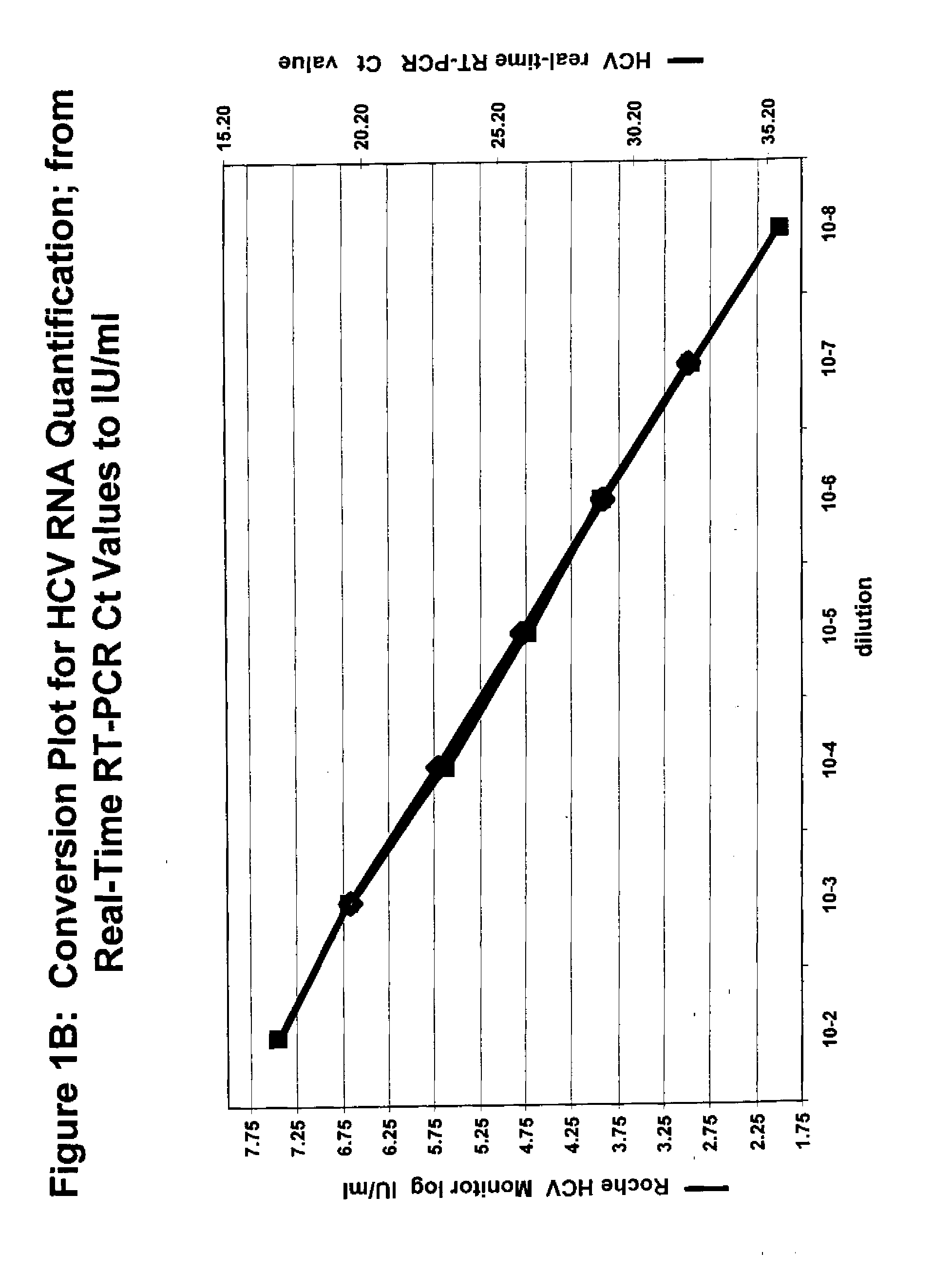
Available tests for HCV infection are limited.
Initial serologic tests for HCV had poor sensitivity.
Second and third-generation serologic tests have improved sensitivity, but are still not completely dependable.

Method used


Image

  • Simultaneous quantification of nucleic acids in diseased cells
  • Simultaneous quantification of nucleic acids in diseased cells
  • Simultaneous quantification of nucleic acids in diseased cells

Examples


example 1

HIV-1 Cell culture

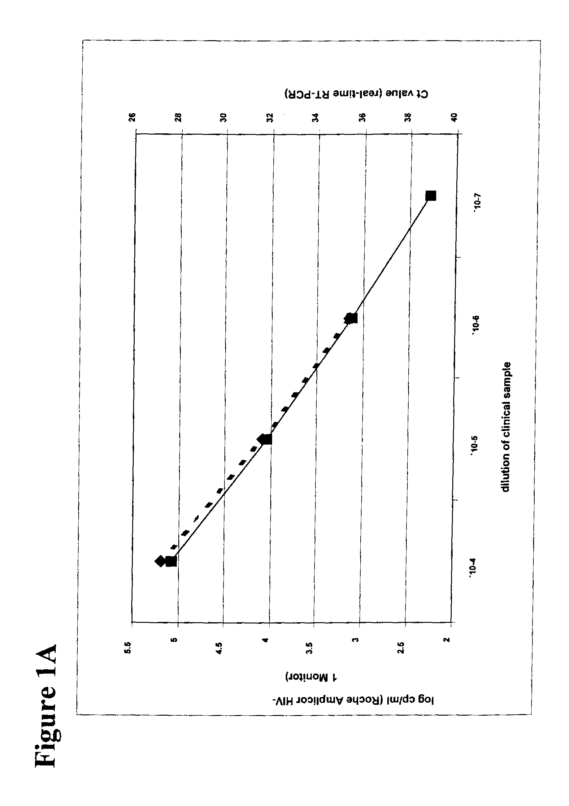
[0234] Human PBMC (1×10⁶ cells / T25 flask) were PHA stimulated for 2 days, and infected with either a sensitive (xxBRU) or a 3TC-resistant (184V) HIV-1 strain at 100 TCID50. The culture was kept for 5 days in presence of test antiviral compounds at serial 1-log dilutions. Subsequently, human PBMC were removed from the culture supernatant by centrifugation (10 min, 400×g, 4° C.). This clarified supernatant was tested either in the RT-assay, or in the real-time RT-PCR assay.

example 2

Reverse Transcriptase (RT) Assay

[0235] Virus particles present in a 1 mL aliquot of culture supernatant were concentrated by centrifugation (2 hr, 20,000×g, 4° C.). After the 2 hour spin, supernatant fluid was removed completely and the virus pellet was dispensed into a 100 μL Virus Solubilization Buffer (VSB: 0.5% Triton X-100; 0.8 M NaCl, 0.5 mM phenylmethylsulfonyl, 20% glycerol, 50 mM Tris.HCl pH 7.8). A 10 μL aliquot of RT-VSB was mixed with 75 μL RT cocktail (60 mM Tris.HCl pH 7.8, 12 mM MgCl2, 6 mM DTT, 6 μg / mL Poly (rA)-Poly (dT), 1.2 mM dATP, and 80 μCi / mL H3-TTP) and incubated for 2 hr at 37° C. Subsequently 100 μL of 10% TCA was added, and the total amount of incorporated H3-TTP was counted.

example 3

RT-PCR Primer and Probe Assessment

[0236] The TaqMan probe and primers were designed by using the Primer Express software (Applied Biosystems, CA) and are covering highly conserved sequences complementary to the DNA sequences present in HIV-1 RNA. By scanning the different genotypes of group M for regions containing only minor variability, the conserved domain was discovered. As a result, the region in the HIV-1 RT domain between codon 200 and 280 fulfilled the required criteria; thus this region was used to design an appropriate set of primers and probe that could work in real time PCR (“RT-PCR”). Primer sequences are as follows: sense 5′-TGGGTTATGAACTCCATCCTGAT-3′ (Sequence ID No.) and 5,-TGTCATTGACAGTCCAGCTGTCT-3′ (Sequence ID No.); the probe sequence is 5′-fluoresent dye-TTTCTGGCAGCACTATAGGCTGTACTGTCCATT-quenching dye-3′ (Sequence ID No.). In this particular case, the probe was labeled with FAM at the 5′ end, and the quencher molecule is TAMARA, provided at the 3′ end. Any othe...


PUM

PropertyMeasurementUnit
temperaturesaaaaaaaaaa
Tmaaaaaaaaaa

Abstract

Processes and methods for the simultaneous quantification of nucleic acids in diseased cells that are based on real-time PCR are provided. The real-time PCR protocol is an excellent tool for reliable quantification of in vitro drug screening and evaluation protocols to determine the efficacy of potential anti-viral agents. Quantification using these simultaneous PCR cycle threshold (Ct) detection techniques during one-step real-time RT-PCR (Applied Biosystems, CA) eliminates the variability resulting from quantification of end-point RT-PCR products. In addition, the mitochondrial toxicity assay is an added tool to assess potential side-effects for these chemotherapeutic agents.

Description

[0001] This application claims priority to U.S. Provisinal Application No. 60 / 241,488, filed on Oct. 18, 2000, U.S. Provisinal Application No. 60 / 256,067 filed on Dec. 15, 2000 and U.S. Provisinal Application No. 60 / 282,156, filed on Apr. 6, 2001.FIELD OF THE INVENTION [0002] This application is in the area of processes for the detection and analysis of viral infections and mitochondrial toxicity, and for processes for the identification of active compounds for the treatment of viral infections and processes to measure mitochondrial toxicity resulting from drug therapies. BACKGROUND OF THE INVENTION [0003] The detection and quantification of nucleic acid sequences is of importance for a wide range of applications. The most widely used method to detect nucleic acids are based on the polymerase chain reaction (PCR). PCR is used to amplify a segment of DNA flanked by stretches of known sequences. Two oligonucleotides binding to these known flanking sequences are used as primers for a s...

Claims


Application Information

Patent Timeline
Patent Type & Authority Applications(United States)
IPC IPC(8): C12Q1/70C12Q1/68C12N15/09C07H19/048C07H19/06C07H19/10C07H19/16C07H19/20C07H21/04
CPCC07H19/048C07H19/06C12Q1/701C12Q1/6895C12Q1/689C07H19/10C07H19/16C07H19/20C07H21/04C12Q1/6809C12Q1/6876C12Q2561/101C12Q2545/101C12Q2537/143C12Q2600/136C12Q2600/142C12Q2600/158Y10S435/81
Inventor STUYVER, LIEVENOTTO, MICHAEL J.
Owner GILEAD PHARMASSET LLC
Learn More