Estrogen receptor-related receptor alpha (ERRalpha) and cartilage formation
- Summary
- Abstract
- Description
- Claims
- Application Information
AI Technical Summary
Benefits of technology
Problems solved by technology
Method used
Image
Examples
example 1
Expression of ERRα in Chondrocyte Lineage Cells Throughout Development
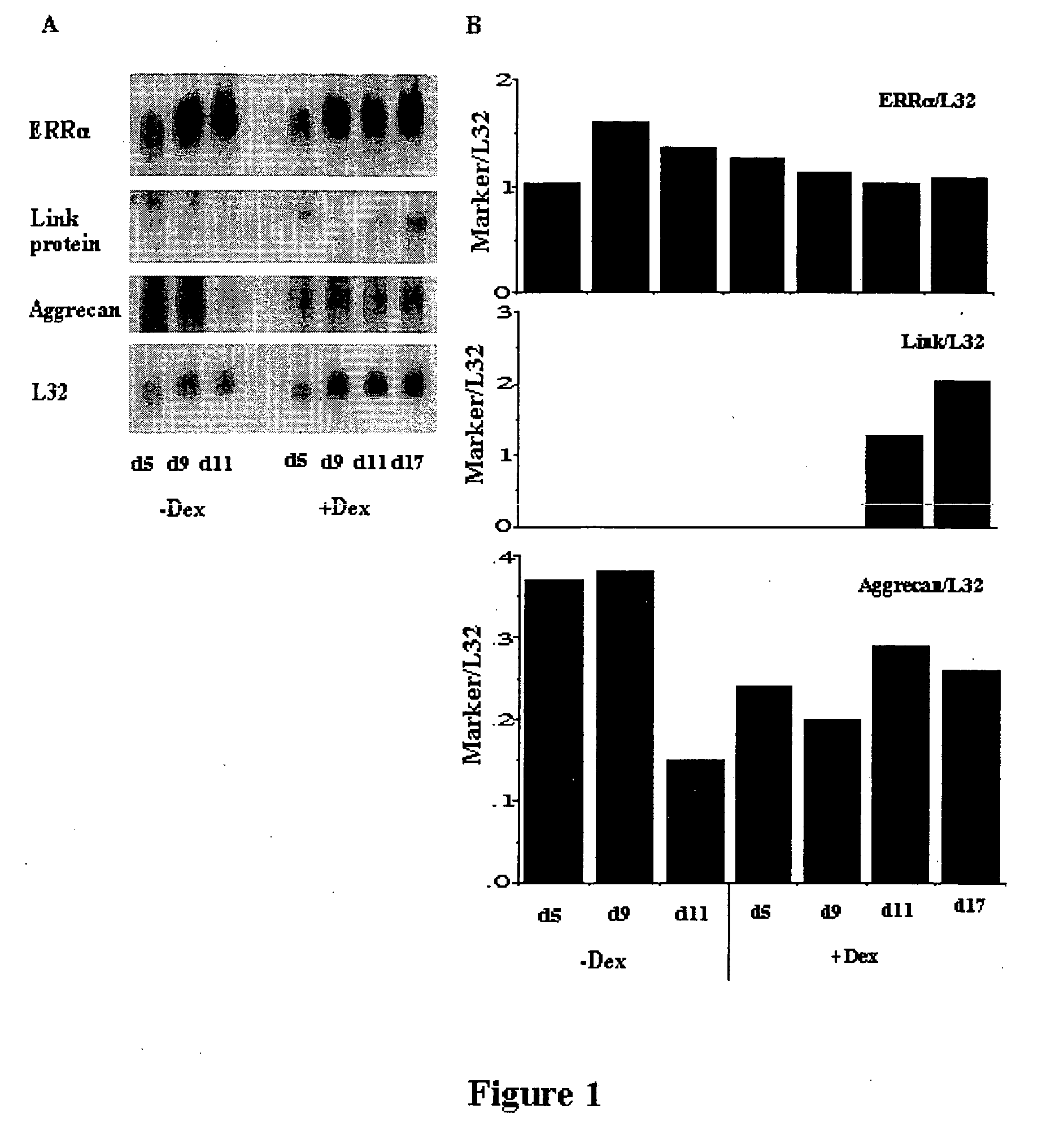
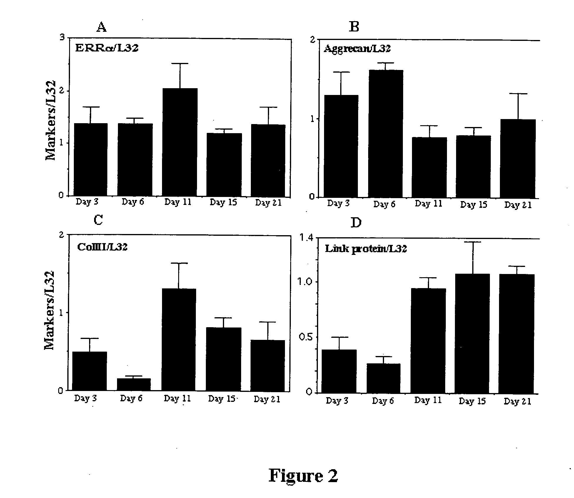
[0133] In order to assess ERRα expression, RCJ.1C5.18 (C5.18) cells were grown as described by Grigoriadis, 1996. This cell line is a fetal rat cell line which undergoes differentiation into cartilage-producing chondrocytes; it is widely used as a model system for the study of chondrogenesis and the regulation of chondrocyte activity. Cells were maintained in α-MEM containing 15% heat-inactivated FBS (Flow Laboratories, McLean, Va.), antibiotics comprising 100 μg / ml penicillin G (Sigma Chemical Co., St. Louis, Mo.), 50 μg / ml gentamycin (Sigma), and 0.3μg / ml fungizone (Flow Laboratories) and 10−8M dexamethasone (Merck, Sharp, and Dohme, Canada, Ltd., Kirkland, PQ). Dexamethasone (Dex) stimulates chondrogenesis and cartilage formation in these cultures. For differentiation studies, cells were grown in the same medium, with or without dexamethasone, and with the addition of 50 μg / ml ascorbic acid and 10 mM sodium β-...
example 2
In Vivo Expression of ERRα
[0139] To determine the in vivo expression of ERRα protein, immuno-cytochemistry was performed on sections of 21 day fetal rat tibiae and metatarsals and on sections of adult rat tibiae and femurs. The sections were rinsed in PBS and incubated for 1 h at room temperature with secondary antibody CY-3-conjugated anti-rabbit (Jackson immunoresearch Lab, West Grove, Pa., USA; 1 / 300 final dilution) for ERRα . After rinsing, samples were mounted in Moviol (Hoechst Ltd, Montreal, PQ, Canada) and observed by epifluorescence microscopy on a Zeiss Photomicroscope III (Zeiss, Oberkochen, Germany).
[0140] ERRα protein was already highly expressed in the chondrocytes of the growth plates of term-pregnant rat fetuses and continued to be expressed in the cartilage of adult animals. In fetal growth plate cartilage, intense label for ERRα was seen in perichondrial precursors and proliferating chondrocytes, while staining in hypertrophic chondrocytes was low or absent. In ad...
example 3
Antisense and Sense Oligonucleotide Treatment
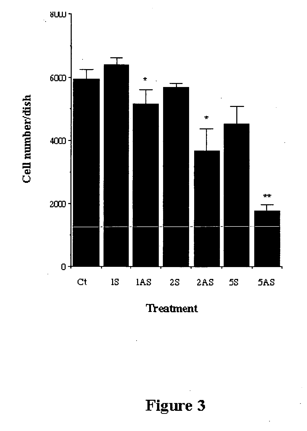
[0141] Antisense oligonucleotides form DNA:RNA duplexes with specific mRNA species, thereby blocking binding of the mRNA to the 40S ribosomal subunit and preventing translation [Jen, 2000]. To examine the involvement of ERRα in chondrocyte differentiation and cartilage formation, C5.18 cells were treated either during the proliferation phase or during the differentiation and cartilage nodule formation phase. Preliminary experiments were done to determine effective oligonucleotide concentrations that were not toxic (not shown) and the efficacy of the antisense was confirmed by immunocytochemistry and Western blots.
[0142] C5.18 cells were plated in 24 wells plates at 104 cells / well and treated with antisense or sense oligonucleotides. Antisense oligonucleotide inhibition of ERRα expression was accomplished with a 20-base phosphorothioate-modified oligonucleotide, localized to the A / B domain. The ERRα antisense oligonucleotide sequence was...
PUM
 Abstract
Description
Claims
Application Information
 - R&D
- Intellectual Property
- Life Sciences
- Materials
- Tech Scout
- Unparalleled Data Quality
- Higher Quality Content
- 60% Fewer Hallucinations
Browse by: Latest US Patents, China's latest patents, Technical Efficacy Thesaurus, Application Domain, Technology Topic, Popular Technical Reports.
© 2025 PatSnap. All rights reserved.Legal|Privacy policy|Modern Slavery Act Transparency Statement|Sitemap|About US| Contact US: help@patsnap.com


