
Medical device with coating that promotes endothelial cell adherence and differentiation

a technology of endothelial cells and medical devices, applied in the field of medical devices, can solve the problems of permanent opening of the affected coronary artery, affecting the actual incidence of the disease in the population, and ischemic damage to the tissues supplied by the artery, so as to improve the prognosis of patients, prevent restnosis, and inhibit intimal hyperplasia

Inactive Publication Date: 2007-02-22
ORBUSNEICH MEDICAL PTE LTD
View PDF99 Cites 54 Cited by
  • Summary
  • Abstract
  • Description
  • Claims
  • Application Information

AI Technical Summary

Benefits of technology

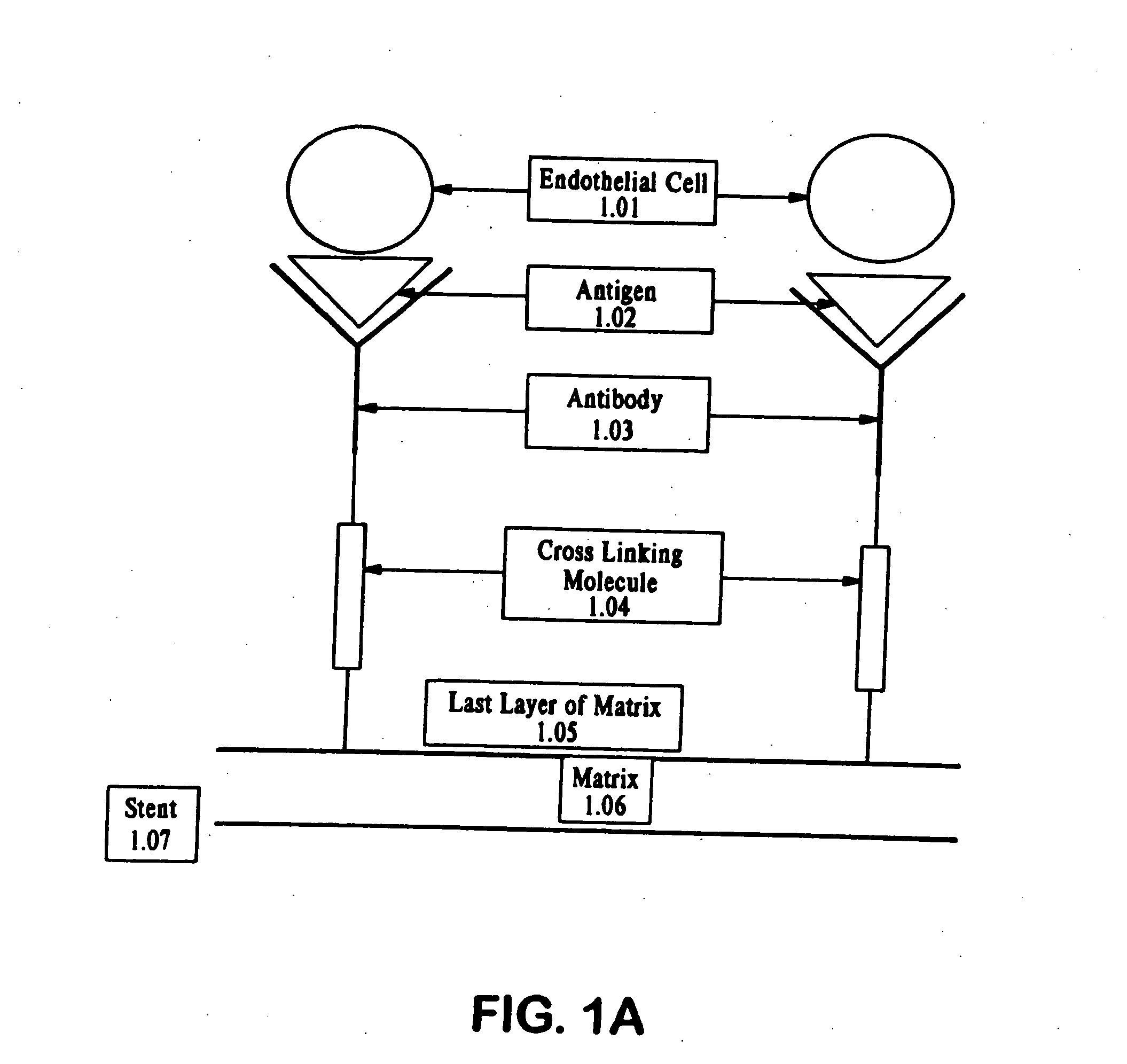
[0019] It is an object of the invention to provide coated medical devices such as stents, stent grafts, heart valves, catheters, vascular prosthetic filters, artificial heart, external and internal left ventricular assist devices (LVADs), and synthetic vascular grafts, for the treatment of vascular diseases, including restenosis, artherosclerosis, thrombosis, blood vessel obstruction, and the like. In one embodiment, the coating on the present medical device comprises a biocompatible matrix, at least one type of antibody or antibody fragment, or a combination of antibody and fragments, and at least a compound such as a growth factor, for modulating adherence, growth and differentiation of captured progenitor endothelial cells on the surface of the medical device to induce the formation of a functional endothelium to inhibit intimal hyperplasia in preventing restenosis, thereby improving the prognosis of patients being treated with vascular disease.
[0020] In one embodiment, the biocompatible matrix comprises an outer surface for attaching a therapeutically effective amount of at least one type of antibody, antibody fragment, or a combination of the antibody and the antibody fragment. The present antibody or antibody fragment recognizes and binds an antigen on a the cell membrane or surface of progenitor endothelial cells so that the cell is immobilized on the surface of the matrix. Additionally, the coating comprises a therapeutically effective amount of at least one compound for stimulating the immobilized progenitor endothelial cells to accelerate the formation of a mature, functional endothelium on the surface of the medical device.
[0029] The invention also provides an engineered method for inducing a healing response. In one embodiment, a method is provided for rapidly inducing the formation of a confluent layer of endothelium in the luminal surface of an implanted device in a target lesion of an implanted vessel, in which the endothelial cells express nitric oxide synthetase and other anti-inflammatory and inflammation-modulating factors. The invention also provides a medical device which has increased biocompatibility over prior art devices, and decreases or inhibits tissue-based excessive intimal hyperplasia and restenosis by decreasing or inhibiting smooth muscle cell migration, smooth muscle cell differentiation, and collagen deposition along the inner luminal surface at the site of implantation of the medical device.

Problems solved by technology

Ultimately, this deposition blocks blood flow distal to the lesion causing ischemic damage to the tissues supplied by the artery.
The therapy, however, does not usually result in a permanent opening of the affected coronary artery.
Despite their success, stents have not eliminated restenosis entirely.
However, this measure may vastly underestimate the actual incidence of the disease in the population.
However, the post-operative results obtained with medical devices such as stents do not match the results obtained using standard operative revascularization procedures, i.e., those using a venous or prosthetic bypass material.
Excessive intimal hyperplasia and thrombosis, however, remain significant problems even with the use of bypass grafts.
Irradiation of the treated vessel can cause severe edge restenosis problems for the patient.
In addition, irradiation does not permit uniform treatment of the affected vessel.
Although heparin and phosphorylcholine appear to markedly reduce thrombosis in animal models in the short term, treatment with these agents appears to have no long-term effect on preventing restenosis.
Additionally, heparin can induce thrombocytopenia, leading to severe thromboembolic complications such as stroke.
Therefore, it is not feasible to load stents with sufficient therapeutically effective quantities of either heparin or phosphorylcholine to make treatment of restenosis in this manner practical.
However, none of these approaches has significantly reduced the incidence of thrombosis or restenosis over an extended period of time.
This technique is not desirable since it has demonstrated that the efficiency of a single dose delivery is low and produces inconsistent results.
Therefore, this procedure cannot be reproduced accurately every time.
Synthetic grafts have also been seeded with endothelial cells, but the clinical results with endothelial seeding have been generally poor, i.e., low post-operative patency rates (Lio et al.

Method used


Image

  • Medical device with coating that promotes endothelial cell adherence and differentiation
  • Medical device with coating that promotes endothelial cell adherence and differentiation
  • Medical device with coating that promotes endothelial cell adherence and differentiation

Examples


experimental examples

[0105] This invention is illustrated in the experimental details section which follows. These sections set forth below the understanding of the invention, but are not intended to, and should not be construed to, limit in any way the invention as set forth in the claims which follow thereafter.

example 1

Endothelial Progenitor Cell Phenotyping

[0106] Endothelial Progenitor Cells (EPC) were isolated either by CD34+Magnetic Bead Isolation (Dynal Biotech) or enriched medium isolation as described recently (Asahara T, Murohara T, Sullivan A, et al. Isolation of putative progenitor endothelial cells for angiogenesis. Science 1997;275:964-7). Briefly, peripheral venous blood was taken from healthy male volunteers and the mononuclear cell fraction was isolated by density gradient centrifugation, and the cells were plated on human fibronectin coated culture slides (Becton Dickinson) in EC basal medium-2 (EBM-2) (Clonetics) supplemented with 5% fetal bovine serum, human VEGF-A, human fibroblast growth factor-2, human epidermal growth factor, insulin-like growth factor-1, and ascorbic acid. EPCs were grown up to seven days with culture media changes every 48 hours. The results of these experiments are shown in FIGS. 2A and 2B. FIGS. 2A and 2B show that the anti-CD34 isolated cell appear more...

example 2

[0109] Endothelial Cell Capture by anti-CD34 coated Stainless Steel Disks: Human Umbilical Vein Endothelial Cells (HUVEC) (American Type Culture Collection) are grown in endothelial cell growth medium for the duration of the experiments. Cells are incubated with CMDX and gelatin coated samples with or without bound antibody on their surface or bare stainless steel (SST) samples. After incubation, the growth medium is removed and the samples are washed twice in PBS. Cells are fixed in 2% paraformaldehyde (PFA) for 10 minutes and washed three times, 10 minutes each wash, in PBS, to ensure all the fixing agent is removed. Each sample is incubated with blocking solution for 30 minutes at room temperature, to block all non-specific binding. The samples are washed once with PBS and the exposed to 1:100 dilution of VEGFR-2 antibody and incubated overnight. The samples are subsequently washed three times with PBS to ensure all primary antibody has been removed. FITC-conjugated secondary ant...


PUM

PropertyMeasurementUnit
Biocompatibilityaaaaaaaaaa

Abstract

Compositions and methods are provided for producing a medical device such as a stent, a stent graft, a synthetic vascular graft, heart valves, coated with a biocompatible matrix which incorporates antibodies, antibody fragments, or small molecules, which recognize, bind to and / or interact with a progenitor cell surface antigen to immobilize the cells at the surface of the device. The coating on the device can also contain a compound or growth factor for promoting the progenitor endothelial cell to accelerate adherence, growth and differentiation of the bound cells into mature and functional endothelial cells on the surface of the device to prevent intimal hyperplasia. Methods for preparing such medical devices, compositions, and methods for treating a mammal with vascular disease such as restenosis, artherosclerosis or other types of vessel obstructions are disclosed.

Description

[0001] This application is a divisional of U.S. patent application Ser. No. 10 / 360,567, filed Feb. 6, 2003, which is a continuation-in-part of U.S. patent application Ser. No. 09 / 808,867, filed Mar. 15, 2001, which claims benefit of U.S. Provisional Application Ser. Nos. 60 / 189,674, filed on Mar. 15, 2000, Ser. No. 60 / 201,789, filed on May 4, 2000, and claims benefit from U.S. Provisional Patent Application Ser. No. 60 / 354,680, filed on Feb. 6, 2002, the contents of which are incorporated by reference herein in their entirety.FIELD OF INVENTION [0002] The present invention relates to the field of medical devices implanted in vessels or hollowed organs within the body. In particularly, the present invention relates to artificial, intraluminal blood contacting surfaces of medical devices such as coated stents, stent grafts, synthetic vascular grafts, heart valves, catheters and vascular prosthetic filters. The coating on the implanted medical device promotes progenitor endothelial cel...

Claims


Application Information

Patent Timeline
IPC IPC(8): A61F2/06A61L27/08A61L27/30A61L27/44A61L27/54A61L29/10A61L29/12A61L29/16A61L31/08A61L31/12A61L31/16C12N5/08
CPCA61L27/08A61L27/303A61L27/44A61L27/54A61L29/103A61L29/126A61L29/16A61L31/084A61L31/125A61L31/16A61L2300/258A61L2300/45B82Y30/00A61L31/088A61L31/10
Inventor KUTRYK, MICHAEL J.B.COTTONE, ROBERT J. JR.ROWLAND, STEPHEN M.KULISZEWSKI, MICHAEL A.
Owner ORBUSNEICH MEDICAL PTE LTD
Learn More