Microwave imaging assisted ultrasonically

a technology of ultrasonography and microwave radiation, applied in mammography, medical science, diagnostics, etc., can solve the problems of high failure rate of mammography for women in the 25 to 40 year old age group, failure to provide qualitative images of electric properties, i.e. permittivity and conductivity of objects with high contrast, and no design generally encompasses all desired characteristics. , to achieve the effect of early detection of breast cancer

Inactive Publication Date: 2007-01-18
CLEMSON UNIVERSITY
View PDF10 Cites 15 Cited by
  • Summary
  • Abstract
  • Description
  • Claims
  • Application Information

AI Technical Summary

Benefits of technology

[0011] Multiple antennas surround the object under investigation. The antenna design, their position and coupling medium assure a uniform electric field pattern in coronal plane of object under investigation. At a certain instant one of the antennas plays the role of the emitter and the other antennas operate as receivers. The amplitude and phase of electric field are measured for each location and for each configuration of individual emitter and multiple receivers. A second microwave measurement is performed without a target (homogeneous case) and the results of this measurement are used to calibrate the microwave data obtained during the object investigation (nonhomogeneous case). This calibration is needed to minimize several differences existing between the experimental system and the numerical models used, including: 1) the 3D imaging system and the 2D numerical models; 2) differences in transmission efficiency of antennas and connection cables; and 3) differences in the antenna positioning.
[0013] In an exemplary embodiment of the present subject matter, the reconstruction algorithm was implemented using a Newton iterative method with combined Marquardt-Tikhonov regularizations. The electric field at each iteration was computed using a finite element formulation of Bayliss and Turkel radiation boundary conditions. The approach using a hybrid regularizations-based Newton method to update an initial dielectric property distribution iteratively in order to minimize an object function composed of a weighted sum of the squared difference between computed and measured data may provide a more stable inverse solution than previously available.
[0015] In accordance with certain aspects of other embodiments of the present subject matter, methodologies have been developed to make possible effective imaging of the radiologically dense breasts of younger women thereby providing greater opportunity for early detection of breast cancer in younger patients.

Problems solved by technology

Presently available information indicates the following: 1) More than 211,000 American women will be diagnosed with breast cancer this year; 2) Ninety two percent of late-stage breast cancer cases in the United States could have been treated if they had been detected earlier; 3) About 70 percent of women over 40 years of age had a mammogram in the last two years; 4) Mammography, in spite of its unpleasant procedure for women (breast constriction) and its radiation exposure problems, remains the only method available for breast cancer detection; and 5) Mammography shows a high rate of failure for women in the 25 to 40 year old age group who tend to have radiological dense breast.
Recently there has been considerable interest in developing alternative methods for breast cancer detection due to the inability of x-ray mammography to image radiological dense breasts as well as the low positive predictive value in such circumstances.
Microwave images obtained based on diffraction algorithms are able to provide qualitative information about objects under investigation but they fail to provide qualitative images of electric properties, i.e. permittivity and conductivity, of object with high contrast.
This failure results from an initial assumption that does not take into consideration multiple scattering but rather assumes the reconstruction problem to be a linear problem.
While various implementations of microwave image reconstruction have been developed, and while various data refining algorithms have been devised, no design has emerged that generally encompasses all of the desired characteristics as hereafter presented in accordance with the subject technology.

Method used


Image

  • Microwave imaging assisted ultrasonically
  • Microwave imaging assisted ultrasonically
  • Microwave imaging assisted ultrasonically

Examples


Embodiment Construction

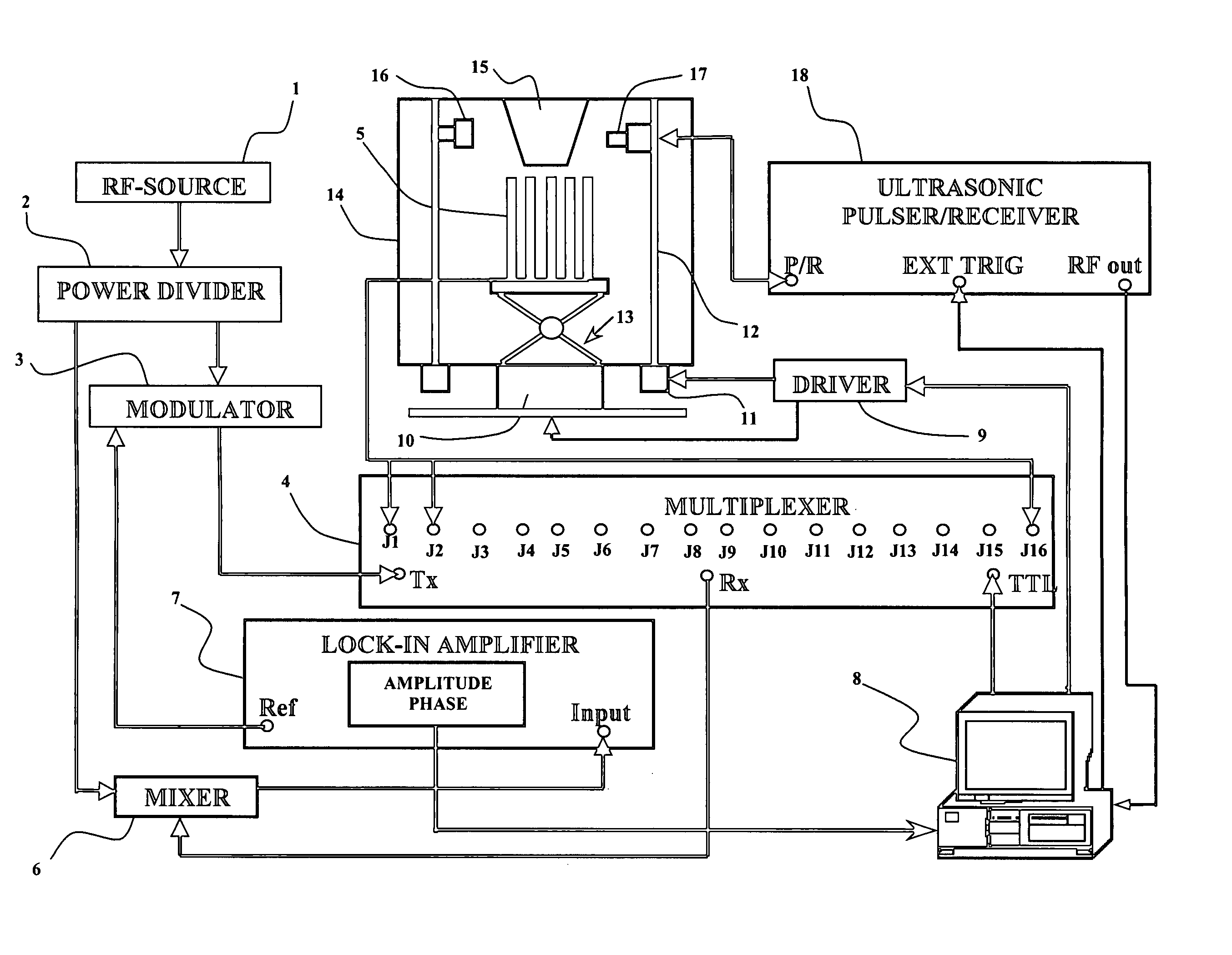
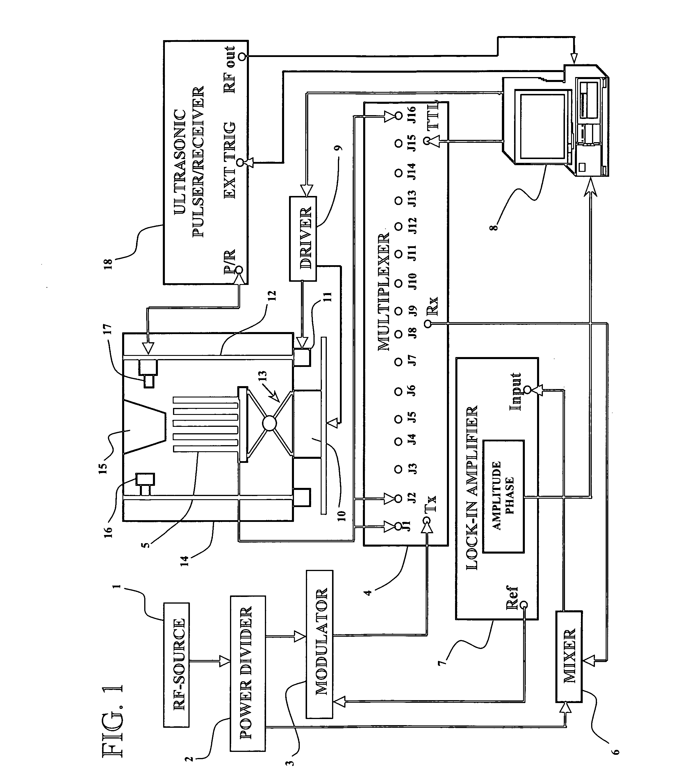
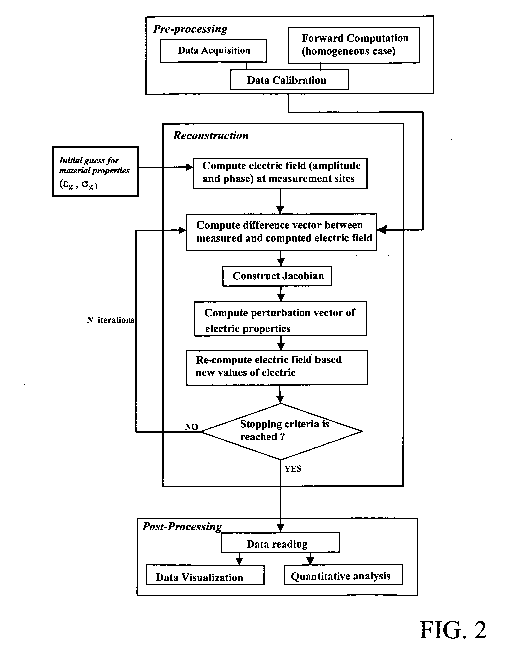
[0027] As discussed in the Summary of the Invention section, the present subject matter is particularly concerned with an improved methodology for microwave image reconstruction of primarily, but not exclusively, breast tissue for medical evaluation of the possible presence of cancer. More particularly, the present subject matter concerns the provision and use of an initial evaluation point or “guess” for a microwave investigation based on the results of an initial ultrasound investigation.

[0028] Selected combinations of aspects of the disclosed technology correspond to a plurality of different embodiments of the present invention. It should be noted that each of the exemplary embodiments presented and discussed herein should not insinuate limitations of the present subject matter. Features or steps illustrated or described as part of one embodiment may be used in combination with aspects of another embodiment to yield yet further embodiments. Additionally, certain features may be ...


PUM


Abstract

Disclosed is an apparatus and methodology for imaging objects, more particularly radiologically dense objects. The currently disclosed technology has particular applicability in the medical field as a tool and methodology for investigation of radiologically dense breast tissue of young patients by using microwave energy in concert with an ultrasonic initial investigation of the tissue. The use of an ultrasonic initial investigation operates as an initial evaluation point for the subsequent microwave investigation.

Description

FIELD OF THE INVENTION [0001] The present subject matter relates to imaging using microwave radiation. More particularly, the present subject matter relates to the production of a representation of electric permittivity and conductivity in a test object, the production of such representation being assisted through the use of an ultrasonic initialization technique. The present subject matter has applicability in the medical arts as an adjunct to, or replacement for, x-ray mammography. BACKGROUND OF THE INVENTION [0002] Presently available information indicates the following: 1) More than 211,000 American women will be diagnosed with breast cancer this year; 2) Ninety two percent of late-stage breast cancer cases in the United States could have been treated if they had been detected earlier; 3) About 70 percent of women over 40 years of age had a mammogram in the last two years; 4) Mammography, in spite of its unpleasant procedure for women (breast constriction) and its radiation expo...

Claims


Application Information

Patent Timeline
IPC IPC(8): A61B5/05
CPCA61B5/05A61B5/0507A61B8/0825A61B5/0035A61B8/4416A61B5/0037A61B8/14
Inventor CIOCAN, RAZVANJIANG, HUABEI
Owner CLEMSON UNIVERSITY
Why Eureka
Try Eureka