Tissue engineered scaffolds and mehtods of preparation thereof
a technology of tissue engineered scaffolds and scaffolds, which is applied in the field of tissue grafts, can solve the problems of unproduced ideal implants and associated tissue transplantation, and achieve the effects of improving cellularity, reducing fibrosis, and improving cellularity
- Summary
- Abstract
- Description
- Claims
- Application Information
AI Technical Summary
Benefits of technology
Problems solved by technology
Method used
Image
Examples
example 1
ACM Preparation
[0059] Fresh bladder are provided and washed in sterile phosphate buffer saline (PBS). The bladder tissue is then extracted with solution A consisting of 10 mM Tris HCl pH 8.0, 5 mM EDTA, 1% Triton X-100, Petabloc Plus™ (protease inhibitor) 0.1 mg / ml and Antibiotics / Antimycotic. The extraction is performed at 4° C. for 24 hrs with stirring.
[0060] The bladder tissue is then extracted with solution B consisting of 10 mM tris HCl pH8.0, 5 mM EDTA, 1% Triton X-100 and 1.5M KCl. The extraction is performed at 4° C. for 24 hrs with stirring.
[0061] The second extraction is followed by two washes in Hanks' Balanced Salt soluiton for 1 h each at room temperature and the tissue is then enzymatically digested with DNAse / RNAse solution at 37° C. with shaking for 6 hrs.
[0062] A third extraction is performed using solution C consisting of 50 mM Tris HCl pH 8.0, 0.25% CHAPS, 1% Triton X-100, and Antibiotics / Antimycotic with shaking at 4° C. for 24 hrs.
[0063] The resulting ACM i...
example 2
ACM Characterization
[0064] The integrity of the ACM resulting from the ACM preparation method described above was evaluated by studying the spatial distribution of different extracellular matrix components in comparison to normal bladder. Histochemical and immunohistochemical analyses of the modified ACM (M-ACM) showed that the glycosaminoglycans (GAGs) were absent but the contents of collagen I, III, IV (collagen), elastin, laminin, and fibronectin were preserved in a similar spatial topographic distribution to normal bladder (Table 1 supra). This result indicated that the retained components were appropriately located for cell attachment and proliferation.
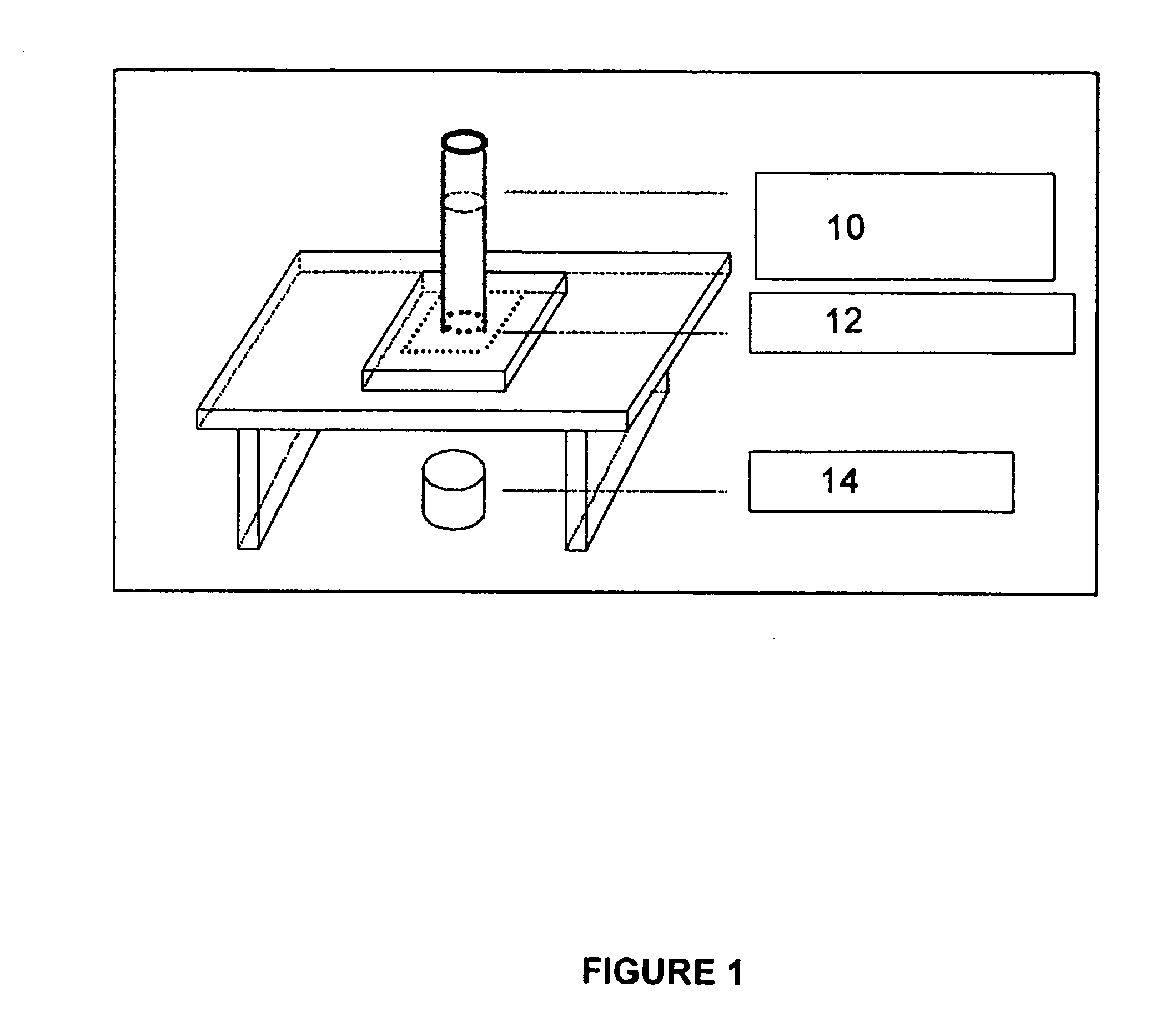
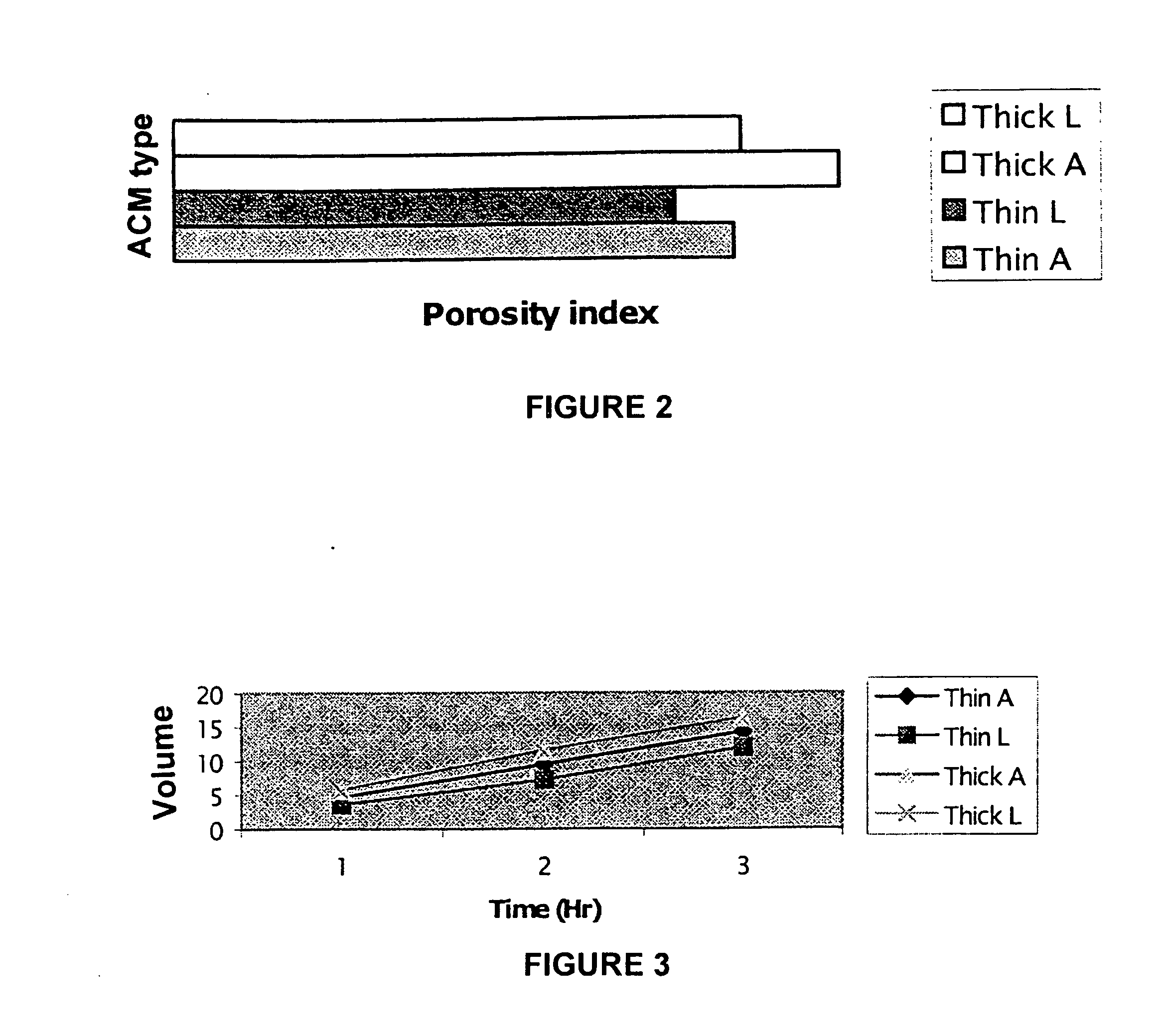
[0065] M-ACM Porosity: The apparatus to measure porosity of the bladder tissue is generally described in FIG. 1 in which there is shown a liquid pressure column 10, placed on top of a tissue holder 12 on which the tissue is placed and reservoir 14 to measure liquid volume going through the tissue. Since bladder permeability bar...
example 3
Experimental Model for Testing Bladder Augmentation
[0083] The domestic pig bladder offers the closest approximation to the human bladder in terms of size, shape, tissue histology and compliance, furthermore, being a growing animal model, pig would be comparable to children as a suitable model. Operative procedures on pigs resemble those performed on humans with respect to instruments and suture materials. In addition, we have developed protocols for isolation of porcine bladder urothelial, smooth muscle, and endothelial cells. Furthermore we have phenotypically characterised these cell types respectively, cytokeratin 7 for urothelium, smooth muscle actin for smooth muscle cells, and factor VIII for endothelial cells.
[0084] Protocol for Urinary bladder acellularization: Pigs averaging 20-28 Kg can be used for bladder M-ACM processing and implantation. Whole porcine urinary bladders are cut in half and extracted in detergent containing solutions as described above. Tissue samples fr...
PUM
| Property | Measurement | Unit |
|---|---|---|
| Volume | aaaaa | aaaaa |
| Volume | aaaaa | aaaaa |
| Volume | aaaaa | aaaaa |
Abstract
Description
Claims
Application Information
 - R&D
- Intellectual Property
- Life Sciences
- Materials
- Tech Scout
- Unparalleled Data Quality
- Higher Quality Content
- 60% Fewer Hallucinations
Browse by: Latest US Patents, China's latest patents, Technical Efficacy Thesaurus, Application Domain, Technology Topic, Popular Technical Reports.
© 2025 PatSnap. All rights reserved.Legal|Privacy policy|Modern Slavery Act Transparency Statement|Sitemap|About US| Contact US: help@patsnap.com


