Isolation of sperm cells from other biological materials using microfabricated devices and related methods thereof

a technology of microfabricated devices and sperm cells, which is applied in the direction of isotope separation, ion-exchanger regeneration, electrolysis, etc., can solve the problems of consuming and laborious procedures, ensuing preparative/analysis steps, and resulting cost-ineffectiveness, etc., to facilitate the evaluation of sperm

Inactive Publication Date: 2006-07-06
LANDERS JAMESP +2
View PDF14 Cites 15 Cited by
  • Summary
  • Abstract
  • Description
  • Claims
  • Application Information

AI Technical Summary

Benefits of technology

[0015] The speed and efficiency of the conventional differential extraction procedure warrants improvement by the micro-miniaturization of cell sorting. A stand-alone microdevice for rapidly sorting sperm cells from epithelial cells and extracting DNA would improve DNA analysis in the crime laboratories by reducing the cost of analysis through improved speed, reduced reagent consumption, decreased technician time, reduced sample handling-induced contamination, and ease of automation. The present invention provides a novel method and device for separation of sperm and epithelial cells on a microdevice. A separation method that does not require a high affinity interaction with the cells but, instead, one that exploits electrophoretic mobility, electroosmotic flow, pressure-based flow and / or combination thereof is exploited. This present invention utilizes the differential physical and biological properties of the cells, such as their propensity for adhesion, specific gravity, cell surface charge, and size. Two important aspects of such a cell separation mechanism separation are, but not limited thereto, the magnitude of the flow, which can be controlled by a number of mechanisms, such as electrophoretic mobility, electroosmotic flow, pressure gradient (pump as well as gravity), and / or combinations thereof, as well as the surface properties of the channel walls. The present invention provides techniques for the isolation of sperm cells from biological materials, preferably other cells and molecular species, most preferably epithelial cells, for forensic applications using microfabricated devices.
[0016] In a further embodiment, the present invention is used to isolate sperm from either other cells or other biologically-derived molecular species enables sperm to be concentrated in smaller volumes. This effect could find utility with in vitro fertilization applications. Beebe et al. has shown that human eggs (oocytes) can be manipulated in microfabricated devices in processes pertinent to in vitro fertilization. The device described herein for sperm cell isolation could be utilized to enhance the concentration (number) of sperm in the collection chamber. One could envision how the presence of an oocyte in the collection chamber where an enhanced concentration of sperm are collected, might improve the efficiency of in vitro fertilization.
[0018] In a further embodiment, the present invention is used to isolate sperm from other cells or other biologically-derived molecular species via some flow-driven separation process presents the possibility of quantitating subpopulations of sperm from the sample. This could facilitate the evaluation of sperm that are dysfunctional with respect to fertilization ability, the separation of sperm subpopulations that are functional relative to those that are dysfunctional due to exposure to toxicants (apoptotic) or cryostorage.

Problems solved by technology

Effective separation of the victim's and perpetrator's DNA, combined with the ensuing preparatory / analysis steps, are time- and labor-intensive processes.
The multistep nature of this current method affords it the same disadvantages from which many conventional isolation methodologies suffer.
First, the time-consuming and labor-intensive procedure translates into cost-ineffectiveness.
Second, extensive sample handling presents opportunities for loss of biological material, which may be problematic if only small amounts of starting material are available.
In addition, extensive sample handling increases the chance of contamination with exogenous DNA, which can complicate interpretation of the results.
There may be numerous problems associated with this approach including: clogging of the column by the large numbers of epithelial cells in a “real-world” sample, inability to integrate the cell separation with other microminiaturization analyses, expense of materials, and numerous steps still required to result in PCR-ready DNA.
A reliable separation may not result using the antibody / magnetic bead approach due to extensive clogging of the column.

Method used


Image

  • Isolation of sperm cells from other biological materials using microfabricated devices and related methods thereof
  • Isolation of sperm cells from other biological materials using microfabricated devices and related methods thereof
  • Isolation of sperm cells from other biological materials using microfabricated devices and related methods thereof

Examples


Embodiment Construction

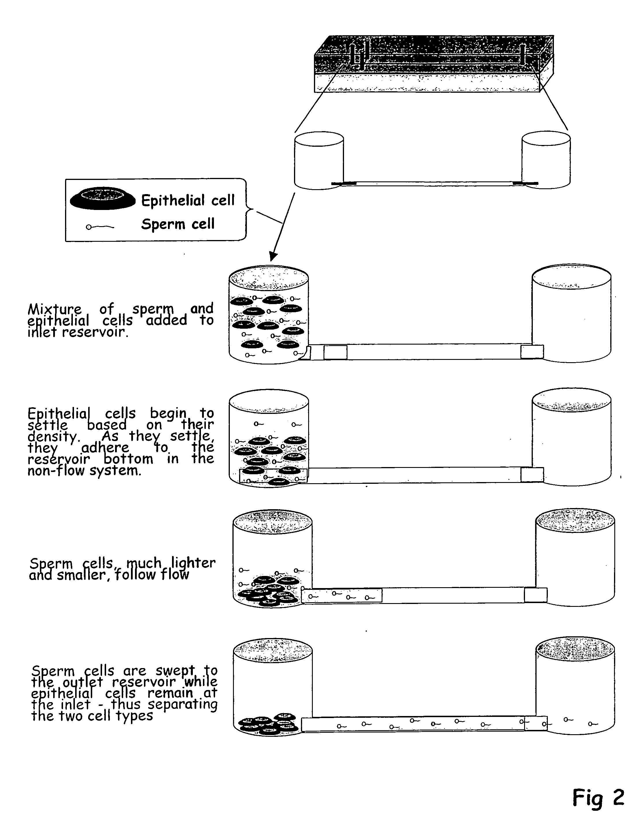
[0024] The present invention exploits physical and / or biological properties of sperm and other biological materials, such as epithelial cells, to effect a robust and reliable separation of the two cell types. Biological materials used herein includes, but is not limited to, other cells, such as epithelial cells, red blood cells, white blood cells, etc.; molecular species, such as nucleic acids (RNA and DNA), proteins, etc.; cell membranes; and organelles. Two separation approaches can be utilized to invoke separation of cells, with a main focus on the separation of sperm from other cells for forensic analysis where both the sperm and the other cells can be important in the forensic process. The first mode amenable to a microfabricated device is a separation driven by an electric field—this inherently involves both an electrophoretic component (mobility of cells based on size and their surface charge) and a flow component in the form of electroosmotic flow (EOF—the flow that results ...


PUM

PropertyMeasurementUnit
Pressureaaaaaaaaaa
Flow rateaaaaaaaaaa
Biological propertiesaaaaaaaaaa

Abstract

The present invention relates to cell separation using microfabricated devices. In particular, the present invention provides methods and devices for separation of sperm from biological materials, such as other cells and molecular species, in a cell mixture in a microfabricated device through the use of electroosmotic flow, electrophoretic mobility, pressure gradient, differential adhesion, and / or combinations thereof.

Description

[0001] This application claims priority to Provisional Patent Application No. 60 / 427,734, filed Nov. 20, 2002.FIELD OF THE INVENTION [0002] The present invention relates to cell separation using microfabricated devices. In particular, the present invention provides methods and devices for separation of sperm from biological materials, such as other cells and molecular species, in a cell mixture in a microfabricated device through the use of electroosmotic flow, electrophoretic mobility, pressure gradient, differential adhesion, and / or combinations thereof. BACKGROUND OF THE INVENTION [0003] The use of DNA typing to verify and often convict suspects in sexual crime cases relies on the separation of the perpetrator DNA from that of the victim. The perpetrator DNA is most easily obtained from sperm cells collected on vaginal swabs, taken in the routine collection of sexual assault evidence. The majority of cells collected on such swabs are epithelial cells from the victim, however, and...

Claims


Application Information

Patent Timeline
IPC IPC(8): B01D57/02B01D15/08B01L3/00B01L7/00C12N5/00C12N5/071C12N5/076G01N27/447
CPCB01D57/02B01L3/502753B01L3/502761B01L7/52B01L2200/0647B01L2200/10B01L2300/0816B01L2300/0864B01L2300/087B01L2400/0409B01L2400/0418B01L2400/0421B01L2400/0457B01L2400/0487C12N5/061C12N5/0612G01N27/44791
Inventor LANDERS, JAMESPFERRANCE, JEROMEPHORSMAN, KATIEM
Owner LANDERS JAMESP
Why Eureka
Try Eureka