
Virtual colonoscopy with radiolabeled phospholipid ether analogs

a technology of phospholipid ether analogs and colonoscopy, which is applied in the field of virtual colonoscopy with radiolabeled phospholipid ether analogs, tumor diagnostic imaging and characterization, can solve the problems of difficult to obtain ct scan functional information during treatment and diagnosis, and the inability to characterize the space occupying lesions as polyps (adenomas) or adenocarcinomas (malignant), etc., to accurately determine if viabl

Active Publication Date: 2006-01-19
CELLECTAR
View PDF9 Cites 16 Cited by
  • Summary
  • Abstract
  • Description
  • Claims
  • Application Information

AI Technical Summary

Benefits of technology

[0020] In particularly preferred embodiments, the radiolabeled tumor-specific agents have therapeutic functions as well as diagnostic functions. Preferably, the same administration of the radiolabeled tumor-specific agents produces both regression and shrinkage of the tumor as well as identification of the tumor. In certain embodiments, the same administration of the radiolabeled tumor-specific agents initially affords accurate detection, localization and characterization of the suspect mass, and when administered in combination with the same or different tumor-specific agent radiolabeled with a therapeutic isotope, also provides the ability to treat a malignant tumor. In other embodiments, the present invention provides agents and methods for monitoring the progress of therapy wherein the agent is avidly taken up by the tumor.

Problems solved by technology

Performed on a traditional multidetector helical CT scanner, virtual colonoscopy allows one to noninvasively scan the intestinal lumen anatomically for tumors but cannot characterize space occupying lesions as either polyps (adenomas) or adenocarcinomas (malignant).
In addition, functional information from CT scans is difficult to obtain during treatment and diagnosis using RF ablation and CT scanning.
With contrast-enhanced helical CT, tumor vascularity may be assessed to some degree, but there is no way of accurately determining if viable tumor cells remain within the RF-ablated lesion.
This decreases the sensitivity and specificity for early detection of recurrent tumor.
Some patients with rectal cancer treated with pre-operative radiation will have a complete response, while others with similar histology (at the light microscopy level) will have a poor response to treatment and will recur.
International Journal of Radiation Oncology, Biology, Physics 2002; 52; 1:14-22), in most cases there is no accurate method of predicting tumor response during actual treatment.
These are investigational and none are in routine clinical use.
Unfortunately, conventional imaging techniques such as computerized tomography (CT) and MRI (magnetic resonance imaging) are limited in their ability to afford a conclusive diagnosis of a suspected lesion, since they are only capable of observing differences in the density or morphology of tissues.
A more invasive and costly biopsy procedure is often necessary to provide a definitive diagnosis.
Unfortunately, the tumor-localizing agents developed thus far for oncological applications have had only limited application.
Unfortunately, 67Ga gallium citrate is taken up by a variety of other non-tumor lesions as well, including inflammatory lesions, and unacceptable amounts of radioactivity can also accumulate in liver and spleen.
The rapid buildup of a radiopharmaceutical in these organs can seriously interfere with the imaging of nearby lesions and also reduces the dosage that can safely be given to a patient.
However, these monoclonal antibodies are specific only to the particular tumor tissue for which they have been produced, and therefore will not localize generally in neoplastic tissue.
Moreover, the use of Mabs for diagnostic imaging has lead to additional problems, including varying degrees of antigen expression, low tumor uptake, non-specific binding and adverse immunogenic reactions and generally high liver localization.
Currently available oncologic PET agents such as 18F-FDG, localize in benign lesions and inflammatory sites as well as in tumors, and are thus not good indicators of malignancy.

Method used


Image

  • Virtual colonoscopy with radiolabeled phospholipid ether analogs
  • Virtual colonoscopy with radiolabeled phospholipid ether analogs
  • Virtual colonoscopy with radiolabeled phospholipid ether analogs

Examples


example i

Synthesis, Radiolabeling, and Formulation of NM404

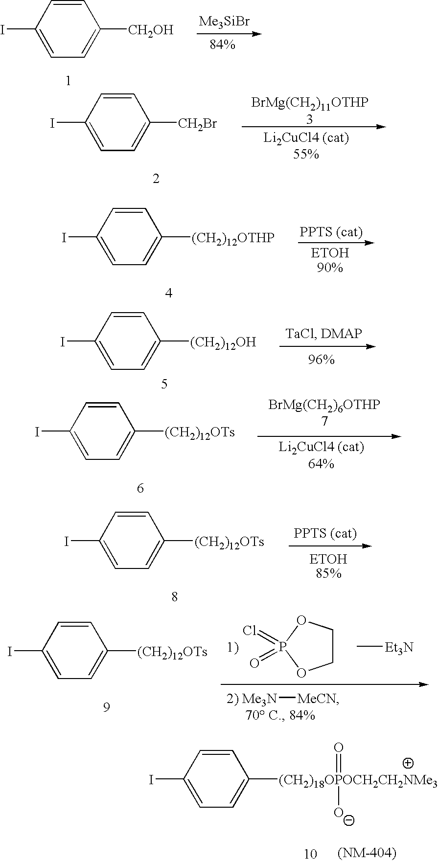
[0112] The synthetic approach was based on the copper-catalyzed cross-coupling reaction of Grignard reagents with alkyl tosylates or halides for the alkyl chain elongation (see the scheme below). The synthesis was started from p-iodobenzyl alcohol 1 which was converted into p-iodobenzyl bromide 2 by reaction with trimethylsilyl bromide. p-Iodobenzyl bromide 2 was further coupled with Grignard reagent 3 in the presence of Li2CuCl4 as a catalyst. 12-(p-Iodophenyl)dodecanol 5 obtained after deprotection of the first coupling product 4 was converted into tosylate 6. In the next step, tosylate 6 was coupled with Grignard reagent 7 containing 6 carbon atoms and this completed the chain elongation process. THP deprotection of 8 gave 18-(p-iodophenyl)octadecanol 9 which was converted into 10 (NM404) by two-step procedure as shown in the scheme.

[0113] Further, rapid high yield synthesis process for labeling NM404 with any isotope if iodine...

example 2

High Specific Activity Synthesis and Radiolabeling

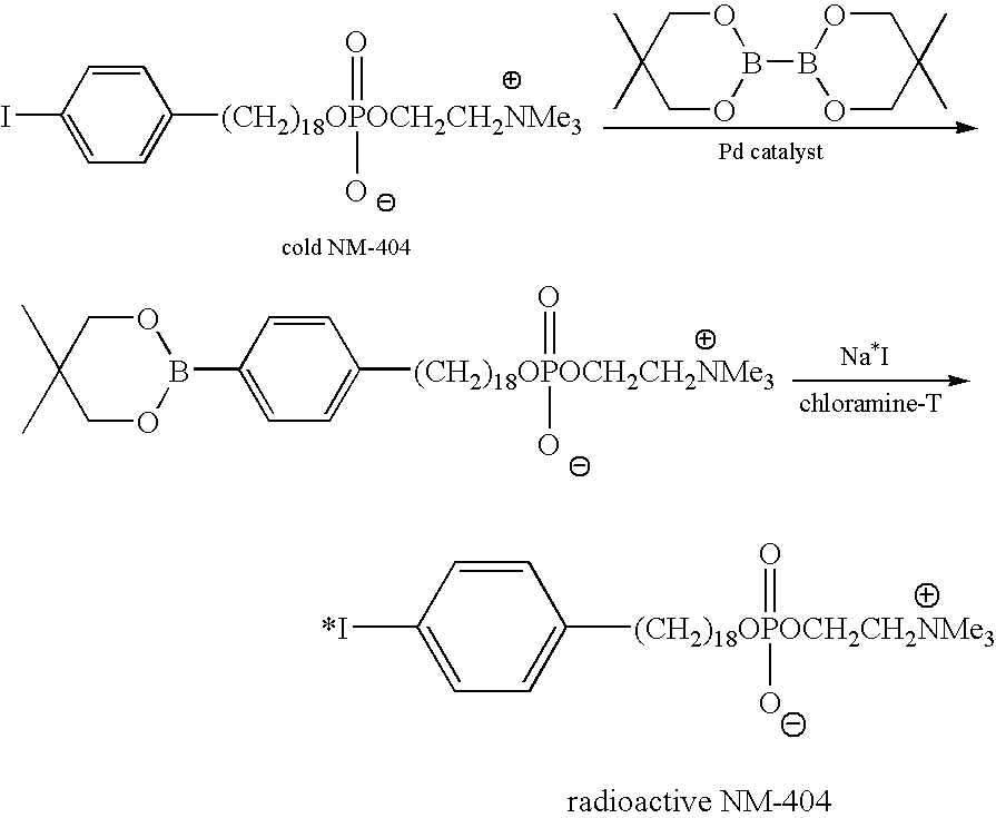
[0129] In the synthesis of high specific activity NM-404, cold NM-404 is first coupled with bis-(neopentyl glycolato)diboron in the presence of Pd catalyst to form arylboronate ester. In the second step, the arylboronate ester is subjected to radioiodo-deboronation using radioactive sodium iodide and chloramine-T to obtain radioiodinated NM-404 with high specific activity.

[0130] Other esters of glycol esters of diboronic acid can also be used in the first reaction, for example: bis-(pinacolato)diboron and bis-(hexylene glycolato)diboron.

[0131] The important criterion of the high specific activity synthesis is that the final labeled compound, for example, 131I-NM404 and its precursor, the boronate compound must be easily separable by HPLC. This ensures that literally every molecule of NM404 that is isolated by HPLC is radiolabeled. Theoretical specific activity of the compound is about 2500curies / mmol. The existing isotope exchan...

example 3

Preclinical Studies with PLE Analogs

[0132] Phospholipid ethers can easily be labeled with iodine radioisotopes using an isotope exchange method described above. The iodophenyl phospholipid ether analogs are specifically designed so that the radioiodine affixed to each molecule is stable to facile in vivo deiodination. Over 20 radiolabeled PLE compounds were synthesized and tested in vitro and in vivo. Two of these, namely NM294 and NM324 [12-(3-iodophenyl)-dodecyl-phosphocholine], initially showed the most promise in animal tumor localization studies. These prototype compounds, labeled with 125I, selectively localized in tumors over time in the following animal tumor models; 1) Sprague-Dawley rat bearing Walker 256 carcinosarcoma; 2) Lewis rat bearing mammary tumor; 3) Copenhagen rat bearing Dunning R3327 prostate tumors; 4) Rabbits bearing Vx2 tumors; and 5) athymic mice-bearing human breast(HT39), small cell lung (NCI-69), colorectal (LS174T), ovarian (HTB77IP3), and melanoma tum...


PUM


Abstract

The present invention provides agents and methods for dual modality virtual colonoscopy that gives both anatomical and functional information using hybrid CT / PET scanning. In preferred embodiments, the present invention provides radiolabeled tumor-specific agents and methods for distinguishing benign polyps from malignant tumors. In further embodiments, the present invention provides compositions and methods useful for distinguishing morphological and functional subregions of a selected region of tissue based on relative levels of phospholipid metabolism. Preferred radiolabeled tumor-specific agents are phospholipid ether analogs labeled with a halogen radiosiotope. In certain preferred embodiments, the compositions including radiolabeled phospholipid ether analogs have therapeutic actions in addition to functionally identifying malignant tissue.

Description

RELATED APPLICATIONS [0001] This application claims benefit of U.S. Provisional Patent Application 60 / 521,831 filed Jul. 8, 2004, the entire contents of which are incorporated by reference for all purposes.FIELD OF THE INVENTION [0002] The invention generally relates to imaging of tumors and specifically relates to diagnostic imaging and characterization of tumors using virtual colonoscopy in combination with a radiolabeled tumor-specific agent, such as a phospholipid ether analog labeled with a radiohalogen. BACKGROUND OF THE INVENTION [0003] The early detection of cancer has been one of the primary goals of modern imaging technology, since the identification of a suspected tumor in a localized stage significantly improves the chances for successful treatment and elimination of the cancerous tissue. A large number of imaging strategies have therefore been designed, using a variety of techniques and modalities, to aid the physician in making an accurate diagnosis as early as possibl...

Claims


Application Information

Patent Timeline
IPC IPC(8): A61K51/00
CPCA61K51/0408A61B6/037A61B6/508A61P35/00A61K51/04
Inventor WEICHERT, JAMEYLONGINO, MARC
Owner CELLECTAR
Learn More