Nonlinear optical detection of fast cellular electrical activity

a technology of cellular electrical activity and optical detection, applied in optical radiation measurement, fluorescence/phosphorescence, instruments, etc., can solve the problems of reducing the effective observed dye response to membrane potential, limiting previous methods, and poor spatial resolution deep in scattering tissues (such as neural tissue), so as to increase the brightness and reduce photodamage, and improve the effect of sensitivity to membrane potential changes

Inactive Publication Date: 2005-11-24
CORNELL RES FOUNDATION INC +1
View PDF3 Cites 19 Cited by
  • Summary
  • Abstract
  • Description
  • Claims
  • Application Information

AI Technical Summary

Benefits of technology

[0015] The methods of the present invention are effective in studying neurons, parts of neurons (e.g., axons, dendrites, etc.), and other types of cellular tissue (e.g., microtubules). For example, axons contain like-polarity microtubule ensembles, but dendrites contain mixed-polarity microtubule ensembles. Because second harmonic generation imaging reveals regions of like-polarity microtubule ensembles and not mixed-polarity regions, the methods of the present invention can be used to locate single axons of neurons or axon-rich areas in the cellular tissue. Membrane potential signals can vary significantly between axons and dendrites; therefore, the ability to locate these regions with second harmonic generation and then image the membrane potential on the specific region should prove useful.
[0017] Due to the molecular alignment requirement of SHG, the present invention has the advantage over all other techniques (including fluorescence, light scattering, birefringence, absorption, etc.) in that the effective response to membrane potential is not attenuated by any background. This allows for an increased signal to noise ratio compared to other techniques that have a similar response to membrane potential, but are adversely affected by background signals. Additionally, this background limits the ability of other techniques to optically quantify the membrane potential. Because SHG is not affected by background, the signal is quantifiable and can be directly related to changes in membrane potential. This ability to optically quantify membrane potential signals deep in scatting tissue has never been demonstrated before (on any spatiotemporal scale).
[0018] The present invention has a number of characteristics that are more advantageous than previously known imaging techniques. For example, the present invention uses changes in second-harmonic generation from a special class of dyes (Molecule A, Molecule B, FM 4-64 and their derivatives) in live cells. The special class of dyes of the present invention can be used in excitable cells (including neurons) during electrical activity. The present invention can be used to record the second-harmonic signal at the necessary temporal resolution (full line scan time of 0.833 milliseconds, and 10 microseconds recording time per measured membrane) to study fast signals, including resolving action potentials, and sub-threshold events. This time scale can be generalized from 1 microsecond to minutes resolution. Other characteristics exhibited by the method of the present invention include, for example, the following: (1) the highest spatial resolution yet achieved for fast membrane potential imaging in live cells with the potential for resolutions <0.1 microns; (2) controllable phototoxicity during recording; (3) the first nonlinear method to record fast cellular electrical signals (including action potentials); (4) no background signal from dye molecules not responding to membrane potential; (5) a single optical signal whose relative intensity change can be quantitatively related to membrane potential changes; and (6) the ability to image hundreds of microns deep in scattering tissue.
[0020] The present invention may also be used for more general applications, including, for example, the following: (1) investigate dye derivatives in this class that should be brighter (larger beta value) and have a larger sensitivity to membrane potential changes in order to avoid the need for signal averaging; (2) illuminate dye derivatives at longer wavelengths to match their resonant frequencies, increase the brightness and sensitivity to membrane potential, and reduce photodamage caused by intrinsic tissue absorption at shorter wavelengths; (3) increase the concentration of dye to increase the second-harmonic signal and to avoid the need for signal averaging; (4) combine with faster whole-frame imaging methods; (5) combine with uniform polarity microtubule second-harmonic signal to identify neurites absolutely as axons or dendrites; (6) intracellularly fill cells with second-harmonic generation membrane dyes with patch pipettes and use in vivo or ex vivo; (7) combine with two-photon fluorescence signal from dyes to increase the signal-to-noise ratio; (8) combine with methods that reduce phototoxicity, such as reducing the oxygen tension or adding free radical scavengers, in order to increase the excitation intensity and therefore the signal-to-noise ratio; (9) combine with two-photon fluorescence signals from ion indicators such as calcium and sodium probes; (10) increasing the spatial resolution two- or three-fold; (11) stain and record membrane potential signals from large populations of neurons; and (12) using the backward propagating SHG signal to image in vivo.

Problems solved by technology

However, in thick preparations high-resolution one-photon techniques are limited to imaging depths of <˜50 μm by light scattering, making the poor spatial resolution deep in scattering tissues (such as neural tissue) the most severe limitation.
Additionally, previous methods are limited by a background signal from dye not bound to the plasma membrane that reduces the effective observed dye response to membrane potential and complicates the optical quantification of membrane potential changes.
To date, there has been no demonstration of the ability to record fast Vm activity in living cells with any form of nonlinear microscopy and therefore ˜1 ms, high spatial resolution optical Vm recording has been limited to thin preparations or superficial regions of thick specimens.
Previous quantitative methods have also been limited to culture dish preparations where background signals can be kept to a minimum, making deep tissue optical quantification of membrane potential unattainable.
Research on the role of MT ensemble polarity in the dynamical development of neuronal processes, growth cones and injury response has been hindered by the lack of suitable techniques.

Method used


Image

  • Nonlinear optical detection of fast cellular electrical activity
  • Nonlinear optical detection of fast cellular electrical activity
  • Nonlinear optical detection of fast cellular electrical activity

Examples


example 1

Uniform Polarity Microtubule Assemblies Imaged in Native Brain Tissue by Second-Harmonic Generation Microscopy

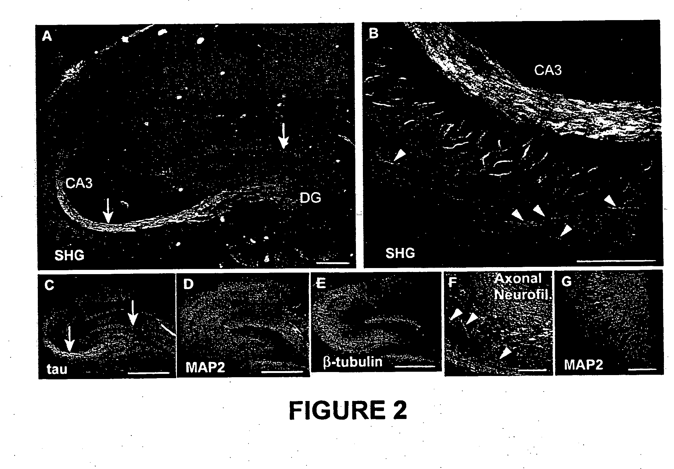
[0047] Microtubule ensemble polarity is a diagnostic determinant of the structure and function of neuronal processes. Polarized microtubule (“MT”) structures are selectively imaged with second-harmonic generation (“SHG”) microscopy in native brain tissue. This SHG is found to colocalize with axons in both brain slices and cultured neurons. Because SHG stems only from non-inversion symmetric structures, the uniform polarity of axonal MTs leads to the observed signal, whereas the mixed polarity in dendrites leads to destructive interference. SHG imaging provides a new tool to investigate the kinetics and function of MT ensemble polarity in dynamic native brain tissue structures and other subcellular motility structures based on polarized MTs.

[0048] As demonstrated in Examples 2-13 (infra), the first intrinsic sources of SHG from cultured neurons and acute slices from the hip...

example 2

Imaging

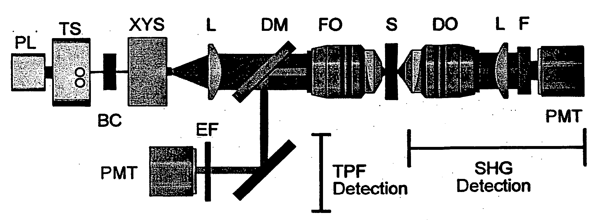
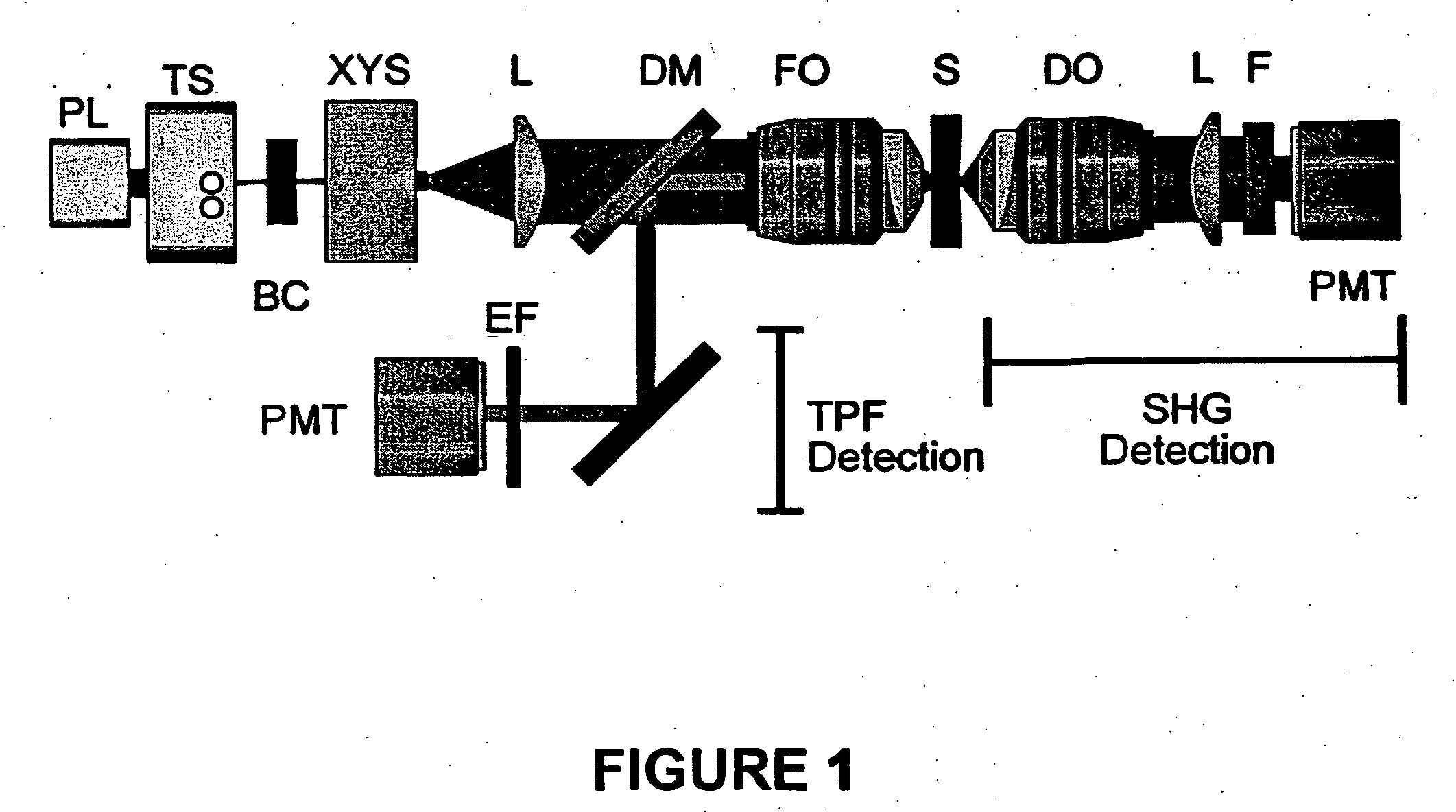
[0049] SHG and TPF microscopy were simultaneously performed on either a Bio-Rad MRC 1024 or Radiance scan head on a modified inverted Olympus IX-70 microscope (FIG. 1). The excitation source was a mode-locked Ti:Sapphire laser (˜100 fs pulses at 80 MHz) (Spectra-Physics) pumped by a 5 W solid state Millennia laser (Spectra-Physics). The laser polarization was controlled via a Berek polarization compensator (New Focus, San Jose, Calif.) and the beam focused into the sample with one of the following (overfilled back aperture) objectives: Zeiss C-Apochromat 10× / 0.45 NA, Olympus UApo 20× / 0.7 NA, Zeiss Fluar 20× / 0.75 NA, Olympus UApo 40× / 1.15 NA, Zeiss Fluar 40× / 1.3 NA. The resultant SHG was collected in the transmitted (forward) direction with an Olympus XLUMPlanFl 20× / 0.95 NA objective, while the TPF was epi-collected through the excitation objective. A combination of dichroic mirrors, band-pass and blue-glass filters (Chroma Technology, Brattleboro, Vt.) and polarization analy...

example 3

Acute Hippocampal Slices

[0051] Transverse hippocampal slices 250-400 μm thick were prepared from 14 to 20 day old Sprague-Dawley rat pups using a vibratome, and were incubated at 34° C. in artificial cerebrospinal fluid (ACSF) containing (in mM): 118 NaCl, 3 KCl, 1 KH2PO4, 1 MgSO4, 20 Glucose, 1.5 CaCl2 and 25 NaHCO3. The ACSF was oxygenated with 95% O2 and 5% CO2. Imaging was performed in ACSF filled glass bottom culture dishes (World Precision Instruments) at room temperature or at 34° C.


PUM


Abstract

The present invention is directed to various methods involving nonlinear microscopy and dyes that are sensitive to fast cellular membrane potential signals and capable of generating nonlinear optical signals. The present invention includes methods of producing high spatiotemporal resolution images of electrical activity in cellular tissue, as well as methods of detecting and investigating disease within a particular cellular tissue of a living organism. The present invention further relates to methods of detecting membrane potential signal changes in a neuron or a part of a neuron, as well as in a population of cells.

Description

[0001] This application claims the benefit of U.S. Provisional Patent Application Ser. No. 60 / 539,380, filed Jan. 27, 2004.[0002] The subject matter of this application was made with support from the United States Government under National Institutes of Health (“NIH”) Grant No. GM08267, NIH Grant No. GM07469, N1H-NIBB Grant No. 9 P41 EB001976-17, and Defense Advanced Research Projects Agency (“DARPA”) Grant No. MDA972-00-1-0021. The U.S. Government may have certain rights.FIELD OF THE INVENTION [0003] The present invention relates to various methods involving nonlinear microscopy and dyes that are sensitive to fast cellular membrane potential signals and capable of generating nonlinear optical signals. BACKGROUND OF THE INVENTION [0004] The investigation of the electrical signaling properties of excitable cells, such as neurons, is predominately accomplished through the use of intracellular microelectrodes. Though these studies are useful for obtaining temporal electrical activity f...

Claims


Application Information

Patent Timeline
IPC IPC(8): G01J3/00G01N1/30G01N21/64
CPCG01N1/30G01N21/6458G01N21/6428
Inventor DOMBECK, DANIEL A.WEBB, WATT W.BLANCHARD-DESCE, MIREILLEMONGIN, OLIVIERMALLEGOL, THOMAS
Owner CORNELL RES FOUNDATION INC
Why Eureka
Try Eureka