Enhanced X-ray imaging system and method
a x-ray imaging and enhanced technology, applied in the field of non-invasive imaging, can solve the problems of non-cartesian coordinate systems (even curved), non-uniform spacing between samples, and general backprojection of parallel beams that do not account for the magnification of cone beams attributable,
- Summary
- Abstract
- Description
- Claims
- Application Information
AI Technical Summary
Benefits of technology
Problems solved by technology
Method used
Image
Examples
example
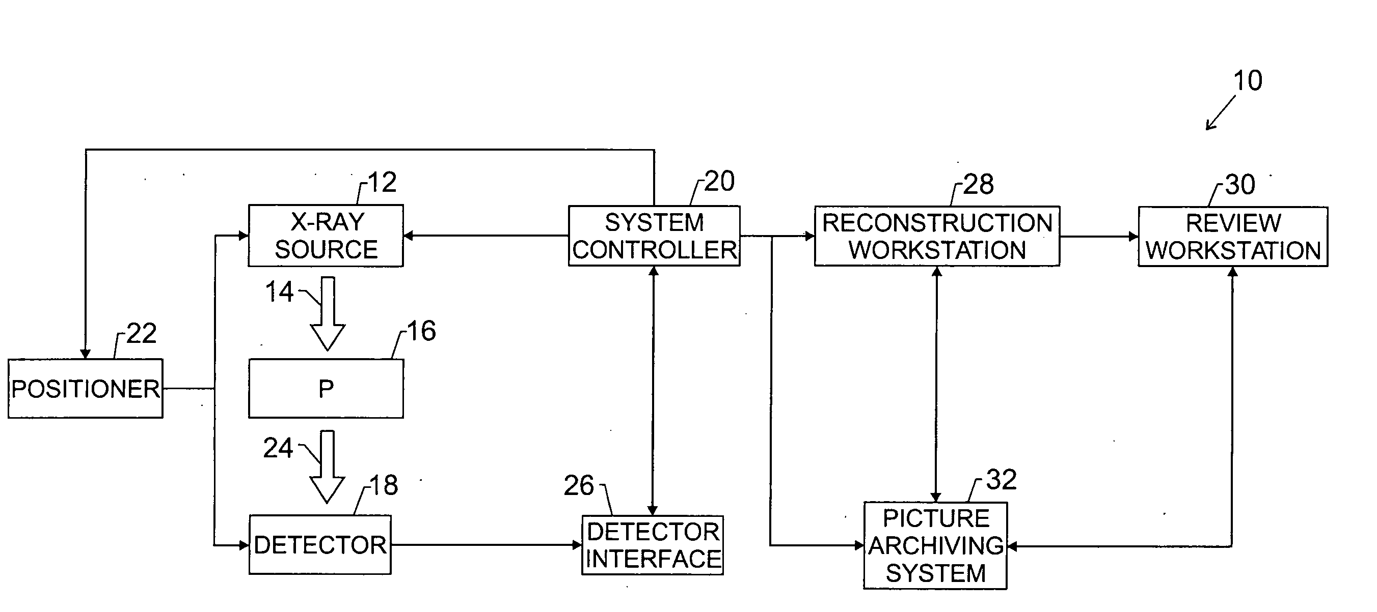
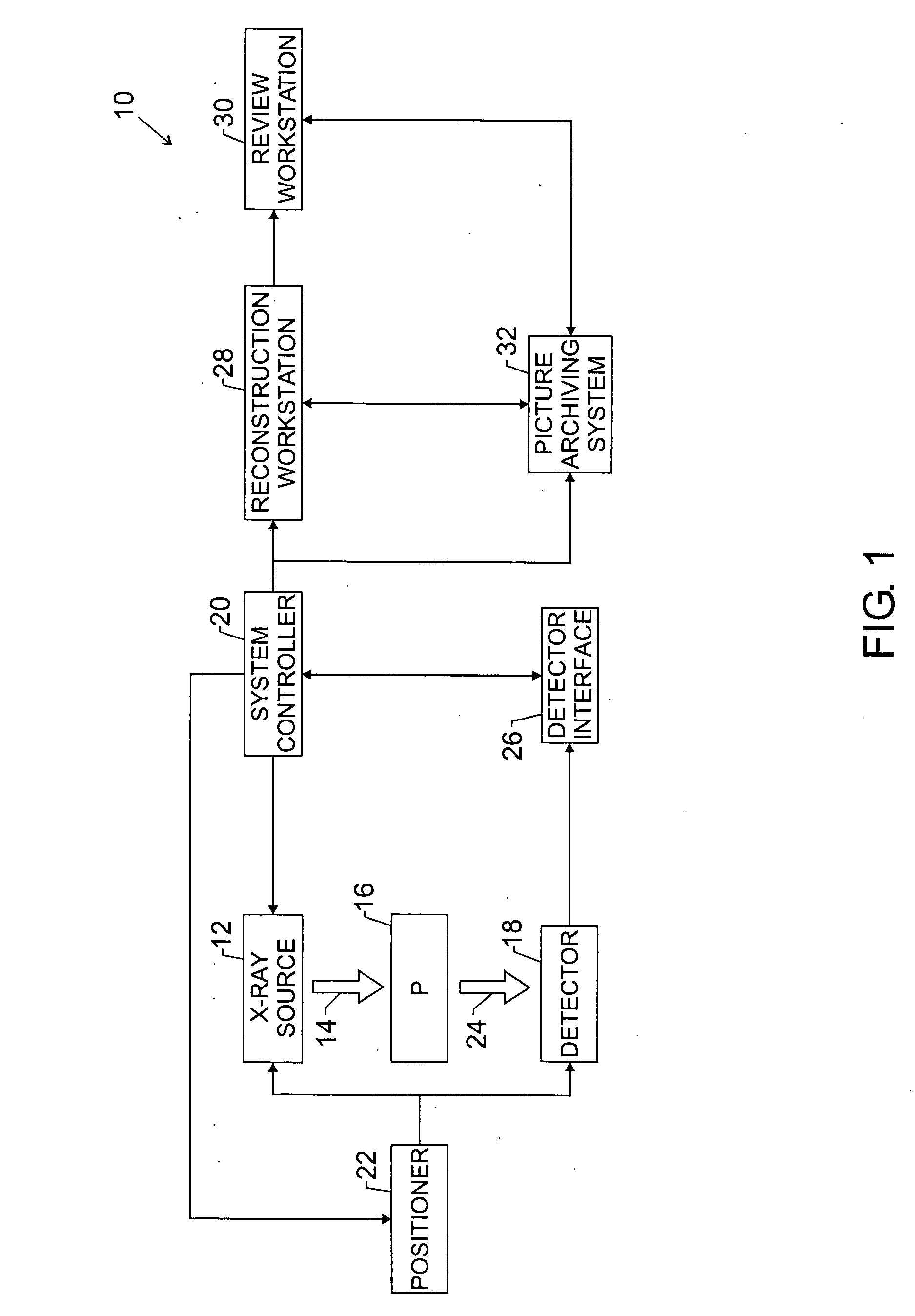
[0078] An exemplary implementation of the foregoing technique is now presented. In this exemplary implementation a three-dimensional imaging system 10 acquires twenty-one projections over a 60° angular range in approximately eight seconds. The X-ray source 12 includes an X-ray tube which moves in a trajectory above the detector 22 and stationary breast. The Source to Image Distance (SID) is 660 mm. For a relatively thick, dense breast (such as 6 cm of compressed thickness), a technique of Rh / Rh at 30 kVp and 160 mAs total may be used. The mAs per tomosynthesis view is 160 / 21=7.62 mAs. The X-ray tube current is approximately 75 mA, for an X-ray “on” time per shot of approximately 0.1 sec. Both the X-ray tube and the detector 22 move during each X-ray exposure. In this example, the X-ray tube moves by approximately 240 microns and the detector 22 moves by approximately 20 microns. The X-ray tube also moves between exposures. Total dose for the tomosynthesis mammogram is approximately ...
PUM
 Abstract
Description
Claims
Application Information
 - R&D
- Intellectual Property
- Life Sciences
- Materials
- Tech Scout
- Unparalleled Data Quality
- Higher Quality Content
- 60% Fewer Hallucinations
Browse by: Latest US Patents, China's latest patents, Technical Efficacy Thesaurus, Application Domain, Technology Topic, Popular Technical Reports.
© 2025 PatSnap. All rights reserved.Legal|Privacy policy|Modern Slavery Act Transparency Statement|Sitemap|About US| Contact US: help@patsnap.com


