
Labeled ligands for lectin-like oxidized low-density lipoprotein receptor (LOX-1)

a low-density lipoprotein receptor and labeling technology, applied in the field of compound labeling with imaging agents, can solve the problems of angina or ischemic sudden death, many unanswered questions, and many unanswered questions

Inactive Publication Date: 2005-04-28
GENERAL ELECTRIC CO
View PDF9 Cites 10 Cited by
  • Summary
  • Abstract
  • Description
  • Claims
  • Application Information

AI Technical Summary

Benefits of technology

[0037] These and other features of the invention will be readily apparent to

Problems solved by technology

Despite these efforts, there are many unanswered questions including how and when atherosclerotic lesions become vulnerable and life-threatening, the best point of intervention, and how to detect and monitor the progression of lesions.
Macrophage-derived foam cells are concentrated in the shoulders of plaques, where their secreted proteinases and collagenases may contribute to plaque rapture, which may lead to a fatal thrombotic event.
These processes, by definition, represent phase 3 (type I lesion), with a subsequent increase in stenosis, possibly resulting in angina or ischemic sudden death.
Phase 2 plaque is not adequately stenotic to create symptoms or even be detected by commonly employed diagnostic techniques such as a cardiac angiogram.
However, these techniques are generally invasive, requiring surgery, insertion of probes, cameras, or other invasive procedures.
It is known, however, that the presence of these substances cannot be used to locate the involved lesion.
These techniques using catheters or devices are invasive, and sometimes may result in or trigger plaque formation or rupture.
An angiogram, however, does not image the vessel wall or the various histopathological components.
One of the major limitations of angiography is that diffuse atherosclerotic disease may narrow the entire lumen of the artery, and as a result, angiography underestimates the degree of stenosis.
However, it views only the lesion surface and is not representative of the internal heterogeneity of the plaque.
As a routine clinical tool, it may not be practical due to the thickness of the catheter and the invasiveness of this technique.
These types of devices differentiate healthy tissue from atherosclerotic plaque, but are not clinically useful for differentiating vulnerable plaque from less dangerous, stable plaque.
In coronary and peripheral arteries of low extremities, however, Duplex scanning is clinically not as useful as the traditional angiography.
A major limitation using EBCT for the characterization of calcium in the plaque is reproducibility (Becker et al., Eur. Radiol. 2000, 10, 629-635).
In addition, coronary calcium screening can not reveal atherosclerotic plaque that has little or no calcification-and such soft, lipid-rich plaques are perhaps the most dangerous of all, vulnerable to rupture as a result of hemodynamic stress or inflammation (Carrington, C., Diagnostic imaging, 2000, (April), 48-53; Doherty et al., Am.
As red blood cells and platelets gather at the site of the rupture, a blood clot forms and blocks the artery, causing a heart attack.
In general, radiolabeled proteins and platelets have shown some clinical potential as imaging agents of atherosclerosis, but due to poor target / background and target / blood ratios, these agents are not ideal for imaging coronary or even carotid lesions.
However, noninvasive imaging of atherosclerosis remains a challenge for nuclear techniques mainly due to their intrinsic shortcomings, such as low resolution, compared to MRI and CT.
However, these diagnostic procedures are either invasive or yield little information on the underlying pathophysiology such as cellular composition of the plaque, and biological characteristics of each component in the plaque at the molecular level.
The presence of large necrotic lipid cores contributes to the vulnerability of plaque to rupture.
The intense macrophage infiltration of the plaque leads to release of cytokines and matrix metalloproteinases and thereby renders the plaque prone to rupture.
None of these technologies have been shown to be entirely satisfactory for the early detection / imaging of vulnerable atherosclerotic plaque.

Method used


Image

  • Labeled ligands for lectin-like oxidized low-density lipoprotein receptor (LOX-1)
  • Labeled ligands for lectin-like oxidized low-density lipoprotein receptor (LOX-1)
  • Labeled ligands for lectin-like oxidized low-density lipoprotein receptor (LOX-1)

Examples


example 1

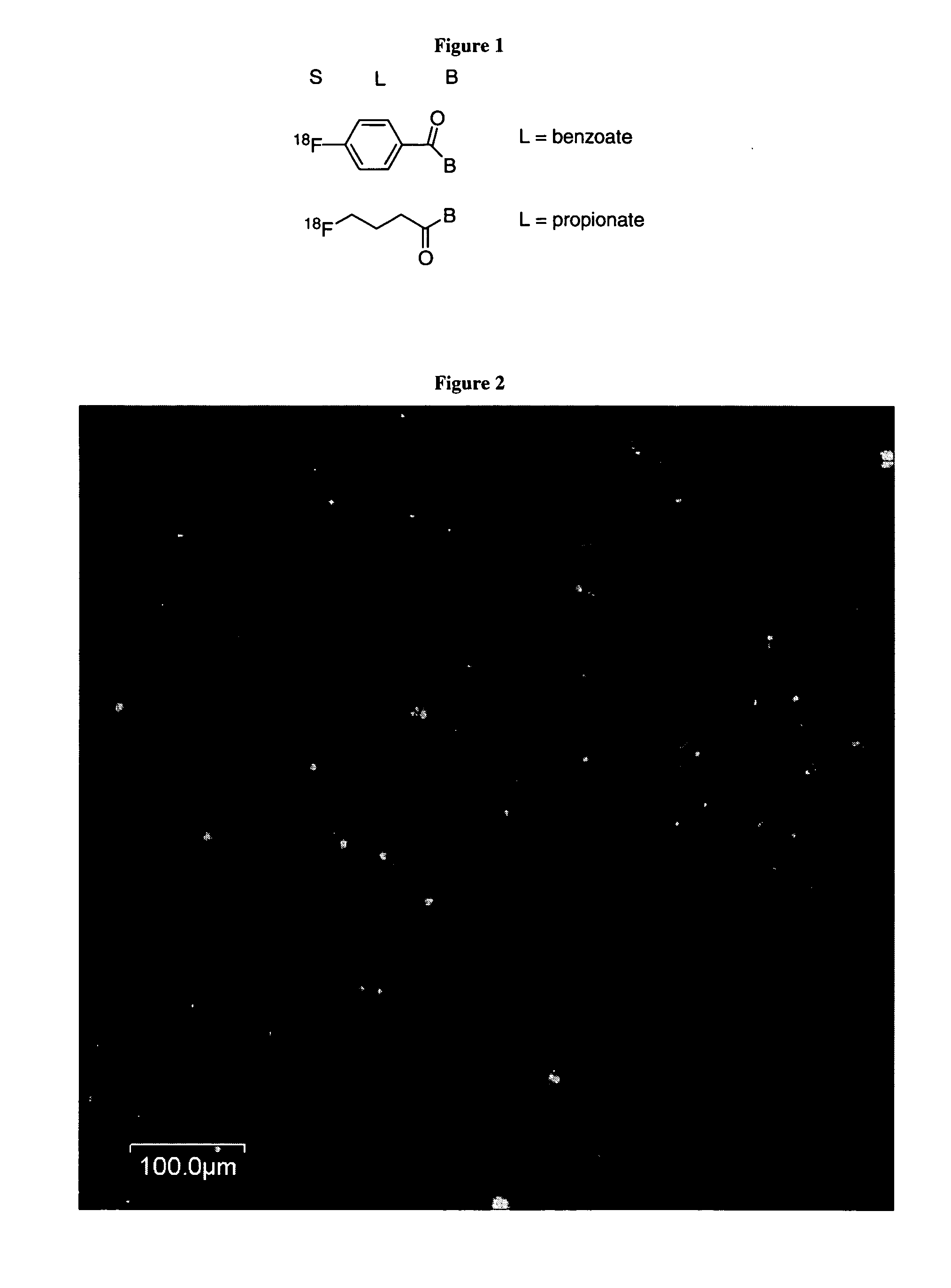
[0115] A peptide was conjugated with fluorescein (Fl-KKGG-FQTPPQL) and was shown to bind to human endothelial coronary artery cells (HCAECs) which are known in the literature to express LOX-1. An image of HCAECs grown in glass slides treated with this peptide obtained using a fluorescent confocal microsope is shown in FIG. 2; the fluorescent image (shows fluorescently tagged peptide as bright green) is overlaid with the transmitted light image (shows outline of cells). The example reveals that the peptide above was localized on the cells. The experimental conditions for imaging the peptide-labeled HCAECs are described previously.

example 2

[0116] A solution of polyclonal antibody (IgG) was produced by Invitrogen Corporation, (Carlsbad, Calif.) against the sequence Arg-Gly-Ala-Val-Tyr-Ala-Glu-Asn-Cys-Ile at a concentration of 1.5 mg / mL. Three aliquots containing 250 μg (166 μL) each were transferred to 1.5 mL Eppendorf tubes and maintained at 0° C. The solutions were treated with NaHCO3 (1M, 20 μL) and gently inverted. In a separate tube, a solution of 5-carboxyfluorescein-N-hydroxysuccinate ester in DMF (1 mg / mL) was prepared. The antibody solutions were treated with 5, 20 or 50 equivalents of the fluorescein / DMF solutions (3.95, 15.8 and 39 μL respectively). The highest concentration of DMF was 17%. The tubes were allowed to warm to room temperature over 1 hour and gently inverted every 15 minutes to assure mixing. During this time PD-10 columns were equilibrated with PBS and eluted until the sorbent bed was exposed.

[0117] The entire reaction mixtures were transferred to the columns and eluted with PBS.

[0118] The f...


PUM

PropertyMeasurementUnit
Lengthaaaaaaaaaa
Lengthaaaaaaaaaa
Electric chargeaaaaaaaaaa

Abstract

Embodiments of the present invention relates to compounds labeled with imaging agents that also are capable of binding lectin-like oxidized low-density lipoprotein (LOX-1). The labeled compounds are useful for the diagnosis and monitoring of diseases in which inflammation plays a role, such as various cardiovascular diseases including but not limited to atherosclerosis, vulnerable plaque, and coronary artery disease, as well as rheumatoid arthritis.

Description

BACKGROUND OF THE INVENTION [0001] 1. Field of the Invention [0002] Embodiments of the present invention relate in general to compounds labeled with imaging agents that also are capable of binding lectin-like oxidized low-density lipoprotein (LOX-1). The LOX-1 receptor compounds are molecules that bind to the LOX-1 receptor, which is over-expressed in atherosclerotic lesions and rheumatoid arthritis. The detectable labels include any detectable label, preferably radionuclides for nuclear scintigraphy or positron emission tomography (PET), paramagnetic metal ions or superparamagnetic particles for magnetic resonance imaging (MRI), heavy metal ions for X-ray or computed tomography (CT), gas-filled microbubbles for targeted ultrasonography (US), or optical dyes for optical imaging, porphyrins or texaphyrins for NMR, fluoresent imaging or photodynamic therapy. The labeled compounds are useful for the diagnosis and monitoring of inflammation and diseases in which inflammation plays a rol...

Claims


Application Information

Patent Timeline
IPC IPC(8): A61K49/00A61K49/14A61K49/16A61K51/10C07B59/00
CPCA61K49/0002A61K49/0043A61K49/0056A61K49/0058C07B59/008A61K49/14A61K49/16A61K51/10A61K49/085
Inventor JOHNSON, BRUCE FLETCHERMONDELLO, FRANK JOHNSYUD, FAISAL AHMEDTORRES, ANDREW SOLIZBROGAN, JOHN BUCKNAM
Owner GENERAL ELECTRIC CO
Learn More