
Photostimulation method and apparatus in combination with glucose determination

Inactive Publication Date: 2005-03-10
GLENN PATENT GROUP
View PDF99 Cites 192 Cited by
  • Summary
  • Abstract
  • Description
  • Claims
  • Application Information

AI Technical Summary

Benefits of technology

A method and apparatus using photo-stimulation to treat or pretreat a sample site prior to analyte concentration determination is presented. More particularly, photo-stimulation at or near at least one sample site is used to enhance perfusion of the sample site leading to reduced errors associated with sampling. Increased perfusion of the sample site leads to increased volume percentages of the target analyte and / or allows the blood or tissue constituent concentrations to more accurately and / or precisely track corresponding sample constituents in more well perfused body compartments or sites such as arteries, veins, or fingertips. In one embodiment, analysis of the photo-stimulated site is used in conjunction with glucose analyzers to determine the analyte concentration with greater ease, accuracy, or precision and allows determination of the analyte concentration of another non-sampled body part or compartment.

Problems solved by technology

Diabetes is a chronic disease that results in improper production and use of insulin, a hormone that facilitates glucose uptake into cells.
Diabetes is a leading cause of death and disability worldwide.
More recently, studies have indicated that testing and control of pre-diabetics leads to a significant delay of the onset of diabetes related complications.
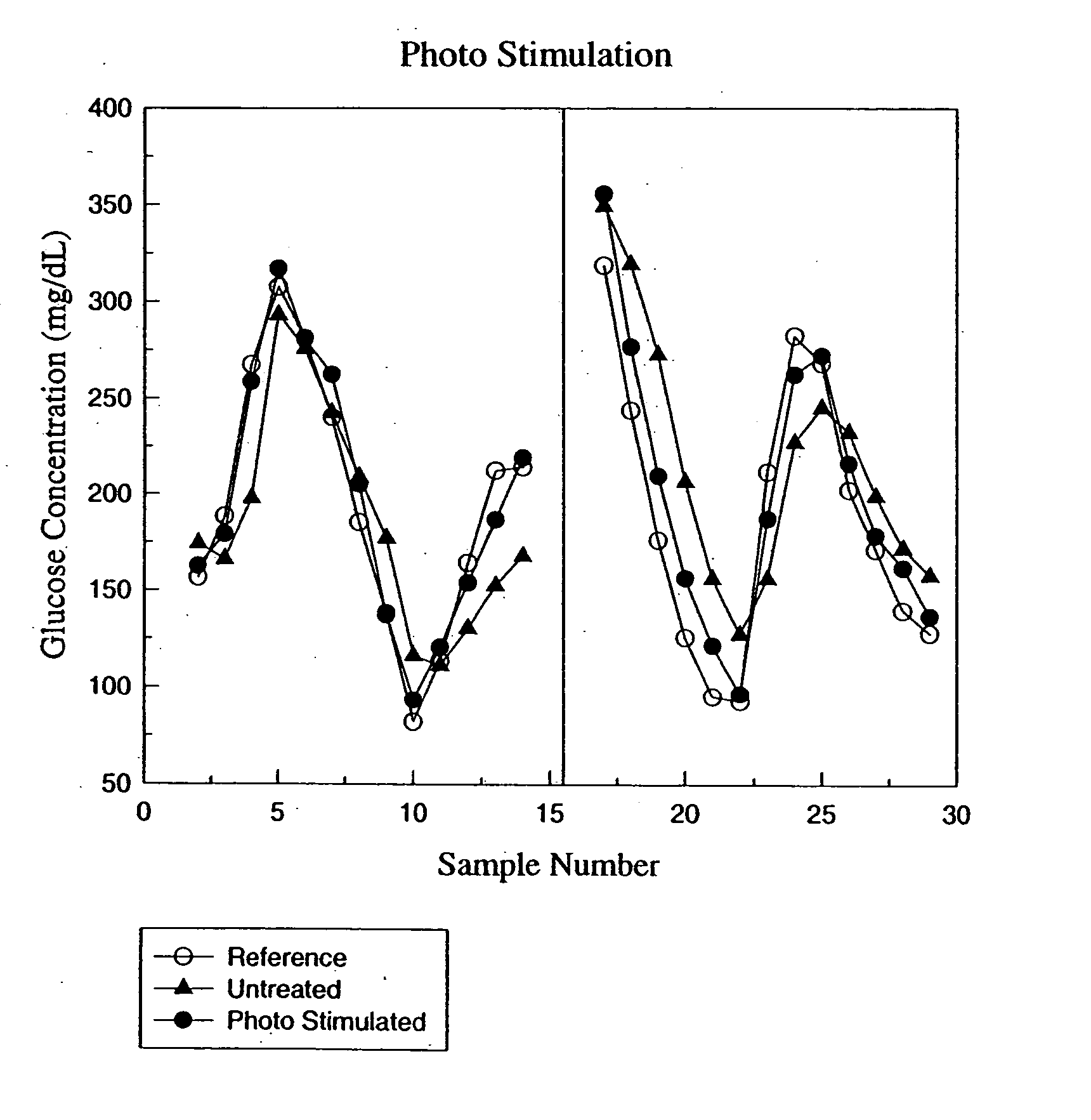
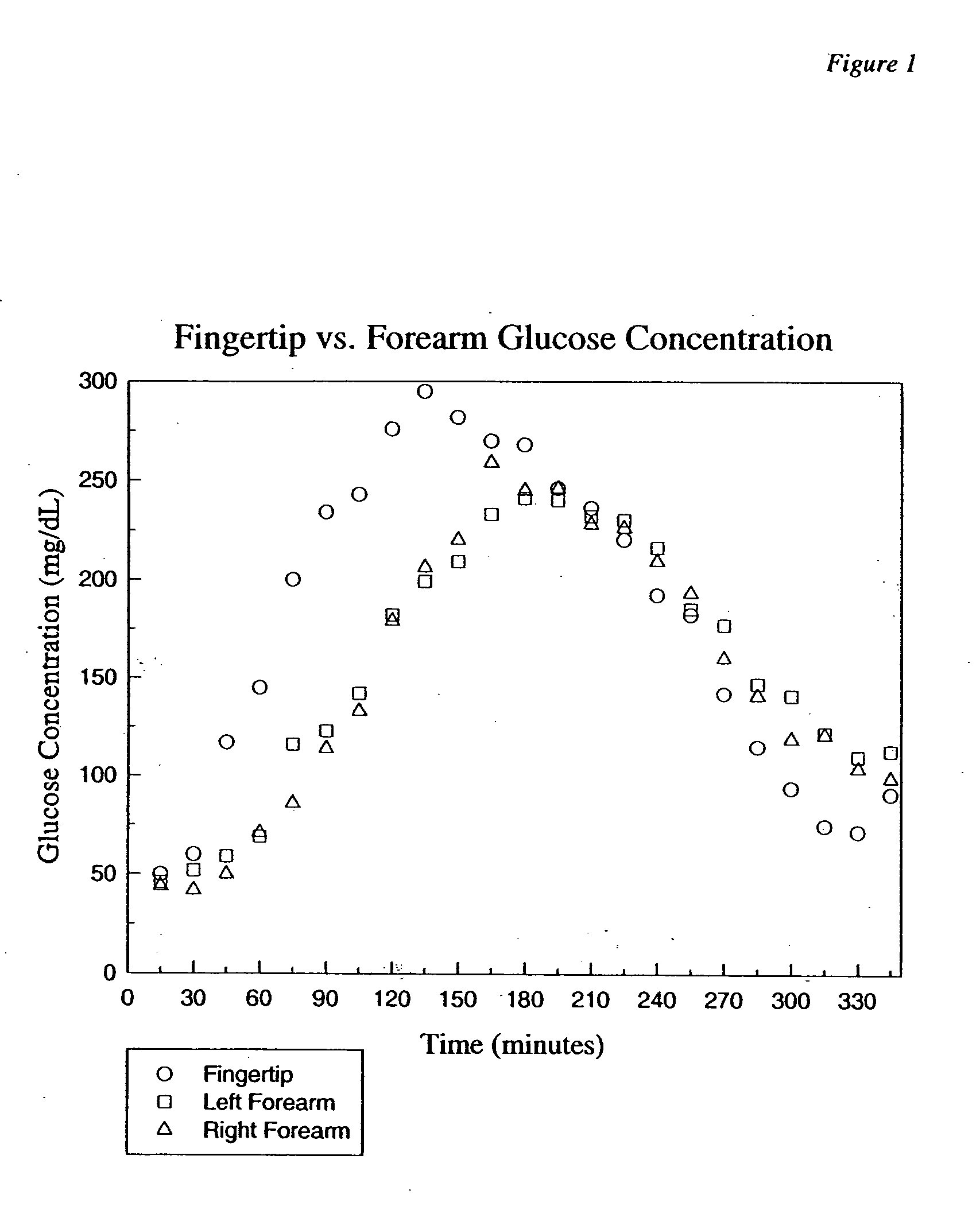
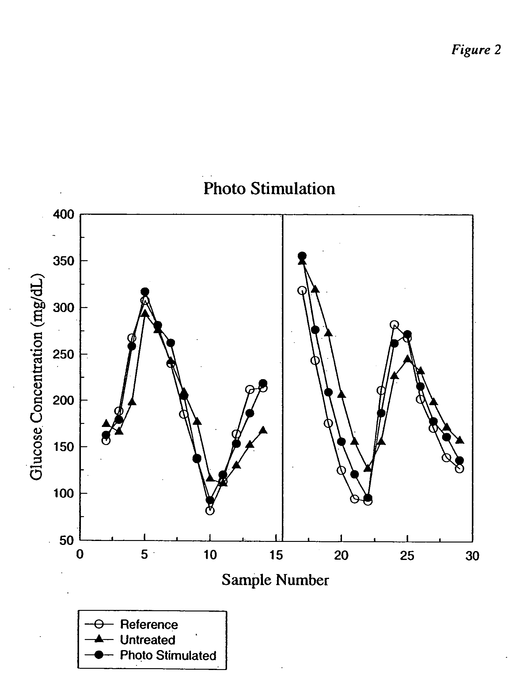
The development of alternative site glucose concentration determinations has somewhat mitigated the pain aspects, but maintains a biohazard issue and may have introduced a difficulty in temporal and spatial differences in glucose concentration between the well perfused fingertip and the less well perfused alternative sites.
However, some of the techniques are applied to the skin in a fashion that draws blood.
This leads to samples being collected with varying analyte and interferent concentrations.
Conversely, devices described above based upon techniques such as a lancet, applied current, laser poration, or suction are referred to as either a traditional invasive or alternative invasive technique as they do not fulfill both the three hour and penetration of skin parameters.
First, at least part of the device penetrates the skin.
The use of intravenous insulin in this study was criticized as creating physiological extremes that influence the observed differences.
Meters with increased error such as 10% are acceptable, though the error of the device being calibrated may increase.
The difference between alternative site glucose concentrations and traditional site glucose concentrations introduces errors associated with sampling into alternative site glucose analyzers.
However, variations in the physiological state and fluid distribution of tissue profoundly affect the optical properties of tissue layers and compartments over a relatively short period of time.
However, this technology has not been suggested for use in combination with noninvasive glucose concentration determination.
Further, minimization of reference glucose concentration differences has not been suggested with the use of photo stimulation.
Finally, to date no FDA device has been approved for use by an individual or a medical professional for noninvasive glucose concentration determination.
This results in certain body constituents constantly being in a state of flux.
Even within the circulatory system, glucose is not always evenly distributed.
Difficulties arise when one portion of the body is sampled to determine or measure a constituent concentration when it is desirable to determine the concentration of that constituent in an alternative body part.

Method used


Image

  • Photostimulation method and apparatus in combination with glucose determination
  • Photostimulation method and apparatus in combination with glucose determination
  • Photostimulation method and apparatus in combination with glucose determination

Examples


example 1

Incident photons, such as those from an LED, are coupled into the skin through air. This has the benefit of not disturbing the sample site by application of pressure. This is beneficial for a noninvasive measurement. However, for an invasive measurement this pressure impact may be minimal. Coupling photons into skin through air is not the most efficient coupling method due to the index of refraction mismatch and the optical roughness of skin.

example 2

Incident photons are coupled to a sample site via coupling optics such as a fiber optic, one or more lenses or flat optics, and / or a coupling fluid. The coupling optics are in direct contact with the skin. This means that the thermal effects of the coupling optics on the sample surface impact the sampling site temperature. This is tolerated or controlled. Similarly, the coupling optics apply at least some pressure to the sampling site that may disturb the sampling site. Again, this may be tolerated or controlled. Those skilled in the mechanical arts will immediately recognize control techniques such as use of thermally stable materials, thermally less or nonconductive materials, temperature controllers, or adjusting the mass in contact with the sample site to achieve desirable thermal control. Those skilled in the mechanical arts will immediately recognize techniques to control pressure effects such as adjusting mass, distributing pressure over an area, use of counter forces, or pe...

example 3

In some instances the photostimulator is not optically attached to the sample site when not in use. In these cases the source is manually turned on or is activated with automatic activation means known to those skilled in the art. For example, activation means include inducement by pressure applied when sampling, by a switch mechanism in a guide, by sensing movement, or by proximity to a magnetic field. Once activated, the duty cycle is continuous or semi-continuous. Photostimulation duration controls include manual and automatically deactivated after a preset time interval. Photostimulation periods include the beginning of a day or operating period, prior to sampling by multiple minutes, just prior to sampling, and during sampling.


PUM


Abstract

A method and apparatus using photo-stimulation to treat or pretreat a sample site prior to analyte concentration determination is presented. More particularly, photo-stimulation at or near at least one sample site is used to enhance perfusion of the sample site leading to reduced errors associated with sampling. Increased perfusion of the sample site leads to increased volume percentages of the target analyte and / or allows the blood or tissue constituent concentrations to more accurately and / or precisely track corresponding sample constituents in more well perfused body compartments or sites such as arteries, veins, or fingertips. In one embodiment, analysis of the photo-stimulated site is used in conjunction with glucose analyzers to determine the analyte concentration with greater ease, accuracy, or precision and may allow determination of the analyte concentration of another non-sampled body part or compartment.

Description

BACKGROUND OF THE INVENTION 1. Technical Field The invention relates generally to biomedical methods and apparatus. More particularly, the invention relates to preparing a tissue sample site for analysis. Still more particularly, the invention relates to the use of photonic stimulation to enhance perfusion of glucose concentrations between body fluid compartments in combination with glucose sampling and / or glucose analysis techniques. 2. Description of the Prior Art Diabetes Diabetes is a chronic disease that results in improper production and use of insulin, a hormone that facilitates glucose uptake into cells. While a precise cause of diabetes is unknown, genetic factors, environmental factors, and obesity appear to play roles. Diabetics have increased risk in three broad categories: cardiovascular heart disease, retinopathy, and neuropathy. Complications of diabetes include: heart disease and stroke, high blood pressure, kidney disease, neuropathy (nerve disease and amputatio...

Claims


Application Information

Patent Timeline
IPC IPC(8): A61B5/00G01N21/27G01N21/35
CPCA61B5/0075A61B5/14532G01N21/359A61B5/1491G01N21/274A61B5/1455A61B2562/146
Inventor BLANK, THOMAS B.MONFRE, STEPHEN L.MAKAREWICZ, MARCYMATTU, MUTUAHAZEN, KEVIN H.HENDERSON, JAMES R.
Owner GLENN PATENT GROUP
Learn More