
Method for rendering digital radiographic images for display based on independent control of fundamental image quality parameters

a digital radiographic image and independent control technology, applied in image enhancement, instruments, applications, etc., can solve the problems of not disclosing a set of parameters that directly and independently control all of the fundamental attributes of image quality, and achieve the control of detail contrast, and noise appearance. , the effect of adjusting the sharpness and noise appearance of the digital imag

Active Publication Date: 2005-01-27
CARESTREAM HEALTH INC
View PDF13 Cites 25 Cited by
  • Summary
  • Abstract
  • Description
  • Claims
  • Application Information

AI Technical Summary

Benefits of technology

1. A method for independently controlling fundamental attributes of image quality of a digital image.
2. A method for controlling detail contrast, sharpness, and noise appearance as a function of log exposure of a digital image is provided.
3. A method for density-dependent control of detail contrast, sharpness and noise appearance of a digital image is provided.

Problems solved by technology

It does not disclose a set of parameters that directly and independently control all of the fundamental attributes of image quality.

Method used


Image

  • Method for rendering digital radiographic images for display based on independent control of fundamental image quality parameters
  • Method for rendering digital radiographic images for display based on independent control of fundamental image quality parameters
  • Method for rendering digital radiographic images for display based on independent control of fundamental image quality parameters

Examples


Embodiment Construction

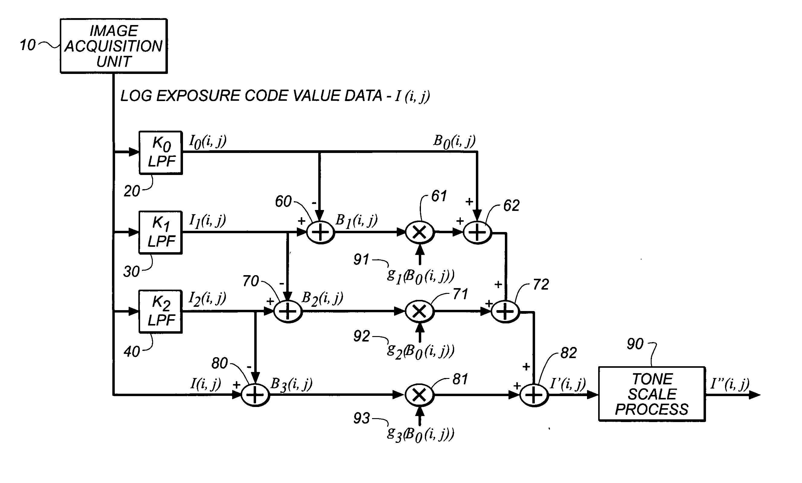
Referring now to FIG. 1, there is shown a block diagram of the present invention. A digital image in which code value is linearly related to log exposure is captured with an image acquisition unit 10. Unit 10 can be for example, a medical image acquisition unit such as, a diagnostic image unit (MRI, CT, PET, US, etc.), a computed radiography or direct digital radiography unit, an x-ray film digitizer, or the like. Any other digital image acquisition unit can also be used). The present invention processes the log exposure code value data, as shown in FIG. 1, accordingly, the digital image data is split into four frequency bands B0(i,j), B1(i,j), B2(i,j), and B3(i,j). The log exposure code value data I(i,j) of the input digital input digital image is first processed by three different low-pass filter operators 20, 30, 40. Each operator uses a square-wave filter. It will be evident to those skilled in the art that other low-pass filter shapes such as a triangle-filter can be used. The...


PUM


Abstract

A method of enhancing a digital image comprising: providing a digital image; decomposing the provided digital image into a multi-frequency band representation including a low frequency band image and multiple different high frequency band images; multiplying each of the high-frequency band images with a gain factor; summing together the unmodified low frequency band image and the modified high-frequency band images to produce a reconstructed digital image; and mapping the reconstructed digital image through a tone-scale look-up-table to map the reconstructed digital image to optical densities

Description

FIELD OF THE INVENTION This invention relates in general to image enhancement in digital image processing and in particular, to a method of image enhancement to be used in a medical radiographic imaging system, such as a computed radiography system. BACKGROUND OF THE INVENTION It is a common desire to enhance images acquired from imaging devices to improve their image quality. There are fundamental attributes that govern the image quality of a grayscale image. These attributes are the brightness of an image, the dynamic range of an image, the contrast of detail in an image (or detail contrast), the sharpness of edges in an image, and the appearance of noise in an image. It is beneficial to provide a system that enables direct and independent control of these attributes of image quality. Further a system, which enables the control of the detail contrast, sharpness, and noise appearance in a density dependent fashion is also beneficial. U.S. Pat. No. 5,805,721, inventors P. Vuylste...

Claims


Application Information

Patent Timeline
IPC IPC(8): A61B6/03G06K9/00A61B5/00G06K9/40G06T1/00G06T5/00G06T5/20H04N1/409
CPCG06T2207/20016G06T5/004G06T5/75
Inventor COUWENHOVEN, MARY E.SENN, ROBERT A.
Owner CARESTREAM HEALTH INC
Learn More