
Method for assay of antibodies and antibody assay device

a technology of antibodies and assay devices, applied in the field of antibodies and antibody assay devices, can solve problems such as false positive tests

Inactive Publication Date: 2003-09-18
OTSUKA PHARM FAB INC
View PDF22 Cites 12 Cited by
  • Summary
  • Abstract
  • Description
  • Claims
  • Application Information

AI Technical Summary

Benefits of technology

[0169] The amount of the E. coli component (LPS) to be incorporated in the sample may for example be generally not less than 5 .mu.g / ml, preferably 5.about.100 .mu.g / ml, more preferably 10.about.50 .mu.g / ml. Although the use of an amount over 100 .mu.g / ml is not prohibitive, the effect of the invention can be accomplished at the addition level of up to 100 .mu.g / ml.
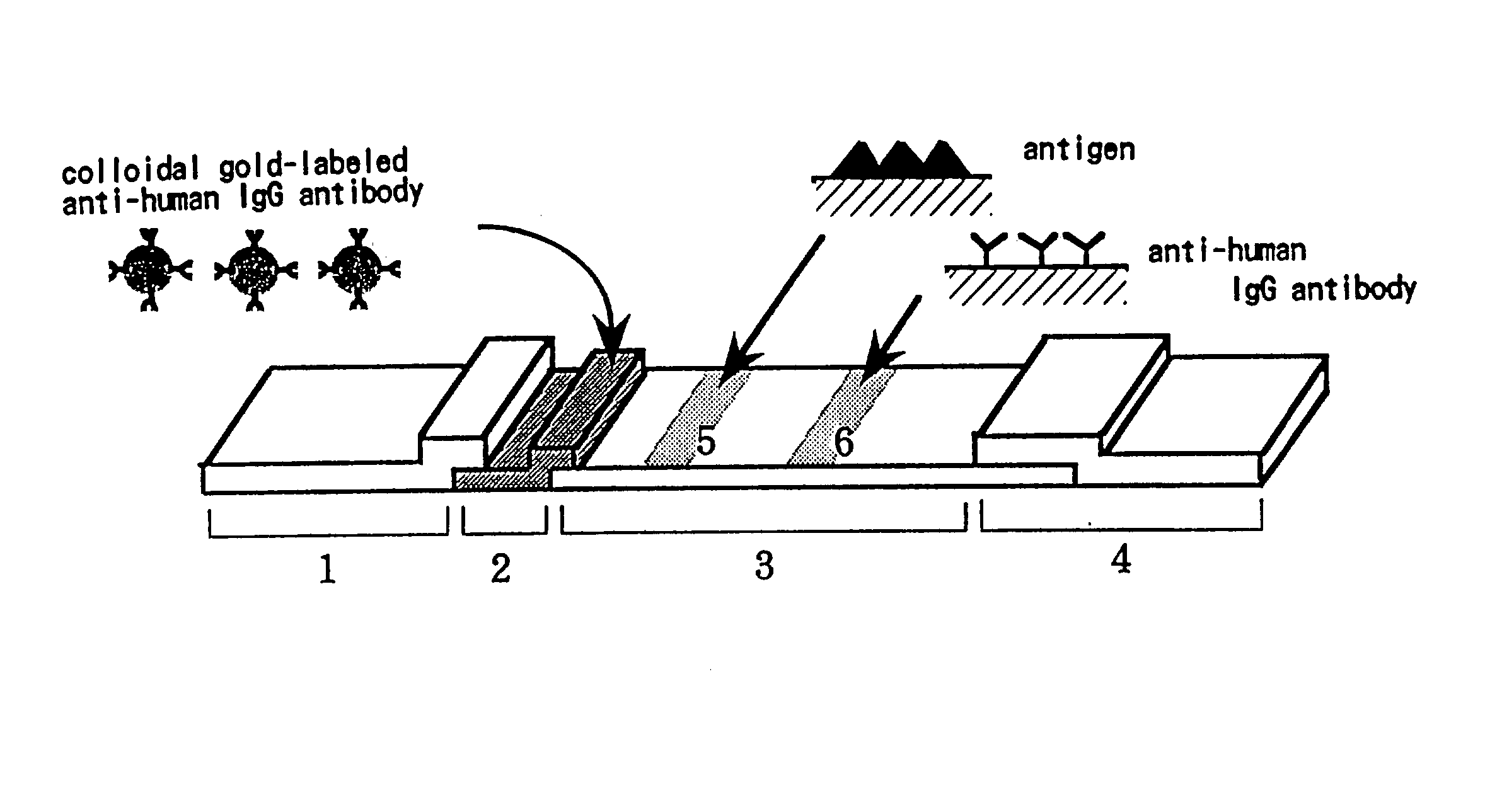
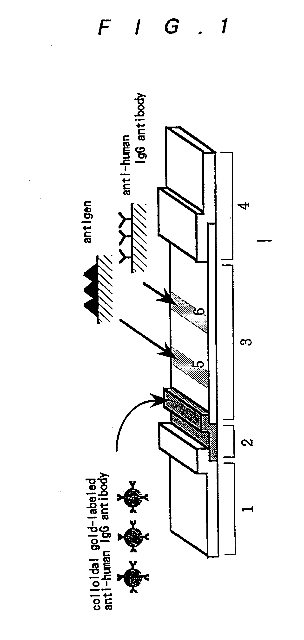
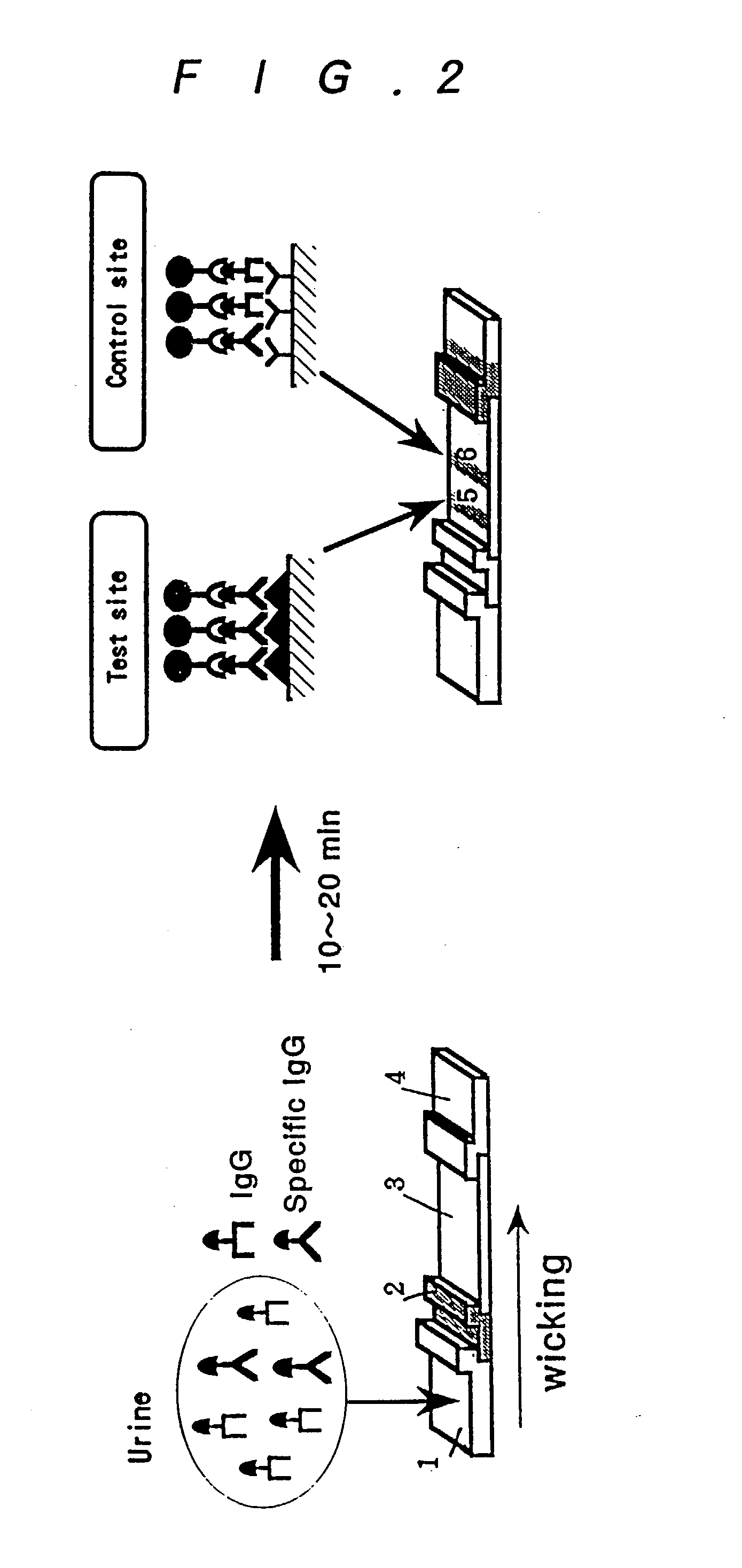
[0170] As the sample is applied to the first region (1), the first region 1 is wetted. The sample applied flows through the first region (1) into the tracer region (2) by capillary action and comes into contact and reacts with the labeled ligand (colloidal gold-labeled anti-human IgG antibody) removably supported in the tracer region.
[0171] When a suitable sample is used and the target antibody is contained in the sample, both the target antibody and arbitrary antibody contained in the sample are coupled to said labeled ligand in the tracer region (2) to form a target antibody / labeled ligand complex and an arbitrary antibody / labeled ligand complex, respectively. After passage of the same through the tracer region (2), the respective complexes or the labeled ligand not forming such complexes are transported together with the sample downstreams of the tracer region (2). In a preferred mode, the labeled ligand not forming a complex yet is given a sufficient time (space) for forming complexes as it travels with the antibody-containing sample from the tracer region (2) to the test site (5) of the second region (3) by capillary action. As the sample reaches the test site (5) in the second region, the target antibody / labeled ligand complex in the sample is coupled to the ligand supported in the test site (5) and immobilized in situ. The sample further migrates downstreams by capillary action to reach the control site (6) where the arbitrary antibody / labeled ligand complex is coupled to the ligand (anti-human immunoglobulin antibody) in that site and immobilized there. Then, by detecting the complexes immobilized in the test site (5) and control site (6) in the second region according to the label component of the labeled ligand, the assay result can be indicated as a positive test. In contrast, when a sample not containing the target antibody is applied, said target antibody / labeled ligand complex to be immobilized in the test site (5) is not formed so that the label is not detected in this test site (5) (a negative test).
[0172] In this connection, the negative test indication in the test site (5) includes both a negative (false negative) test due to a low concentration of the sample (i.e. a small total amount of antibody in the sample) and a negative (true negative) test due to the absence of the target antibody in the sample. These negative tests cannot be differentiated from each other according to the result in the test site (5) alone. However, in the case of a false negative test, said arbitrary antibody / labeled ligand complex to be immobilized in the control site (6) is not formed so that the control site gives a negative indication, while in the case of a true negative test, said arbitrary antibody / labeled ligand complex is formed so that the control site gives a positive indication. Therefore, according to whether the indication at the control site is negative or positive, it is possible to tell whether the negative result in the test site (5) is a false negative test or a true negative test. Furthermore, when a labeled ligand which has been deactivated is used or otherwise a proper assay is not performed in a proper system, the control site (6) gives a negative indication so that the finding of a false negative test owing to such causes can be prevented.
[0173] The sample liquid containing all the unbound antibodies, labeled ligands, etc. continues to migrate further downstreams of the second region (3) to the third region (4). Optionally an indicator zone may be provided and, in this case, the liquid front coveys the dye from the indicator zone to the end-point zone, thus indicating the passage of the liquid and dye through the third region and completion of the assay.
[0174] FIG. 3 shows an example of the antibody assay device of the invention for use in horizontal position. The solid phase support (A) comprising the first region (1), tracer region (2), second region (3) (including the test site (5) and control site (6)) and third region (4) is accommodated in a housing (B) made of a suitable material. The housing material is preferably a moldable plastic material such as polystyrene, although other materials such as glass, metal and paper can also be used. The housing consists of an upper section (7) having several apertures and a lower section (8), and the solid phase support is disposed on the lower section of the housing and covered with the upper section on top thereof. The apertures (9) and (10) in the upper section of the housing are disposed in alignment with the series of regions of the solid phase support and in the positions corresponding to the first region (1) and second region (3), respectively, of the solid phase support.

Problems solved by technology

The inventors further discovered that said nonspecific binding antibody component comprises IgG fragments and / or their denaturation products which retain the antigenicity of the light (L) chain or F (ab) region of the IgG and that this antibody component cross-reacts with the ordinary antibody assay reagents (e.g. secondary antibodies) used in serum antibody assay systems, thus leading to false positive tests.

Method used


Image

  • Method for assay of antibodies and antibody assay device
  • Method for assay of antibodies and antibody assay device
  • Method for assay of antibodies and antibody assay device

Examples


example 2

[0207] Assay of Anti-Hepatitis B Virus (HBc) Antibody

[0208] (1) Assay of Anti-hepatitis B Virus (HBc) Antibody

[0209] An antigen plate was prepared using HBc antigen (Chemicon International) in accordance with the procedure of Example 1 (2) and the assay of anti-hepatitis B (core) (HBc) antibody in urine samples was carried out in accordance with Example 1 (4).

[0210] Thus, 25 .mu.l of first buffer containing the E. coli extract in a varying concentration and 100 .mu.l of sample urine were added to each well of the HBc antigen plate and after 10 seconds' stirring, the plate was allowed to sit at 37.degree. C. for 1 hour. After the plate was washed with 6 portions of PBST, 100 .mu.l of a 11,000-fold dilution of enzyme (HRP)-labeled anti-human IgG antibody in second buffer was added and the plate was allowed to sit at 37.degree. C. for 1 hour and then washed (6 times with PBST).

[0211] Then, 100 .mu.l of a color developer solution was added and the reaction was carried out at room temper...

example 3

[0216] Using urine samples which gave false positive tests in the determination of anti-HIV antibody in urine by a known assay method [(Calypte.TM. HIV-1 Urine EIA: Arch. Pathol. Lab. Med., 119, 139-141 (1995); Clinical Infectious Diseases, 19, 1100-1104 (1994)], an exploratory experiment was carried out to identify the component supposedly responsible for a nonspecific reaction in the same assay system.

[0217] (1) Each of the above urine samples was adjusted to pH 7.4 with 1 M phosphate buffer (pH 7.7) and filtered through 5.0, 0.8 and 0.2 .mu.m-cut filters. A 20 ml portion of the filtrate was concentrated by ultrafiltration (a 10 kDa-cut membrane) to 2 ml. The concentrated urine was subjected to gel permeation chromatography (Sephacryl S-300, Pharmacia) and each fraction was tested for its reactivity to HIV antigen.

[0218] The reactivity to HIV antigen was confirmed by causing each fraction to react with an HIV antigen-immobilized plate prepared by immobilizing HIV antigen (gp160) a...

example 5

[0229] Assay of Anti-H. pylori Antibody in Urine

[0230] (1) Preparation of an H. pylori Antigen Plate

[0231] To a 100 mg / ml suspension of Helicobacter pylori (a clinical isolate) in cold Dulbecco-PBS as prepared in the routine manner [J. Clin. Microbiol., 29: 2587-2589 (1991)], an equal volume of cold 0.2% Triton-X solution was added under constant stirring with a stirrer and the mixture was further stirred for 5 minutes and centrifuged (3,000 rpm, 20 min.). The supernatant was transferred to a new tube for use as the extract (1.about.1.5 mg / ml as protein).

[0232] This extract was diluted with D-PBS (2.5 .mu.g / ml) and the dilution was distributed into a 96-well plate, 100 .mu.l per well, and incubated at 25.degree. C. overnight. After each well was washed, 300 .mu.l of a blocking solution (D-PBS, 0.5% casein, 5% sorbitol, 0.05% NaN.sub.3 (pH 7.4)) was added, followed by incubation at 25.degree. C. overnight. The blocking solution was then discarded and the plate was dried at 25.degree....


PUM

PropertyMeasurementUnit
temperatureaaaaaaaaaa
thickaaaaaaaaaa
thickaaaaaaaaaa

Abstract

The invention relates to a technology by which antibodies directed to sources of infection in body fluids can be assayed with high accuracy, expediency and specificity. More particularly, the invention provides an antibody immunoassay method in which the antigen-antibody reaction between a target antibody in a sample and an assay antigen is conducted in the presence of an E. coli component and an antibody assay method which comprises using a reagent having a specific affinity for the Fc region of an antibody IgG as the antibody assay reagent. The invention further provides an antibody assay device comprising a solid-phase support having at least (a) a first region to which a sample is applied and (b) a second region where the antibody in the sample is reacted as disposed in such an arrangement that the sample is wicked from the first region to the second region and a labeling means for detection of the result of the reaction in the second region, characterized in that the (b) second region is provided with (i) a test site in which a ligand for capturing the target antibody to be assayed is immobilized and (ii) a control site in which a ligand for capturing an arbitrary antibody occurring in the sample is immobilized.

Description

[0001] The present invention relates to a method of detecting or quantitating antibodies in samples and more particularly to a method by which antibodies against sources of infection such as bacteria and viruses as occurring in clinical body fluid samples, particularly urine samples, can be detected or assayed with high accuracy, expediently, and with good specificity.[0002] The present invention in a further aspect relates to a device for detecting or quantitating an antibody in a sample and more particularly to a device with which the antibody against a source of infection as occurring in clinical body fluid samples, particularly urine samples, can be detected or assayed with high accuracy, expediently, and with good specificity.[0003] The invention further relates to an antibody assay reagent kit which is useful for the above antibody assay method and the assay method using said antibody assay device.[0004] Detection of antibodies specific to various sources of infection (pathoge...

Claims


Application Information

Patent Timeline
Patent Type & Authority Applications(United States)
IPC IPC(8): G01N33/53G01N33/543G01N33/558G01N33/569G01N33/68
CPCG01N33/5306G01N33/54393G01N2333/245G01N33/56916G01N33/6857G01N33/558Y10S436/824Y10S435/962Y10S435/849Y10S435/948G01N2474/20G01N33/543
Inventor TACHIKAWA, TETSUYANODA, ATSUNARIKATSURAGI, KIYONORI
Owner OTSUKA PHARM FAB INC
Learn More