Method for manufacturing telencephalon or progenitor tissue thereof
a technology of telencephalon and progenitor tissue, which is applied in the field of manufacturing telencephalon or progenitor tissue thereof, can solve the problem of elusive detail of early human corticogenesis
- Summary
- Abstract
- Description
- Claims
- Application Information
AI Technical Summary
Benefits of technology
Problems solved by technology
Method used
Image
Examples
example 1
Selective Three Dimensional Formation of Cortical Progenitor Tissue from Human Pluripotent Stem Cells
(Method)
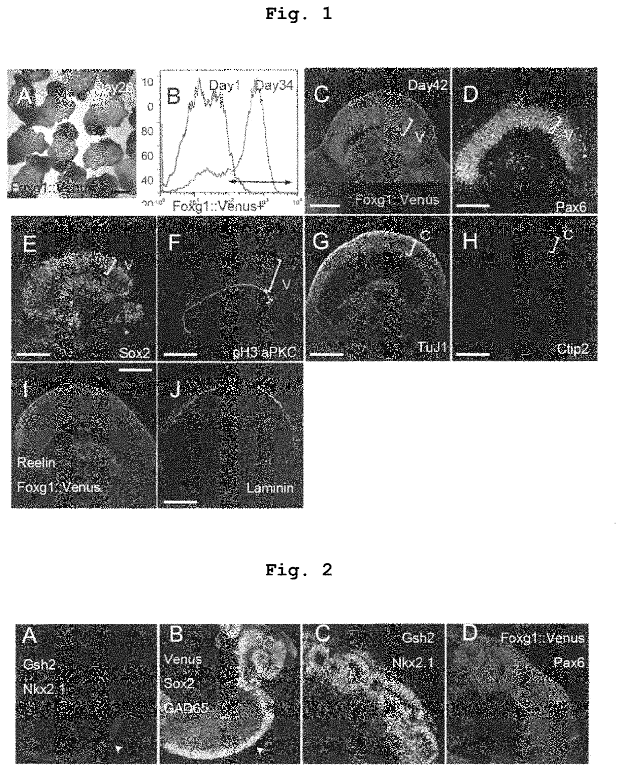
[0249]Human ES cells (KhES-1; a fluorescence protein gene Venus is knocked-in a telencephalon specific gene Foxg1) were dispersed to single cells by a trypsin treatment, and according to the SFEBq method (Nakano et al, Cell Stem Cell, 2012), aggregates were formed and subjected to suspension aggregate culture at 37° C. in the presence of 5% CO2 for differentiation induction. The dispersed 9000 human ES cells were seeded in each well of a V bottom 96 well plate applied with a low cell adsorptive surface coating, and a growth factor-free G-MEM medium (Gibco / Invitrogen) added with 20% KSR (Knockout Serum Replacement), 0.1 mM non-essential amino acid solution (Gibco / Invitrogen), 1 mM sodium pyruvate solution (Sigma), and 0.1 mM 2-mercaptoethanol was used as the medium for differentiation induction. To suppress dispersion-induced cell death, 20 μM of a ROCK inhibitor Y-27632 was a...
example 2
Three Dimensional Formation of Basal Ganglia Progenitor Tissue from Human Pluripotent Stem Cells
(Method)
[0251]Up to day 35 of differentiation induction, cells were cultured under culture conditions similar to those of Example 1. That is, human ES cell aggregates were cultured in a V bottom 96 well plate up to 18 days after differentiation induction, suspended aggregates were transferred to a non-cell adhesive petri dish (diameter 6 cm), and suspension culture was performed at 37° C. in the presence of 5% CO2, 40% O2 from day 18 to day 35 of differentiation induction. However, Sonic hedgehog (Shh) signal agonist SAG was added to the culture medium of Example 1 at a final concentration of 30 nM or 500 nM only in the period of from day 15 to day 21 and allowed to react. The aggregate was analyzed by immunohistostaining on day 35.
(Results)
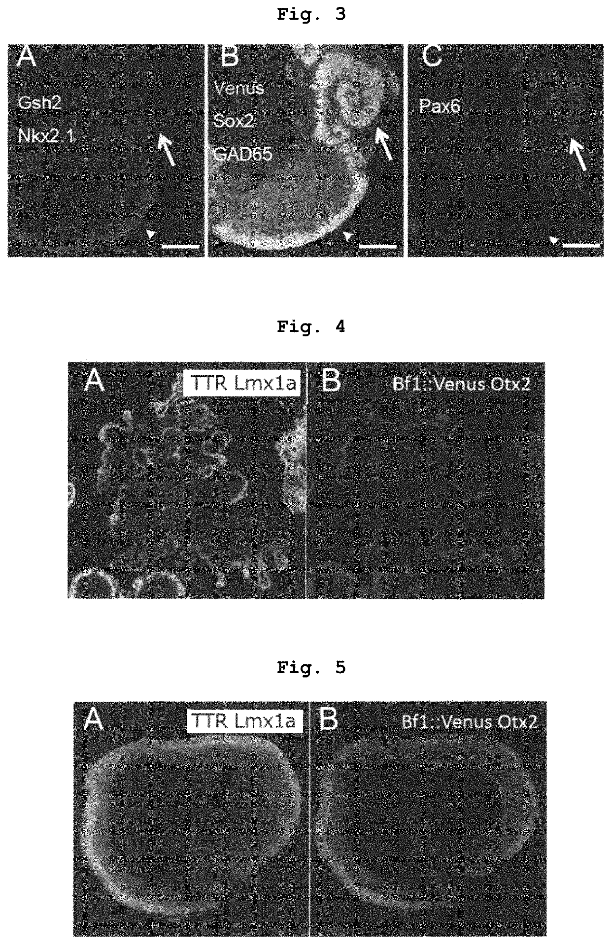
[0252]When 30 nM Shh signal agonist SAG was reacted, lateral ganglionic eminence (LGE) expressing Gsh2 was formed in the Foxg1::venus positive telence...
example 3
Continuous Three Dimensional Formation of Cerebral Cortex and Basal Ganglion
(Method)
[0254]Up to day 35 of differentiation induction, cells were cultured under culture conditions similar to those of Example 2. That is, human ES cell aggregates were cultured in a V bottom 96 well plate up to 18 days after differentiation induction, suspended aggregates were transferred to a non-cell adhesive petri dish (diameter 9 cm), and suspension culture was performed at 37° C. in the presence of 5% CO2, 40% O2, from day 18 to day 35 of differentiation induction. However, Shh signal agonist SAG was added to the culture medium at a concentration of 30 nM only in the period of from day 15 to day 21 of differentiation induction. The aggregate was analyzed by immunohistostaining on day 35.
(Results)
[0255]As shown in Example 2, when 30 nM Shh signal agonist SAG was reacted, the telencephalon neuroepithelium was Foxg1::venus positive, and expressed lateral ganglionic eminence (LGE) markers Gsh2, GAD65 (F...
PUM
| Property | Measurement | Unit |
|---|---|---|
| diameter | aaaaa | aaaaa |
| diameter | aaaaa | aaaaa |
| cleavage angle | aaaaa | aaaaa |
Abstract
Description
Claims
Application Information
 - R&D
- Intellectual Property
- Life Sciences
- Materials
- Tech Scout
- Unparalleled Data Quality
- Higher Quality Content
- 60% Fewer Hallucinations
Browse by: Latest US Patents, China's latest patents, Technical Efficacy Thesaurus, Application Domain, Technology Topic, Popular Technical Reports.
© 2025 PatSnap. All rights reserved.Legal|Privacy policy|Modern Slavery Act Transparency Statement|Sitemap|About US| Contact US: help@patsnap.com


