Deep-learning-based cancer classification using a hierarchical classification framework

a hierarchical classification and deep learning technology, applied in the field of deep learning based magnetic resonance imaging methods, can solve the problems of high false positive rate, difficult task of accurate cancer classification, and confusion in patients, and achieve the effect of avoiding false positives, avoiding overtreatment, and avoiding overtreatmen

Active Publication Date: 2021-03-09
RGT UNIV OF CALIFORNIA
View PDF11 Cites 0 Cited by
  • Summary
  • Abstract
  • Description
  • Claims
  • Application Information

AI Technical Summary

Benefits of technology

[0015]The present technology provides a deep-learning-based computerized cancer classification system and method using magnetic resonance imaging and preferably using multi-parametric magnetic resonance imaging (mp-MRI). The hierarchical classification framework avoids (1) the need to provide handcrafted features (e.g., features predefined by a domain expert) as well as (2) the need for precise segmentation of cancerous lesions (e.g., exact delineation of lesion boundaries by a human or computerized algorithm).
[0017]One preferred embodiment uses a pre-trained convolution neural network (CNN) as a feature extractor to overcome the issue of having limited training samples (e.g., datasets that have been labeled with outcome information), which is often the case in the medical domain. Furthermore, instead of a precise contour of the lesion, the method only requires approximate image patches containing the lesion as input (e.g., versus precise lesion boundaries), which is much more convenient and robust. Finally, the method combines the outputs of classifiers in a layered manner, providing a more efficient way to combine features from deep learning and conventional clinical features. This hybrid approach allows the incorporation of additional features beyond imaging and results from multiple modalities as inputs to generate a final classification: this two-step classification approach yields higher accuracy compared to using a single classification step.
[0019]In one embodiment described herein, a pre-trained CNN method may be used to alleviate the learning requirement. However, the method can incorporate both pre-trained and non-pre-trained CNN methods, depending on the availability of labeled training data that can attain sufficient feature learning. One CNN method known as OverFeat, for example, allows the use of deep learning out-of-the-box when limited training data sets are available as the deep features are pre-trained using a dataset of natural images (a set of 1.2 million natural images).
[0021]It will be appreciated that the model-based approach is capable of accurately classifying the tumor Gleason Score to assist radiologists with staging lesions. The classification framework is capable of improving diagnostic accuracy by learning from the growing amounts of clinical and imaging data that are being collected as part of routine care.
[0023]Another aspect of the technology is to provide a method for classifying lesions that builds a hybrid two-order classification model that combines deep learning and conventional statistical features. Another aspect of the technology is to provide a method for classifying lesions that does not require precise lesion boundaries and anatomical-location-specific training.

Problems solved by technology

Accurate cancer classification is often a difficult task because of the ambiguous appearance of lesions that are regularly observed on diagnostic imaging studies.
Unfortunately, the Gleason grading system can assign the same score to significantly different risk groups and therefore other grading systems have been proposed.
This scoring can cause confusion in patients who assume that their cancer is serious because their cancer is in the middle of the scale and that aggressive treatment may be needed.
However, prostate cancer is challenging to diagnose because biopsies do not provide a uniform and accurate sampling of the prostate tissue, leading to high false positive rates and potential overtreatment.
However, the MRI cannot detect all prostate tumors and has shown poor sensitivity for imaging low volume Gleason (3+3) tumors.
MRI imaging can also be very challenging and often requires a significant amount of post processing and analysis by very experienced radiologists because of low imaging sensitivity.
One well identified limitation of mp-MRI is that the tumors or nodules may not be clearly demarcated or encapsulated because the margins of the nodules are indistinct in the image.
Although multi-parametric MRI is a promising imaging modality for the detection and grading of prostatic carcinoma, current mp-MRI scoring systems, such as PI-RADS, are generally subjective and have a limited ability to distinguish between indolent and clinically significant cancers.

Method used


Image

  • Deep-learning-based cancer classification using a hierarchical classification framework
  • Deep-learning-based cancer classification using a hierarchical classification framework
  • Deep-learning-based cancer classification using a hierarchical classification framework

Examples


example 1

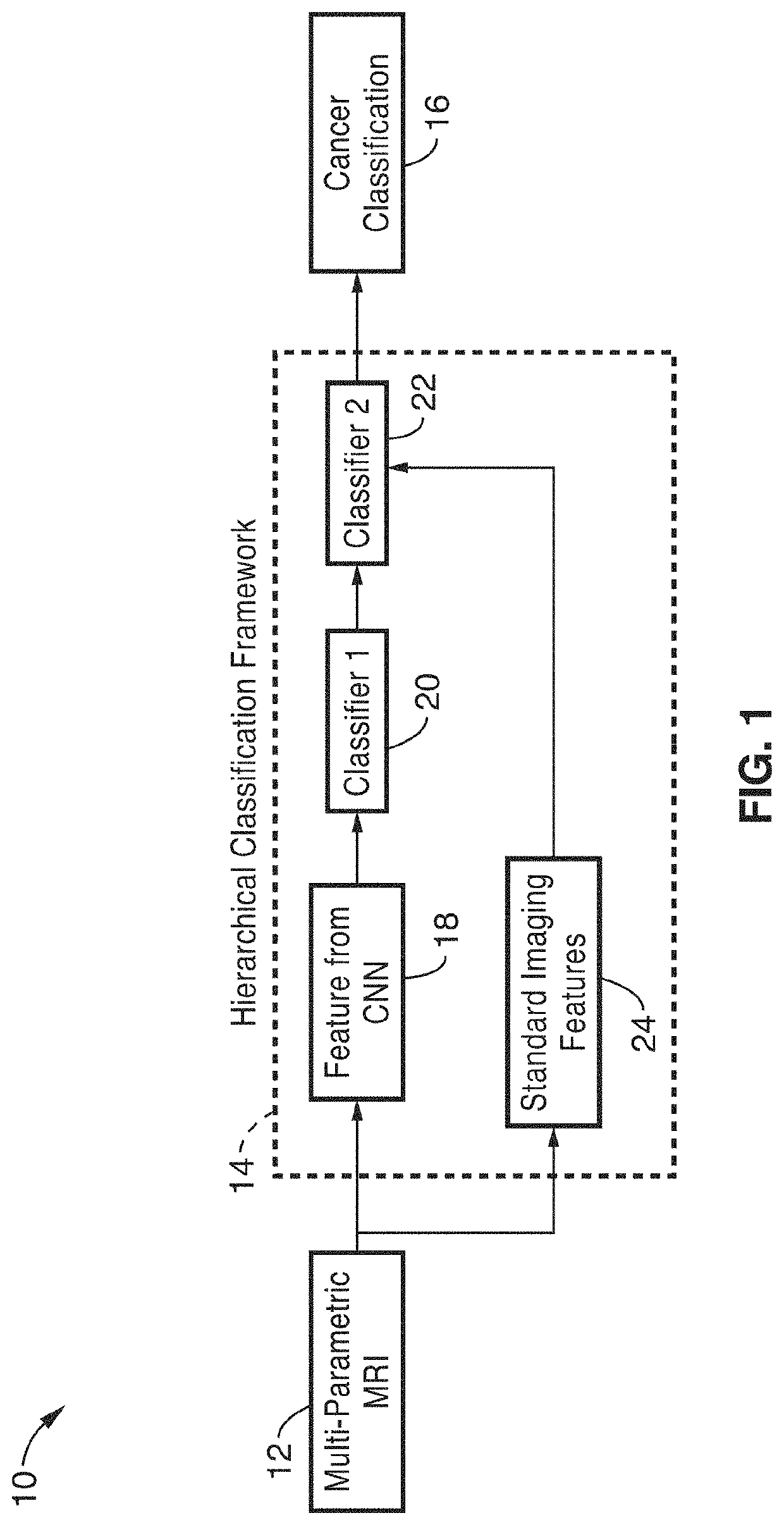
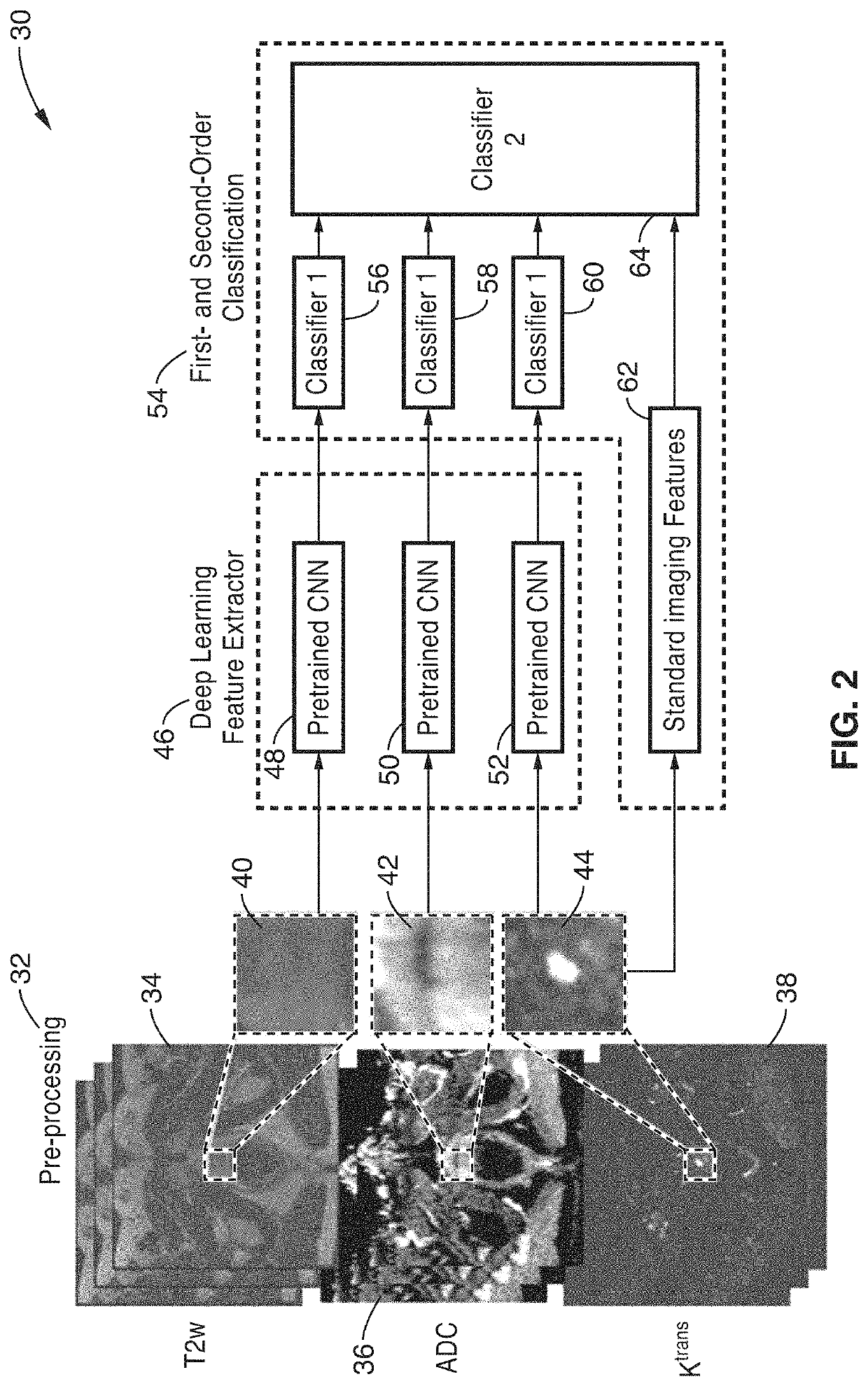
[0043]In order to demonstrate the operational principles of the apparatus and imaging and classification methods 30, a dataset of mp-MRI images were recorded for a total of 68 patients and processed using the processing steps shown generally in FIG. 1 and FIG. 2. A study cohort of 68 consecutive men who underwent 3.0T mp-MRI (Skyra and Trio, Siemens Healthcare) prostate imaging prior to radical prostatectomy was acquired. Each mp-MRI study, including T2-weighted (T2w), DWI and DCE images, was correlated with whole mount histopathology by experienced GU pathologists, and lesions were matched with respect to location, size and Gleason Score (GS). Indolent PCa cases were defined as having a GS smaller than seven (GS≤6) and cancerous (CS) PCa ones were defined has having a GS that was larger or equal to seven (GS≥7). A total of 102 lesions were identified, including 48 indolent and 54 CS sub-cohorts.

[0044]As shown schematically in FIG. 2, the acquired images were initially preprocessed ...

example 2

[0048]To demonstrate the effectiveness of the system, the four classification models were built and compared. Specifically, four different SVMs were built using only fs, fT2, fADC or fK, respectively. The performance of these models was also evaluated with a five-fold cross validation using the whole dataset. The results were measured using the mean areas under curve, mean accuracy, mean sensitivity and mean specificity as shown in Table 1.

[0049]FIG. 3 is a graph depicting the receiver operating characteristic (ROC) curves for the four models in this example. The model of the system (fs+fT2+fADC+fK) shown in a solid line in FIG. 3 achieved the highest performance compared to the other models. It can be seen from FIG. 3 and Table 1 that the standard model using six statistical features (fs) achieved the lowest performance mainly due to lack of accurate lesion contours and anatomical-location-specific training. The results also suggest that deep features significantly contribute to th...


PUM


Abstract

An automatic classification method for distinguishing between indolent and clinically significant carcinoma using multiparametric MRI (mp-MRI) imaging is provided. By utilizing a convolutional neural network (CNN), which automatically extracts deep features, the hierarchical classification framework avoids deficiencies in current schemes in the art such as the need to provide handcrafted features predefined by a domain expert and the precise delineation of lesion boundaries by a human or computerized algorithm. This hierarchical classification framework is trained using previously acquired mp-MRI data with known cancer classification characteristics and the framework is applied to mp-MRI images of new patients to provide identification and computerized cancer classification results of a suspicious lesion.

Description

CROSS-REFERENCE TO RELATED APPLICATIONS[0001]This application is a 35 U.S.C. § 111(a) continuation of PCT international application number PCT / US2017 / 024071 filed on Mar. 24, 2017, incorporated herein by reference in its entirety, which claims priority to, and the benefit of, U.S. provisional patent application Ser. No. 62 / 312,946 filed on Mar. 24, 2016, incorporated herein by reference in its entirety. Priority is claimed to each of the foregoing applications.[0002]The above-referenced PCT international application was published as PCT International Publication No. WO 2017 / 165801 on Sep. 28, 2017, which publication is incorporated herein by reference in its entirety.STATEMENT REGARDING FEDERALLY SPONSORED RESEARCH OR DEVELOPMENT[0003]This invention was made with Government support under 1436827, awarded by the National Science Foundation. The Government has certain rights in the invention.NOTICE OF MATERIAL SUBJECT TO COPYRIGHT PROTECTION[0004]A portion of the material in this pate...

Claims


Application Information

Patent Timeline
Patent Type & Authority Patents(United States)
IPC IPC(8): A61B5/00A61B5/055G06T7/00G06K9/42G06K9/52G06K9/62G06K9/66A61B5/20A61B5/08
CPCA61B5/7267A61B5/055A61B5/4381G06K9/42G06K9/527G06K9/6269G06K9/66G06T7/0012A61B5/08A61B5/201A61B5/4244G06T2207/10088G06T2207/20081G06T2207/20084G06T2207/30096G16H50/70G06F18/2411
Inventor SUNG, KYUNG HYUNHSU, WILLIAMSHEN, SHIWENZHONG, XINRAN
Owner RGT UNIV OF CALIFORNIA
Why Eureka
Try Eureka