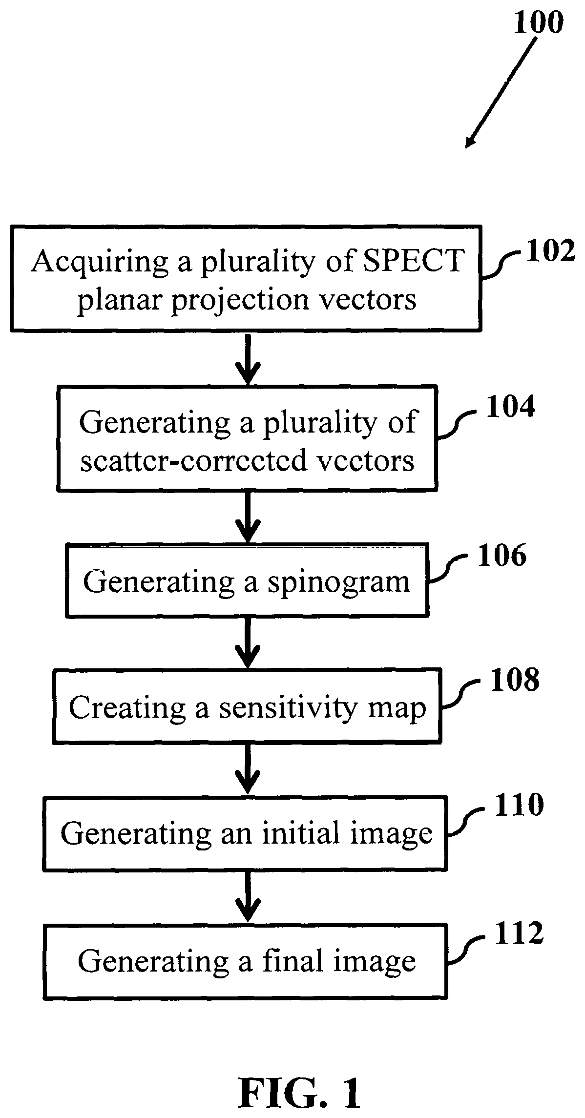
Single photon emission computed tomography imaging with a spinning parallel-slat collimator
a computed tomography and parallel-slat collimator technology, applied in the field of medical imaging, can solve the problems of impeded further development, lack of fast image reconstruction methods and appropriate imaging settings, and long total scan tim
- Summary
- Abstract
- Description
- Claims
- Application Information
AI Technical Summary
Benefits of technology
Problems solved by technology
Method used
Image

Examples
example
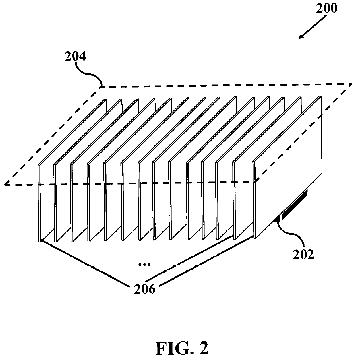
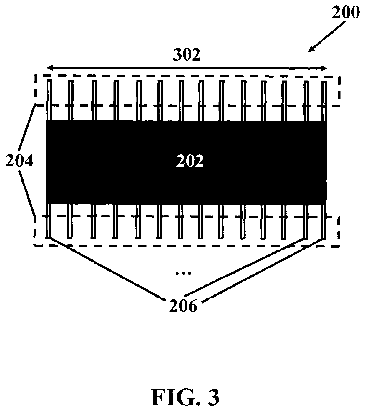
[0059]Table 1 presents the specifications of one example of a heart-dedicated SPECT imaging system. The size of this example system is about 30 cm×15 cm×5 cm. In addition, the example system includes of a series of highly attenuating parallel slats mounted on a monolithic CsI(Na) crystal. Planar projections are acquired at Ns=16 SPECT spin angles, over a 180° span. To adapt the example system for cardiac applications, the system rotates around 180° with 16 stops, at Nr=16 SPECT rotation angles. Therefore, there are a total of 256 planar projections (16 SPECT spin angles×16 SPECT rotation angles). Total scan time is set to 32 min. The radius-of-rotation (ROR) of the system is set to 20 cm. Using the monolithic CsI(Na) crystal along with SiPM readout provides a low-weight, small-footprint, low-cost, and magnetic resonance (MR)-compatible cardiac SPECT imaging system.
[0060]
TABLE 1Specifications of an example heart-dedicated SPECT imaging systemParameterSpecificationDetector typeMonolit...
PUM
 Abstract
Description
Claims
Application Information
 - R&D
- Intellectual Property
- Life Sciences
- Materials
- Tech Scout
- Unparalleled Data Quality
- Higher Quality Content
- 60% Fewer Hallucinations
Browse by: Latest US Patents, China's latest patents, Technical Efficacy Thesaurus, Application Domain, Technology Topic, Popular Technical Reports.
© 2025 PatSnap. All rights reserved.Legal|Privacy policy|Modern Slavery Act Transparency Statement|Sitemap|About US| Contact US: help@patsnap.com