Mobile Molecular Imaging System and Intervention System Including Same

a molecular imaging and mobile technology, applied in the field of medical procedures using molecular imaging techniques, can solve the problems of hardly compatible geometries, imaging techniques, and limited resources of full body equipmen

Inactive Publication Date: 2019-01-17
GENERAL EQUIP FOR MEDICAL IMAGING SL
View PDF0 Cites 2 Cited by
  • Summary
  • Abstract
  • Description
  • Claims
  • Application Information

AI Technical Summary

Benefits of technology

The patent describes a mobile version of a molecular imaging system, such as a PET or SPECT device. It includes a group of detector modules with a rounded shape, each containing multiple detector modules. This design allows for improved image resolution and accuracy. The technical effect of this invention is to provide a portable and efficient tool for molecular imaging that can be easily moved and used in different locations.

Problems solved by technology

A current limitation of nuclear medicine techniques is its unique diagnostic application because the equipment presents geometries hardly compatible with any type of intervention in the patient during the scan.
Neither the full body equipment nor the dedicated organ equipment with closed configurations have the capacity to observe any intervention on the patient in real time.
There are medical imaging techniques that do allow viewing or tracking interventions, but are limited to structural imaging such as CT (computerized tomography), MR (magnetic resonance) or ultrasounds, wherein one can observe the morphology of the organ to be intervened, but not the biological functionality that a nuclear medicine system would be able to envision.
Many of these systems also do not allow real-time monitoring, but only taking pictures at different times of the process to ensure that the intervention is carried out at the desired morphological point.
Ultrasound based systems are an exception, allowing real-time images, but very low resolution and specificity.

Method used


Image

  • Mobile Molecular Imaging System and Intervention System Including Same
  • Mobile Molecular Imaging System and Intervention System Including Same
  • Mobile Molecular Imaging System and Intervention System Including Same

Examples


example

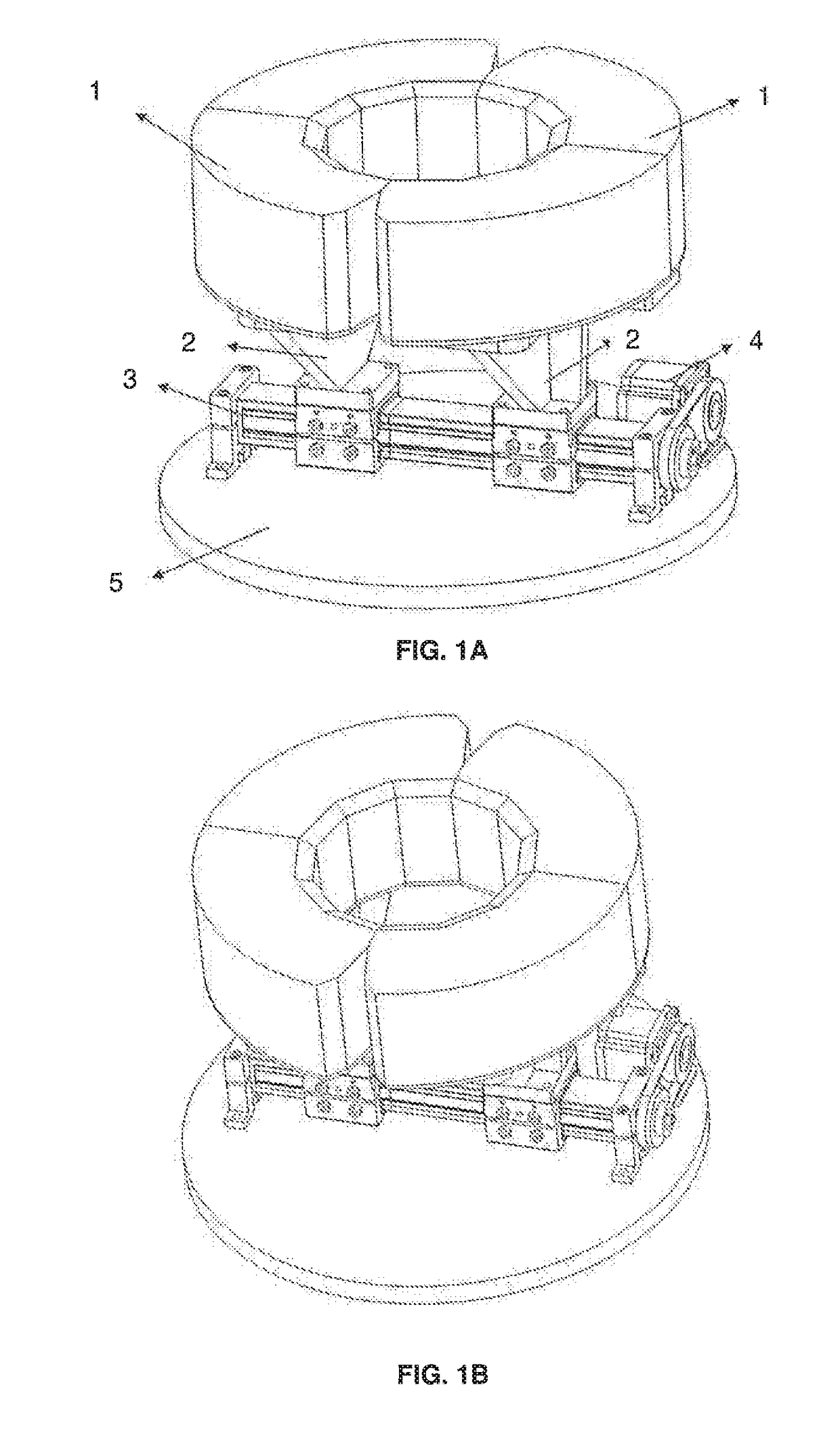
[0099]A particular embodiment of the Molecular Imaging System is a PET device as the one shown in FIG. 7, comprising two superposed sets of semi-rings of detector modules (two upper semi-rings and two lower semi-rings) with the same number of detector modules each semi-ring, preferably 6 detector modules each.

[0100]According to this specific embodiment a PET system is provided consisting of two rings with 12 modules each. Each module contains a single LYSO continuous scintillation crystal coupled to a PSPMT H8500 from Hamamatsu Photonics (Hamamatsu city, Japan) and an electronic readout board. The use of trapezoidal crystals reduces the image compression effect and improves energy, spatial, and depth of interaction (DOI) resolutions especially when considering truncation angles smaller than 60°. The Mobile Molecular Imaging System—here a PET with detector modules forming semi-rings, has an aperture of 186 mm.

[0101]The detector design uses 12 mm thick scintillation LYSO crystals whos...


PUM


Abstract

The present invention refers to a Mobile Molecular Imaging System, for example a PET (positron emission tomography) or SPECT (single photon emission computerized tomography) device, comprising:at least one group of n detector modules conforming detector module sets with a rounded shape comprising m detector modules each, being m<n and said detector module sets being separable from each other,first means to carry out the movement for separating the detector module sets in the transaxial planesecond means to carry out a rotation movement of the Imaging Systema Vertical Actuator that allows the axial movement of the Imaging System.and also refers to an Interventional System comprising the Mobile Molecular Imaging System and use of any of them in medical procedures, such as biopsy, radiotherapy, radiofrequency or ultrasound, among others.

Description

CROSS-REFERENCE TO RELATED APPLICATIONS[0001]This application is a continuation of and claims priority to PCT Application No. PCT / ES2017 / 070099, filed on Feb. 22, 2017, which, in turn, claims priority to Spanish Application No. P201630226, filed on Feb. 26, 2016. The entire contents of each of these applications is incorporated herein by reference.FIELD OF THE INVENTION[0002]The present invention is encompassed in the field of medical procedures using molecular imaging techniques and more particularly to interventional devices associated and guided by a molecular imaging system, such as a biopsy, radiofrequency, ultrasound or radiotherapy guided devices among others.BACKGROUND OF THE INVENTION[0003]Imaging techniques applied in medicine envision the inside of living organisms allowing to perform accurate and early diagnosis that substantially affect the strategies in therapy, what improves the outcome of treatment and reduces the mortality and morbidity of multiple diseases. Its sig...

Claims


Application Information

Patent Timeline
IPC IPC(8): A61B6/00A61B6/03G06T11/00A61B10/04A61N5/10A61N7/00A61B34/20
CPCA61B6/4405A61B6/037A61B6/4266G06T11/003A61B10/04A61N5/1001G01T1/2985A61B34/20A61B6/4476G06T2211/428A61N2005/1087A61N1/40A61N7/00A61B6/0414A61B6/4275A61B6/4452A61B6/4458A61B6/502A61N2005/1052
Inventor LAMO VALENZUELA, JORGECORRECHER SALVADOR, CARLOSBARBER BALLESTER, JULIOMART NEZ BENEDICTO, JOSE
Owner GENERAL EQUIP FOR MEDICAL IMAGING SL
Why Eureka
Try Eureka