Methods and devices for imaging large intact tissue samples
a tissue sample and large-scale technology, applied in the field of methods and devices for imaging large-scale intact tissue samples, can solve the problems of slow imaging speed of confocal microscopes and damage to the signal-emitting capabilities of samples
- Summary
- Abstract
- Description
- Claims
- Application Information
AI Technical Summary
Benefits of technology
Problems solved by technology
Method used
Image
Examples
example 1
Ultrafast Imaging of Whole Mouse Brain Using COLM
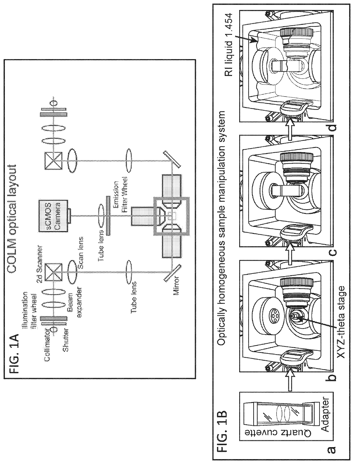
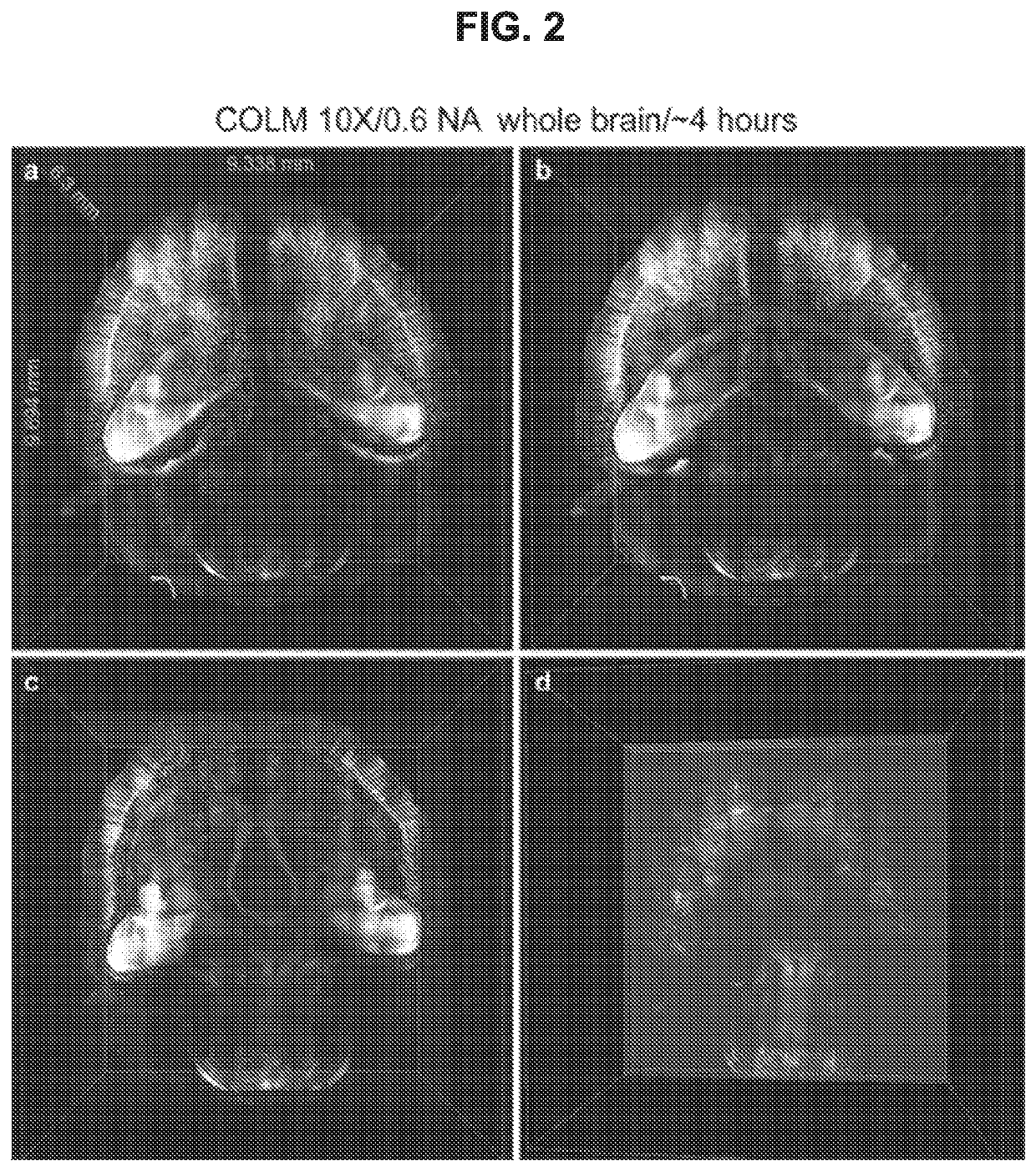
[0101]Thy1-eYFP mouse brain was perfused with 0.5% acrylamide monomer solution, and clarified passively at 37° C. with gentle shaking Camera exposure time of 20 ms was used, and the refractive index liquid 1.454 was used as immersion media. The entire dataset was acquired in ˜4 hours using a 10×, 0.6 NA objective. Internal details of the intact mouse brain volume were visualized by successive removal of occluding dorsal-side images (FIG. 2, Panels b, c and d). FIG. 10 Panels b,c and d present an example of imaging entire central nervous system (Brain and Spinal cord attached) using two independent detection arm COLM implementation.
example 2
Fast High-Resolution Imaging of Clarified Brain Using COLM
[0102]Thy1-eYFP mouse brain was perfused with 0.5% acrylamide monomer solution. A 3.15 mm×3.15 mm×5.3 mm volume was acquired from an intact clarified brain using COLM with 25× magnification. The complete image dataset was acquired in ˜1.5 hours; for optimal contrast the LUT of zoomed-in images was linearly adjusted between panels. Magnified views from FIG. 3 panel c region defined by rectangles were obtained (FIG. 3, Panels a, b). Maximum-intensity projections of over a 50 micron-thick volume were obtained (FIG. 3, Panels d-i, shown by the progression of boxes and arrows). Camera exposure time of 20 ms was used; refractive index liquid 1.454 was used as the immersion medium.
example 3
Molecular Interrogation of Clarified Tissue
[0103]Whole mouse brain was perfused with 4% acrylamide monomer solution and clarified passively, and immunostained to label all parvalbumin (PV) positive neurons. The intact brain was imaged using COLM with a 25× objective. Labeled cells at different depths in the sample were observed (FIG. 4B).
[0104]CLARITY optimized objectives were used to image clarified mouse brain tissue blocks with a confocal microscope (FIGS. 4A and C). The mouse brain was perfused with 4% acrylamide monomer solution and immunostained with anti-PV antibody. Confocal microscopy with CLARITY optimized water-immersion objectives (10×, 0.6 NA, 3 mm) was used to acquire high quality images of a 1 mm thick tissue block, and the images were processed to generate maximum intensity projection images of PV positive neurons (FIG. 4A).
[0105]It was also possible to perform multiple rounds of immunostaining and confocal imaging with CLARITY optimized water-immersion objectives (1...
PUM
| Property | Measurement | Unit |
|---|---|---|
| wavelength | aaaaa | aaaaa |
| wavelength | aaaaa | aaaaa |
| wavelength | aaaaa | aaaaa |
Abstract
Description
Claims
Application Information
 - R&D
- Intellectual Property
- Life Sciences
- Materials
- Tech Scout
- Unparalleled Data Quality
- Higher Quality Content
- 60% Fewer Hallucinations
Browse by: Latest US Patents, China's latest patents, Technical Efficacy Thesaurus, Application Domain, Technology Topic, Popular Technical Reports.
© 2025 PatSnap. All rights reserved.Legal|Privacy policy|Modern Slavery Act Transparency Statement|Sitemap|About US| Contact US: help@patsnap.com


