
Immunochromatography, and detection device and reagent for the same

a detection device and reagent technology, applied in the field of immunochromatography, can solve the problems of insufficient sensitivity of immunochromatography using such colored particles, and achieve the effects of reducing device cost, space saving, and improving the efficiency of immunochromatographic tests

Active Publication Date: 2018-07-31
FURUKAWA ELECTRIC CO LTD
View PDF39 Cites 0 Cited by
  • Summary
  • Abstract
  • Description
  • Claims
  • Application Information

AI Technical Summary

Benefits of technology

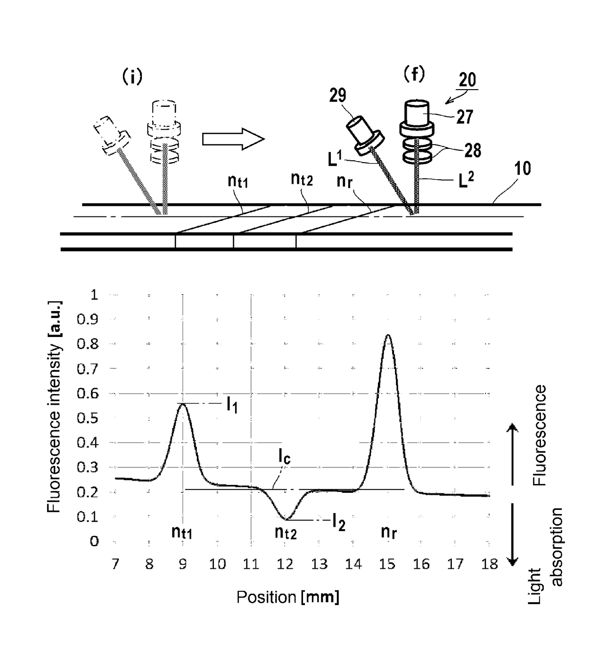
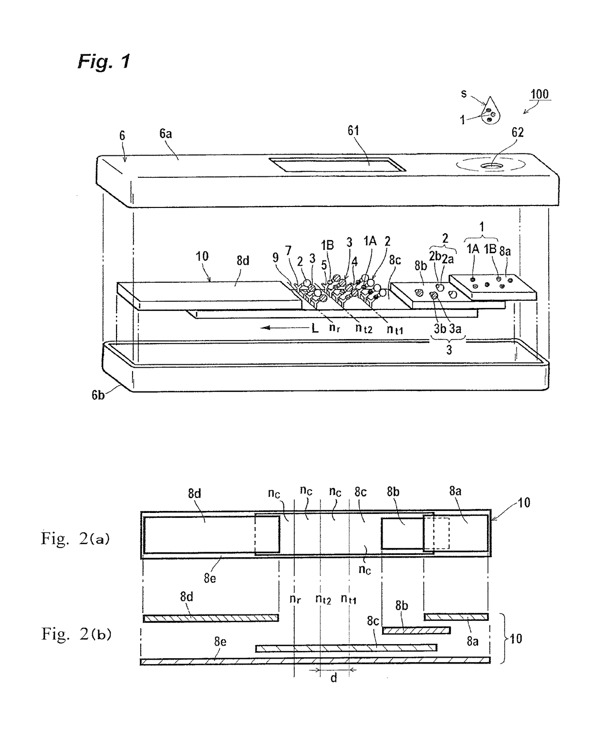
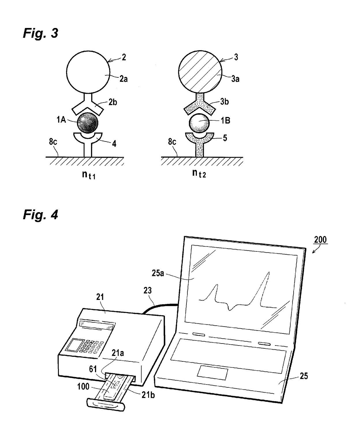
[0053]In immunochromatography using a combination of fluorescent particles and light absorbing particles for capturing different target substances, respectively, the immunochromatography and detection device of the present invention makes it possible to detect and measure the fluorescence and the light absorption at once. The detection and measurement of the fluorescence and the light absorption at once allows efficient detection and measurement of a plurality of target substances, which makes it possible to significantly improve the efficiency of immunochromatographic tests for diagnosis and treatment. The detection device of the present invention makes it possible to detect fluorescence and light absorption at once with a single detection unit, so that it can contribute to space saving and device cost reduction for immunochromatography.
[0054]In the immunochromatography of the present invention, silica particles or latex particles can be used as reagent particles (specifically fluorescent particles). Silica is a material having a light transmittance higher than that of latex particles. When silica particles are used for fluorescent particles, light from a light source can efficiently enter and pass through the inside of the silica particles, so that a dye in the silica particles can be efficiently excited. In the case of fluorescent particles, light can efficiently excite the dye in the silica particles. In the case of light absorbing particles, a dye in the silica particles can efficiently absorb light from a light source. Therefore, silica can produce more advantageous effects.
[0055]Other and further features and advantages of the invention will appear more fully from the following description, appropriately referring to the accompanying drawings.

Problems solved by technology

In some cases, the sensitivity of immunochromatography using such colored particles is insufficient, depending on the condition or amount of the analyte or the measurement item.
Moreover, the result may be false positive or false negative, which can be a cause of misdiagnosis and the like.

Method used


Image

  • Immunochromatography, and detection device and reagent for the same
  • Immunochromatography, and detection device and reagent for the same
  • Immunochromatography, and detection device and reagent for the same

Examples


preparation example 1 (

Preparation of Fluorescent Silica Nanoparticles)

[0149]2.9 mg of 5-(and-6)-carboxyrhodamine 6G.succinimidyl ester (trade name, manufactured by EMP Biotech GmbH) was dissolved in 1 mL of dimethyl formamide (DMF). Then, 1.3 μL of APS was added thereto and the reaction was carried out for 1 hour at room temperature (25° C.).

[0150]600 μL of the resulting reaction liquid was admixed with 140 mL of ethanol, 6.5 mL of TEOS, 35 mL of distilled water, and 15.5 mL of 28% by mass ammonia water, and the reaction was allowed to occur at room temperature for 24 hours.

[0151]The reaction solution was centrifuged at a gravitational acceleration of 15,000×g for 30 minutes, and the supernatant was removed. 4 mL of distilled water was added to the precipitated silica particles for dispersion, and the dispersion was centrifuged again at a gravitational acceleration of 15,000×g for 20 minutes. Further, this washing operation was repeated twice additionally, for removal of the unreacted TEOS, ammonia and o...

preparation example 2 (

Preparation of Complex Particles of Fluorescent Silica Particles and an Antibody)

[0152]To 100 μL of dispersion solution (dispersion medium: distilled water) of the fluorescent silica particles (average particle diameter; 197 nm) containing rhodamine 6G at a concentration of 5 mg / mL, which had been used in Preparation Example 1, 775 μL of distilled water, 100 μL of an aqueous solution of sodium alginate with a concentration of 10 mg / mL (weight average molecular weight: 70,000), and 25 μL of an aqueous ammonia solution with a concentration of 28% by weight were added. Then, the mixture was slowly stirred for 1 hour at room temperature (24° C.). The obtained colloid was subjected to centrifugation for 30 minutes with a gravitational acceleration of 12,000×g, and then the supernatant was removed. Distilled water (875 μL) was added thereto and the particles were re-dispersed. Subsequently, 100 μL of an aqueous solution of sodium alginate with a concentration of 10 mg / mL was added thereto...

preparation example 3 (

Preparation of Complex Particles of Gold Colloid Particles and an Antibody)

[0156]To 0.5 mL of gold colloid (particle diameter: 40 nm), 100 μL of 50 μg / mL antibody to nucleoprotein of influenza B (Anti-Human Influenza B, Monoclonal (Clone 9D6), manufactured by Takara Bio Inc.) was added, and then the mixture was allowed to stand at room temperature for 10 minutes. Subsequently, 100 μL of a phosphate buffer (pH 7.5) containing 1% by weight of bovine serum albumin was added, and the mixture was further allowed to stand at room temperature for 10 minutes. The mixture was then centrifuged at 8,000×G for 15 minutes, and the supernatant was removed. Then, 100 μL of a phosphate buffer (pH 7.5) containing 1% by weight of bovine serum albumin was added to disperse the particles.


PUM

PropertyMeasurementUnit
wavelengthaaaaaaaaaa
distanceaaaaaaaaaa
absorption wavelengthaaaaaaaaaa

Abstract

An immunochromatography for multi-item detection is provided which contains detecting and measuring fluorescence and light absorption respectively at once with a detection device. The immunochromatography contains using fluorescent particles and light absorbing particles, wherein the fluorescence excitation wavelength of the fluorescent particles and the absorption wavelength of the light absorbing particles are in the same wavelength region; and detecting and measuring, at once, the intensity of reflected light from a test area, the intensity of reflected light from another test area, and the intensity of reflected light from a non-test area other than the test areas.

Description

CROSS-REFERENCE TO RELATED APPLICATIONS[0001]This application is a Continuation of PCT International Application No. PCT / JP2013 / 081913 filed on Nov. 27, 2013, which claims priority under 35 U.S.C. § 119 (a) to Japanese Patent Application No. 2012-260227 filed on Nov. 28, 2012. Each of the above applications is hereby expressly incorporated by reference, in its entirety, into the present application.TECHNICAL FIELD[0002]The present invention relates to an immunochromatography, and a detection device and a reagent for the same.BACKGROUND ART[0003]An immunochromatography is a diagnostic method using nanoparticles. Being simple to operate and requiring relatively short time of approximately 5 to 30 minutes for determination and no expensive devices, it is widely used as an excellent simple diagnostic method in actual clinical scenes. For example, for determination of influenza virus infection, by having a pharynx cotton swab or nasal cavity cotton swab collected from a patient as an ana...

Claims


Application Information

Patent Timeline
Patent Type & Authority Patents(United States)
IPC IPC(8): G01N33/558G01N21/64G01N33/58G01N21/84G01N21/59G01N33/569B82Y30/00
CPCG01N33/56983G01N21/59G01N21/6428G01N21/8483G01N33/558G01N33/582G01N2469/10G01N21/6489G01N2021/6439G01N2021/8488G01N2333/08G01N2333/11B82Y30/00G01N21/78G01N33/543G01N33/54388
Inventor AIZAWA, HIDEKIOHKUBO, MICHIOMIYOSHI, KAZUTOMI
Owner FURUKAWA ELECTRIC CO LTD
Learn More