
Nuclear magnetic resonance machine requiring long waiting time between pre-pulses and imaging pulse train

A nuclear magnetic resonance, pre-pulse technology, used in the measurement of magnetic variables, medical science, diagnosis, etc.

Inactive Publication Date: 2004-11-03
TOSHIBA MEDICAL SYST CORP
View PDF0 Cites 10 Cited by
  • Summary
  • Abstract
  • Description
  • Claims
  • Application Information

AI Technical Summary

Problems solved by technology

[0013] For this reason, as mentioned above, only images during diastole can be obtained in conventional methods

Method used


Image

  • Nuclear magnetic resonance machine requiring long waiting time between pre-pulses and imaging pulse train
  • Nuclear magnetic resonance machine requiring long waiting time between pre-pulses and imaging pulse train
  • Nuclear magnetic resonance machine requiring long waiting time between pre-pulses and imaging pulse train

Examples


Embodiment Construction

[0036] Refer below Figure 4-6 and Figure 7A , 7B An embodiment of the present invention is described.

[0037] Figure 4 The structure of the nuclear magnetic resonance imaging apparatus of this embodiment is schematically shown.

[0038] The nuclear magnetic resonance imaging device includes: a lying part of the subject, on which the imaged object P (the imaged person) lies; a static magnetic field generating part for generating a static magnetic field; a gradient magnetic field generating part for It is used to add position signals to the static magnetic field; the sending / receiving part is used to send and receive high-frequency signals; the control and operation part is used to reliably control the whole system and image reconstruction; the electrocardiogram measurement part is used to measure as a representation an ECG (electrocardiogram) signal of a signal of the instantaneous phase of the heart of the imaging subject P; and a breath-hold command portion for instru...


PUM


Abstract

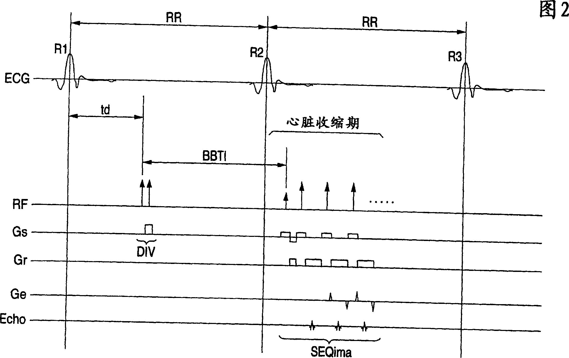
In the black blood method using a double inversion pulse, images in the systole of the cardiac cycle can be captured in a reliable manner even in the presence of a cycle-to-cycle variance in the heart beat cycle. A pulse sequence of the black blood method composed of a double inversion pulse DIV and an imaging pulse train SEQima is used. This sequence is applied in sync with an ECG signal of a subject to be imaged, and magnetic resonance imaging is thereby performed. The double inversion DIV is applied in sync with an R-wave:R1 appearing on the ECG signal at a given timing, with a first delay time td1 (fixed value), and the imaging pulse train SEQima is applied in sync with the following R-wave:R2 with a second delay time td2 (fixed value: set in accordance with the systole). A variance of the cardiac cycle is absorbed in an inversion time BBTI.

Description

technical field [0001] The present invention relates to a nuclear magnetic resonance machine (MRI) for imaging the interior of an object with an imaging sequence comprising a pre-pulse, and more particularly to MR imaging using an imaging sequence in which after the pre-pulse is applied, the imaging sequence is started Like the pulse train, there is a waiting time, and this waiting time is longer than the cardiac cycle as in the black blood method and the like. Background technique [0002] At present, nuclear magnetic resonance imaging is widely used as a medical imaging method. Magnetic resonance imaging is an imaging method that uses a high-frequency signal at the Larmor frequency to induce magnetic excitation of nuclear spins in an object to be imaged in a static magnetic field, and then uses the excitation-correlated The resulting MR signals are used to reconstruct an image of the interior of the imaged object. NMR machines include various types and are distinguished ...

Claims


Application Information

Patent Timeline
Patent Type & Authority Applications(China)
IPC IPC(8): A61B5/055G01R33/561G01R33/563G01R33/567
CPCA61B5/7285G01R33/56308G01R33/567A61B5/055G01R33/563
Inventor 金泽仁
Owner TOSHIBA MEDICAL SYST CORP
Learn More