Detection reagent and detection method for Zika virus
A detection method and virus technology are applied in the field of analysis and detection to achieve the effects of sensitive on-site detection and improved detection sensitivity
- Summary
- Abstract
- Description
- Claims
- Application Information
AI Technical Summary
Problems solved by technology
Method used
Image

Examples
Embodiment 1
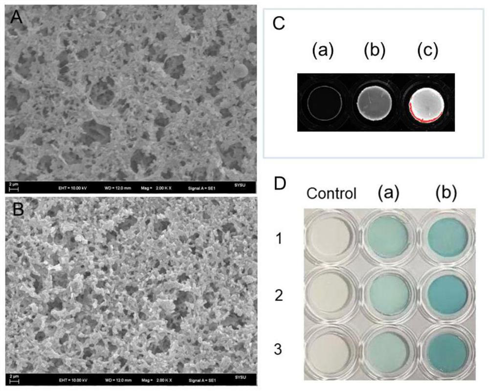
[0047] The construction of biotin-SA scaffold on the membrane of embodiment 1
[0048] The nylon membrane is first cut into circles using a custom steel die. The circular membranes were then rinsed three times in 0.1 M NaOH and deionized water, respectively. Subsequently, with NH 2 - The capped surface-modified membrane was gently shaken for 20 minutes in freshly prepared EDCI solution (150 mg / L) containing 15 mg / L biotin. Thus, biotin was immobilized on the membrane. Next, the biotin-labeled membrane was washed twice with deionized water and once with PBST, and incubated with SA (10 mg / L in PBST) for 30 minutes at 4°C with gentle shaking. After three washes with PBST to remove excess SA, the membrane was incubated with 40 μL cApt for 30 min at 4 °C. To reduce non-specific adsorption on the membrane, wash the membrane three times with PBST, then incubate with fatty acid-free BSA dissolved in PBST (1%, m / v) for 1 h at 4 °C, wash three times in PBST to remove residual blocki...
Embodiment 2
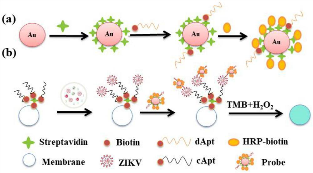
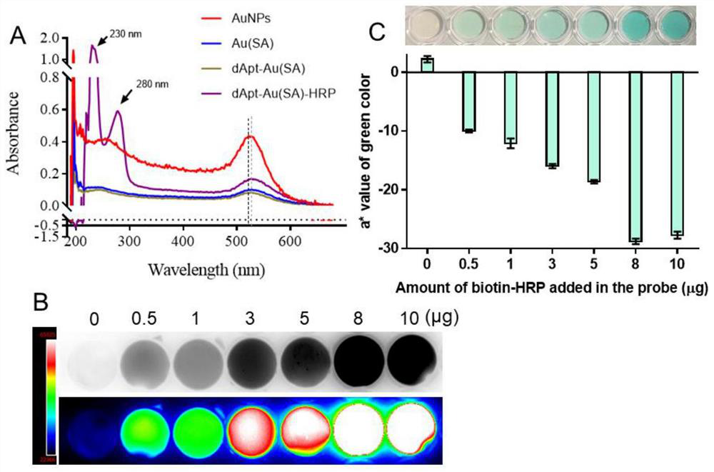
[0049] The preparation of embodiment 2 probe
[0050] The preparation of the probes can be briefly summarized as the surface of AuNPs was first immobilized with SA, where dApt and biotin-HRP molecules were immobilized sequentially.
[0051] First, 100 μL of SA (1 mg / mL) was added to 1 mL of AuNP, followed by gentle shaking at 4 °C for 1 h to immobilize SA molecules on the AuNP surface through electrostatic interaction. Then, the mixture was centrifuged at 8000 g for 10 min at 4°C to remove excess reagents and dissolved in deionized water (pH 7.5). Subsequently, 10 μL aliquots of dApt and 8 μL aliquots of biotin-HRP were added to the mixture and incubated at 4° C. for 30 minutes, respectively. Then, the mixture was centrifuged at 8000g for 10 minutes at 4°C to remove reagents. Finally, the centrifuged mixture was dissolved in deionized water (pH 7.5) containing 1% BSA (m / v) and stored at 4°C.
Embodiment 3
[0052] Example 3 Immunoassay and colorimetric reaction
[0053] The sensors prepared by the above method were incubated in PBST and real samples containing different concentrations of ZIKV-NS1 at room temperature for 30 min with slight shaking, and then rinsed three times with PBST. Subsequently, the antigen-conjugated sensor was incubated with the probe for 10 minutes, which was used as a detection probe to form a sandwich immunoassay. Finally, the reacted membrane-based sensor was rinsed three times with PBST and immersed in SA, TMB one solution (containing TMB and H 2 o 2 ) for 10 minutes for a sufficient colorimetric reaction. With the catalysis of the HRP loaded on the probe, the TMB molecule in the H 2 o 2 oxidized to TMB under the combined action of 2+ , indicating the presence of ZIKV-NS1.
PUM
 Abstract
Description
Claims
Application Information
 - R&D
- Intellectual Property
- Life Sciences
- Materials
- Tech Scout
- Unparalleled Data Quality
- Higher Quality Content
- 60% Fewer Hallucinations
Browse by: Latest US Patents, China's latest patents, Technical Efficacy Thesaurus, Application Domain, Technology Topic, Popular Technical Reports.
© 2025 PatSnap. All rights reserved.Legal|Privacy policy|Modern Slavery Act Transparency Statement|Sitemap|About US| Contact US: help@patsnap.com