Nuclear magnetic resonance image reconstruction method and terminal
A nuclear magnetic resonance image and terminal technology, which is applied in the field of medical image processing, can solve problems such as image artifacts and affect image quality, and achieve the effect of stable and highly robust nuclear magnetic resonance image reconstruction.
- Summary
- Abstract
- Description
- Claims
- Application Information
AI Technical Summary
Problems solved by technology
Method used
Image

Examples
Embodiment 1
[0079] Please refer to figure 1 , Embodiment 1 of the present invention is:
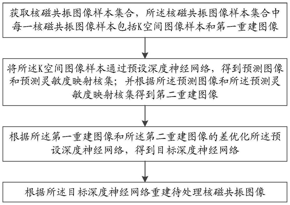
[0080] A nuclear magnetic resonance image reconstruction method, comprising steps:
[0081] S1. Acquire a nuclear magnetic resonance image sample set, where each nuclear magnetic resonance image sample in the nuclear magnetic resonance image sample set includes a K-space image sample and a first reconstructed image;
[0082] Wherein, the K-space image represents a frequency domain image, such as an image in the Fourier domain;
[0083] S2. Pass the K-space image sample through a preset deep neural network to obtain a predicted image and a predicted sensitivity map kernel set; and obtain a second reconstructed image according to the predicted image and the predicted sensitivity map kernel set, specifically:
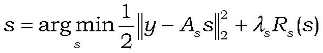
[0084] S21. Establish an overall optimization target according to the K-space image samples
[0085] Wherein, y represents the K-space image sample, A m Represents a linear operator compo...
Embodiment 2
[0103] A nuclear magnetic resonance image reconstruction method, which differs from Embodiment 1 in that S4 includes:
[0104] S41. Receive the nuclear magnetic resonance image to be processed, and perform preprocessing on the nuclear magnetic resonance image to be processed:
[0105] Pre-generate the sampling mask, the sampling mask is equivalent to a mask, and image data is processed within this range;
[0106]Obtain relevant parameters: MRI (Magnetic Resonance Imaging, nuclear magnetic resonance imaging) sample list index, number of MRI slices nums, number of MRI center slices center_slice_idx, downsampling factor, sampling mask, direction y; and predict sensitivity map maps;
[0107] Convert the data corresponding to the MRI image to be processed in the MRI sample list into a digitized tensor;
[0108] Separate slices and samples; map always starts counting from zero, and the initial value of the counting parameter count=0;
[0109] Load the MRI image to be processed;
...
Embodiment 3
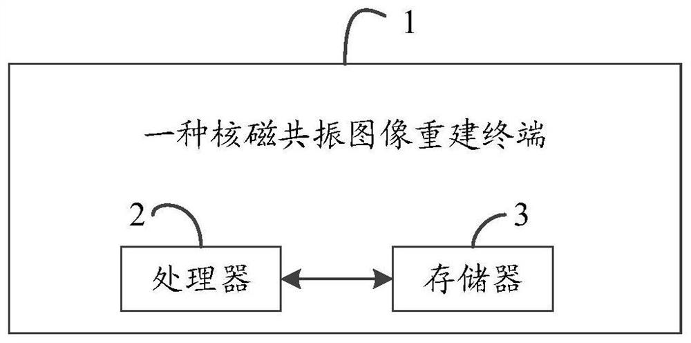
[0134] Please refer to figure 2 , Embodiment three of the present invention is:
[0135] A nuclear magnetic resonance image reconstruction terminal 1, comprising a processor 2, a memory 3, and a computer program stored on the memory 3 and operable on the processor 2, and the embodiment is realized when the processor 2 executes the computer program Each step in one.
[0136] To sum up, this application provides a method and terminal for nuclear magnetic resonance image reconstruction. Through a deep learning method, the reconstruction of nuclear magnetic resonance images is realized on the basis of unfolding and alternating minimization, and the robustness of the reconstruction process is enhanced. Interleaving a learnable model with an optimization step, the entire system is end-to-end trained with a supervised loss. Using a calibration-free approach to structure in parallel nuclear magnetic resonance (MRI) models, sensitivity maps are spatially varied smoothly and low-rank...
PUM
 Abstract
Description
Claims
Application Information
 - Generate Ideas
- Intellectual Property
- Life Sciences
- Materials
- Tech Scout
- Unparalleled Data Quality
- Higher Quality Content
- 60% Fewer Hallucinations
Browse by: Latest US Patents, China's latest patents, Technical Efficacy Thesaurus, Application Domain, Technology Topic, Popular Technical Reports.
© 2025 PatSnap. All rights reserved.Legal|Privacy policy|Modern Slavery Act Transparency Statement|Sitemap|About US| Contact US: help@patsnap.com