Medical radiography device for radiology department and otolaryngology department

A technology of ENT and radiology, applied in the field of ENT, can solve the problems that affect the work efficiency of medical staff, increase the workload of medical staff, and cannot rotate and adjust.

Inactive Publication Date: 2022-02-08
胡楠
View PDF0 Cites 0 Cited by
  • Summary
  • Abstract
  • Description
  • Claims
  • Application Information

AI Technical Summary

Problems solved by technology

[0003] However, the existing medical imaging devices for radiology and otolaryngology have poor protection performance and are not easy to adjust. During the inspection process, the device may be displaced or tilted due to external forces or other factors, which will cause the inspection data to appear. The...

Method used


Image

  • Medical radiography device for radiology department and otolaryngology department
  • Medical radiography device for radiology department and otolaryngology department
  • Medical radiography device for radiology department and otolaryngology department

Examples


Embodiment Construction

[0022] The following will clearly and completely describe the technical solutions in the embodiments of the present invention with reference to the accompanying drawings in the embodiments of the present invention. Obviously, the described embodiments are only some, not all, embodiments of the present invention. Based on the embodiments of the present invention, all other embodiments obtained by persons of ordinary skill in the art without making creative efforts belong to the protection scope of the present invention.

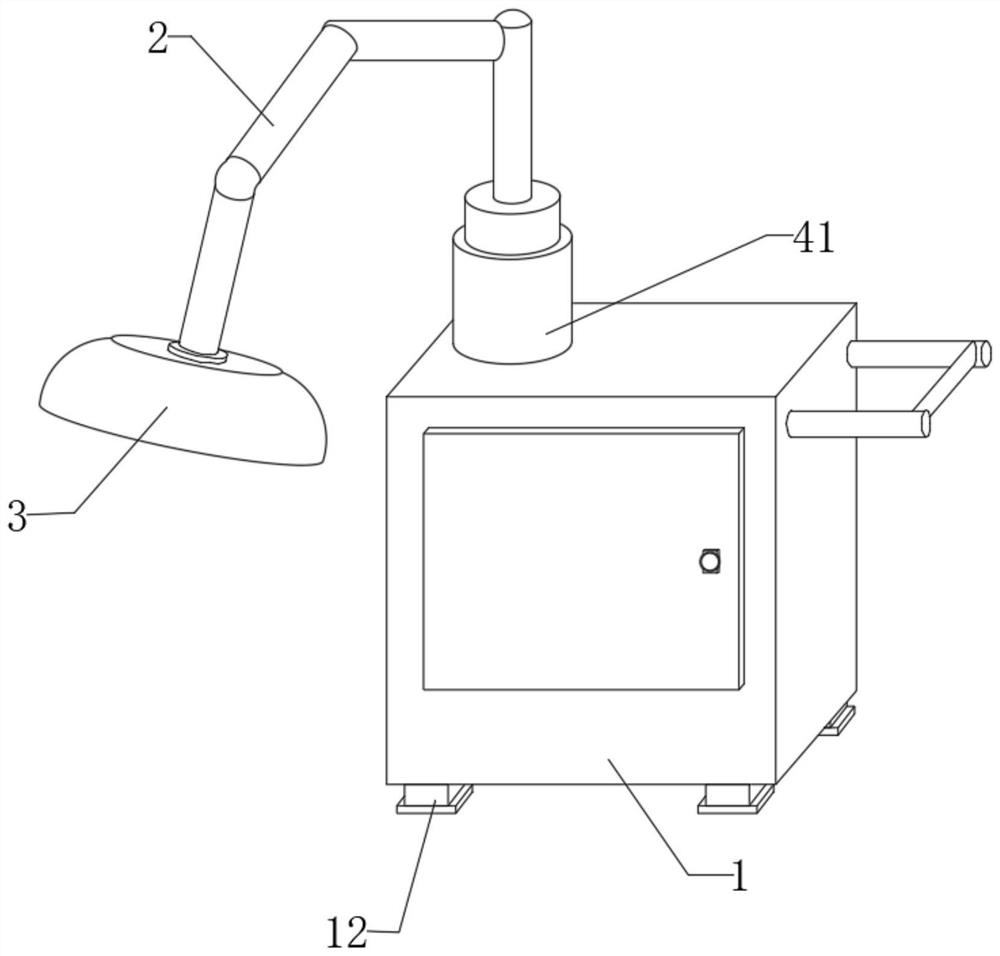
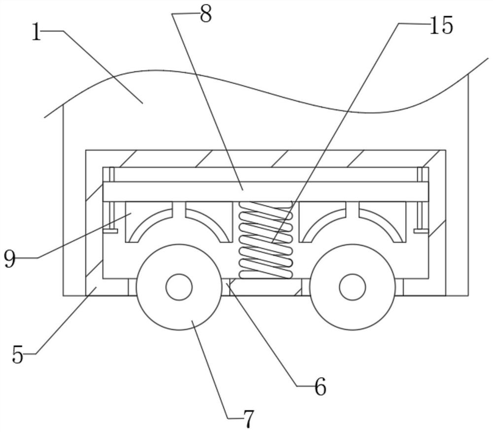
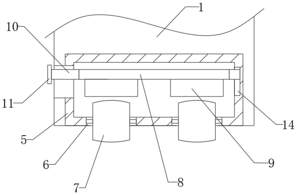
[0023] see Figure 1-6 , the present invention provides a technical solution of a medical imaging device for radiology and otolaryngology: it includes an equipment box 1, a mechanical arm 2 and an imaging equipment 3, and the left side of the top of the equipment box 1 is provided with an adjustment piece 4, and the adjustment piece 4 Including the fixed shell 41, the bottom of the inner cavity of the fixed shell 41 is provided with a deceleration micromotor 4...


PUM


Abstract

The invention discloses a medical radiography device for radiology department and otolaryngology department. The medical radiography device comprises an equipment box, a mechanical arm and radiography equipment, an adjusting part is arranged on the left side of the top of the equipment box, the adjusting part comprises a fixed shell. Before use, an inserting rod is moved out of a push plate, then a moving block is driven by the push plate to move upwards, the moving block drives the moving plate to move upwards, the moving plate drives arc-shaped clamping blocks to be separated from a rolling wheel to move upwards through assistance of a sliding sleeve, a sliding rod, a guide groove and a guide block, and a user pushes the equipment box through a grab rail and a handle to move the equipment box and the radiography equipment to a designated area. Then the arc-shaped clamping blocks are moved to the two sides of the rolling wheel through operation opposite to the steps, the two sides of the rolling wheel are fixed, and the situation that the radiography equipment cannot work normally, data errors are caused, and judgment of medical staff is affected due to the fact that the radiography equipment is displaced and inclined due to external force or other factors during working is prevented.

Description

technical field [0001] The invention relates to the related technical field of otolaryngology, in particular to a medical imaging device used in radiology and otolaryngology. Background technique [0002] "Otorhinolaryngology" is a surgical discipline that diagnoses and treats the ear, nose, pharynx, larynx, and related head and neck areas. Microsurgery, Ear Neurosurgery, Lateral Skull Base Surgery, Audiology and Balance Science, Nasal Endoscopic Surgery, Nasal Neurosurgery (Nasal Skull Base Surgery), Head and Neck Surgery, Laryngeal Microsurgery, Voice and Speech Diseases, Pediatric Otolaryngology The emergence of the Department of Throat and Throat has greatly enriched the content of the Department of Otorhinolaryngology. The Department of Otolaryngology needs to use some imaging devices to examine patients during diagnosis. [0003] However, the existing medical imaging devices for radiology and otolaryngology have poor protection performance and are not easy to adjust. ...

Claims


Application Information

Patent Timeline
IPC IPC(8): A61B6/00A61B6/10
CPCA61B6/4435A61B6/44A61B6/102
Inventor 胡楠肖子朋
Owner 胡楠
Why Eureka
Try Eureka